iMAgENS EM REUMATOLOgiA
ÓRgÃO OFiCiAL dA SOCiEdAdE PORTUgUESA dE REUMATOLOgiA
269
Exuberant plantar calcifications after
corticosteroid injections
ACTA REUMATOL PORT. 2016;41:269-270
IntRodUctIon
Plantar fasciitis is a common cause of heel pain, usual-ly a self-limiting condition. The aetiology of plantar fas-ciopathy is likely to be multifactorial, several risk fac-tors have been reported (anatomic, biomechanical and environmental), but also inflammatory rheumatic dis-eases and neurologic conditions must be consi dered1.
The studies showed that corticosteroid injections (CI) result in improvement of plantar fasciitis, redu cing the heel pain and plantar fascia thickness2. Plantar cal-cifications are a rare secondary consequence after CI while plantar fascia rupture and the heel fat pad atro-phy are the most feared complications, generally asso-ciated with multiple injections2. Plantar fascia rupture ranged from 2.4% to 6.7% in two retrospective studies, with an average of 2.67 vs 2.1CI in the sample2.
cAsE REpoRt
A 54-year-old woman was referred to the Physical Medicine and Rehabilitation department for plantar fasciitis, with 15 years of duration, refractory to physi -cal therapy, oral anti-inflammatory treatment and CI.
A previous plain X-ray (Figure 1-A), four years be-fore, revealed a heel spur. She had been submitted to six CI with betamethasone (5) and methylprednisolone acetate (1) suspensions, with poor clinical response.
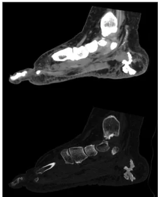
During the last two years the patient complained of increasing heel pain while walking and with prolonged weight bearing. A new X-ray (Figure 1-B) of her foot showed a calcification near the plantar fascia insertion and the computedtomography (Figure 2) an exube -rant conglomerate of calcifications in the medial aspect of plantar fat pad associated with subcutaneous
oede-Raposo A1, Santos R2, Matias C2
1. Serviço de Reumatologia, Unidade Local de Saúde do Alto Minho, Ponte de Lima;
2. Serviço de Medicina Física e de Reabilitação, Hospital de Braga
FIGURE 1.X-Ray image of A) 2009; and B) 2015, with a new
area of calcification near the plantar fascia insertion
FIGURE 2.Computed-tomography image with an exuberant conglomerate of calcifications in the medial aspect of plantar fat pad associated with subcutaneous oedema
A
ÓRgÃO OFiCiAL dA SOCiEdAdE PORTUgUESA dE REUMATOLOgiA
270
ExubErant plantar calcifications aftEr corticostEroid injEctions
coRREspondEncE to
Ana Raposo
Serviço de Reumatologia
Unidade Local de Saúde do Alto Minho, Ponte de Lima, Portugal
E-mail: [email protected]
REFEREncEs
1. Rosenbaum AJ, DiPreta JA, Misener D. Plantar heel pain. Med Clin North Am. 2014;98(2):339-352.
2. Ang TW. The effectiveness of corticosteroid injection in the treatment of plantar fasciitis. Singapore Med J. 2015;56(8):423--432.
3. Conti RJ, Shinder M. Soft tissue calcifications induced by local corticosteroid injection. J Foot Surg. 1991;30(1):34-37. 4. Fox TP, Oliver G, Wek C, Hester T. Plantar fascia calcification
a sequelae of corticosteroid injection in the treatment of recal-citrant plantar fasciitis. BMJ Case Rep. 2013;16;2013. 5. Raghavendran RR, Peart F, Grindulis KA. Subcutaneous
calci-fication following injection of triamcinolone hexacetonide for plantar fasciitis. Rheumatology (Oxford). 2008;47(12):1838.
ma suggesting steatonecrosis. The patient was also refrac tory to posterior extracorporeal shockwave thera py.
dIscUssIon
Very few case-reports published plantar calcifications following CI3-5. It was suggested that the accumulation of insoluble steroid acts as a foreign body and induces a chronic inflammatory process with subsequent cal-cification3.
Incorrect injection of corticosteroids can also in-duce necrosis and atrophy of the plantar fat pad, asso-ciated with increased morbility4.