Isolation of
Trichophyton mentogrophytes var mentogrophytes
from
naturally infected laboratory albino rats: experimental infection and
treatment in rabbits
N. A. Issa and I. K. Zangana
Department of Internal and Preventive Medicine, College of Veterinary Medicine, University of Dohuk, Dohuk, Iraq
(Received September 21, 2008; Accepted May 20, 2009)
Abstract
The present study demonstrated for the first time the occurrence of dermatophytosis in naturally infected rats and from asymptomatic and from breeding boxes of white rats kept in animal housing of college of Veterinary Medicine, University of Dohuk, Iraq. The prevalence rate of infection was (28%), clinically infected rats characterized by appearance of scaly ovoid type lesions with crusty edge and patch of hair loss mostly seen on the back, neck and face of the infected rats, itching was reported in some rats. Only one species of the trichophyton, T. mentogrophytes var mentogrophytes was isolated with growth rate (85.71%) of samples collected from clinically infected rats, and (28.57%) from asymptomatic and from breeding cages, the growth was observed within the 21 days at 25ºC on Sabouraud's Dextrose Agar. Lacto phenol cotton blue staining slides of
T. mentogrophytes var mentogrophytes revealed both microconidia and macroconidia. Microconidia found in numerous numbers often in dense cluster which were hyaline, smooth walled and predominantly spherical to sub spherical in shape, varying numbers of chlamydoconidia. Spiral hyphae and smooth, thin walled clavate shaped multicelled macroconidia were also present. The study also dealt with experimental infection in rabbits with T. mentogrophytes var mentogrophytes and treated by two drugs, natural herbal preparation of acidic pomegranate (Punica granatum) fruit and synthetic nystatine ointment. The complete recovery of lesions was recorded after 14 days and 21 days of topical application of a pomegranate and nystatine ointment for 5 successive days respectively.
Keywords: Dermatophytosis, Trichopyton mentogrophytes var mentogrophytes, Nystatin Available online at http://www.vetmedmosul.org/ijvs
Trichophyton mentogrophytes var mentogrophytes
!
"
#
$
% !$
&
! " #
$
%
%
& % '
(
% )
* + * ,
, -"
%
#
. / 0
# 1(
.&
2
3
("
$
45
6
( 7
) # 8*.
9
$
(
% )
' .
! - # 8.
% )
:
" ,
;
(
3
'- <.
=
%
>
Trichophyton
$
5?@?A
6
%
&
% #)
$
(
% )
45@?A
6
%
$
$ % )
%
. / 0
3
>B&
0
4C
<
4?
o
<
- ' $
' .$
$
< &.$
3
. > 8 ! - # 1(
#
# 1(
F
08 #
.
0
> 8
) %
-%
- ;
: 8
'
0
)
B
. #
%
)
B& #
0
-3
$
9.
G
("
#
B-
"
> '
0
.
?
. ! - <
# 8 %
9
9 .$
F
%
9
H
Punica granatum
I
- ( % . .$ <
@
+ 08
CJ
<
9 .$
< &.$ %
4C
% . .$ < &.$ % <
3
Introduction
Dermatophytosis is a fungal infection of the skin caused by dermatophytes-filamentous fungi (Keratiniphylic fungi) which have ability to invade the epidermis and keratinized structure derived from it such as hair or nails (1). They have a very wide range hosts, including rodents, rabbits, carnivores and people (2). It has been reported that animals housed in close proximity to each other for long periods and presence of infected debris in building account for both the higher incidence and the greater infection rates mainly in winter (3,4). The main causes of ringworm in white rats are: Trichophyton mentogrophytes (5). However other isolated that has been seen are T.ajello, T.schoenleini ,T.terrester, Microsporum gallinae,M.gypseum and M. cookie (5,6). The aims of present study include: comparing the efficacy between a natural prepared herbal treatment ointments and nystatine ointment in experimentally infected rabbits. Isolating and identifying of the common cause of dermatophytosis in laboratory white rats.
Materials and Methods
Animal sampling
The study was conducted on 75 small white rats kept in laboratory breeding boxes in the college of Veterinary Medicine, University of Dohuk. Twenty one samples were taken from rats showed skin lesions and seven samples from asymptomatic rats and seven from breeding boxes.
Skin scraping
Skin scarping was taken from edge of the lesions with a surgical blade and some hairs were taken from symptomatic rats by plucking them off with forceps. Direct microscopic and concentration methods examination were done by using methods described by the (7). Each positive sample by direct methods was cultured on mycobiotic agar containing Sabouraud's Dextrose Agar (65g/L), cycloheximid (0.5g/L),chloramphenicol (250mg/L), gentamycin (0.65 ml/L); yeast extracts (5g /L). All samples Incubated at 25 to 30 °C for at least three weeks.
To identify the growth of the pathogenic fungi, macroscopic and microscopic examination was performed daily then the time of appearance of the growth, colony morphology, and color, shape, size and colony reverse side
morphology were recorded. Microscopic examination of positive fungi cultures was done using the Lacto phenol cotton blue (LPCB) wet mount method as described by (8). For differentiation this species of the fungi from others, different cultures media were used as: cultivation on subouraud's dextrose agar with 5% of Nacl, 1% peptone agar ands urea agar (hydrolysis of urea).
Experimental infection
For experimental infection, nine rabbits one month age were used in this study, the area of the flank and shoulder of one side and the belly of the alternative side aseptically prepared, the areas were scarified by ten days subculture growth of T. mentogrophytes var mentogrophytes isolated from naturally infected white rats. Inoculation was carried out by a wet swab wiped from the surface of the culture and rubbed on the surface of the scarified areas. Daily inspection was carried out for detection of the appearance of the lesions, and two week later re isolation of the fungi was performed this method was described by (9,10).
Treatments
In this study two types of treatment were used for five successive days, the first one was a natural herbal preparation composed from (100 g of the Vaseline, 10 g of grinding powder of wall part (cortical wall) of acidic pomegranate (punica granatum) fruit and 10 g of aluminum hydroxide applied locally on the lesions of the right flanks, while the second one was a nystatine ointment (100000 IU/g) manufactured by HAYAT drug production CO. Baghdad – Iraq, which was applied on the shoulder area at the same side. While lesions at left side belly area was left without treatment to serve as control. All animals kept under observation following the treatment for one month.
Results
Clinical Signs
Fig 1: Scaly ovoid type lesions with crusty edge, mostly seen on the back, neck and face of the infected rats.
Diagnosis
All examined skin scraping samples which positive by direct and concentration methods were revealed the presence of small ectothrix spores of fungi arranged as longitudinal chains out site of the shaft of the hairs Fig 2.
Fig 2: Small ectothrix spores of fungi arranged as longitudinal chains out site of the shaft of the infected hair (1000x).
The cultured samples revealed that 18 (85.71%) of naturally infected rats and 2 (28.57%) of asymptomatic rats and 2 (28.57%) of samples collected from contaminated fomities were gave typical growth on Sabouraud's Dextrose Agar.
Only one species of trichphyton was isolated in this study from all collected samples which was Trichphyton mentogrophytes var mentogrophytes. The colony of this species appeared on cultured media after 21 days from cultivation at 25 ºC. The colonies found as flat, white to cream in color, with a powdery to granular surface, some cultures showed central folding or developed raised central tufts or polymorphic suede like to downy area. Reverse pigmentation is usually reddish- brown color Fig 3, 4.
Fig 3: Colony of T.mentogrophytes var mentogrophytes on Sabouraud's Dextrose Agar after 21 days at 25 ºC.
Fig 4: Reddish- brown color reverse side of T. mentogrophytes var mentogrophytes on Sabouraud's Dextrose Agar after 21 days at 25 ºC.
On Sabouraud's Dextrose Agar with 5% of Nacl, the colonies appeared after 14 days at 25 ºC as heaped with buff to brown in color with suede like surface texture and characteristically have very dark reddish brown reverse pigmentation Fig 5, 6.
Fig 6: Darkreddish brown pigmentation on reverse side of
T.mentogrophytes var mentogrophytes on Sabouraud's Dextrose Agar with 5% Nacl after 14 days at 25 ºC.
Colonies on 1% peptone agar revealed as flat cream colored powder to granular colony with no reverse pigment Fig 7, 8.
Fig 7: Flat cream colored powder Colony of
T.mentogrophytes var mentogrophytes on 1% peptone agar after 7 days at 25 ºC.
Fig 8: Reverse side of T.mentogrophytes var mentogrophytes on 1% peptone agar after 7 days at 25ºC revealed no pigmentation.
As well as the present study revealed that this specie was positive for hydrolysis of the urea with in 4 days as shown in figure 9.
Fig 9: Showed the growth of the fungi on urea agar after 4 days with hydrolysis of the urea.
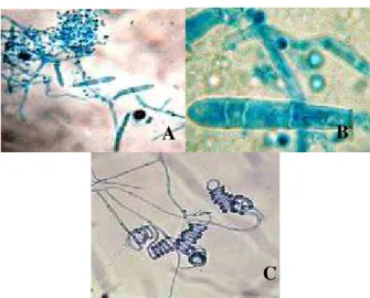
Microscopically, lacto phenol cotton blue staining slides of T.mentogrophytes var mentogrophytes revealed both microconidia and macroconidia. Microconidia found in numerous numbers often in dense cluster which are hyaline, smooth walled and predominantly spherical to sub spherical in shape with varing number of chlamydoconidia. Spiral hyphae and smooth, thin walled clavate shaped multicelled macroconidia were also revealed. As Fig 10.
Fig 10: A- Smooth, thin walled clavate shaped multicelled macroconidia with spherical Microconidia LPCB stain. (400X). B- Macroconidia of
T.mentogrophytes var mentogrophytes Slide stained with LPCB stain. (1000X). C- Spiral hyphae of T.mentogrophytes var mentogrophytes
Slide stained with LPCB stain. (400X).
Experimental infection
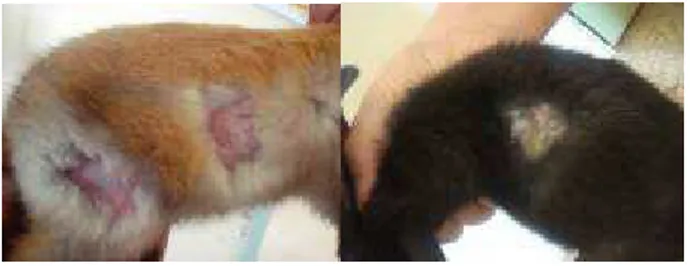
Experimentally infected rabbits with T. mentogrophytes var mentogrophytes had showed appearance of cutaneous lesion at the site of inoculation after 7 days of the infection. The lesions appeared as inflammatory sparse, erythematic, oedema, followed by scaly ovoid with crusty edge which mostly seen on the back, neck and face. Fig 11.
A
B
Fig 11: Cutaneous lesions of experimentally infected rabbit after 7 days from inoculation with T. mentogrophytes var mentogrophytes.
Treatment
The present study revealed that the lesions which treated with natural herbal preparation were effectively cured following the topical application of the treatment for five successive days and complete recovery were recorded after 14 days. While complete recoveries of lesions were recorded after 21 days from topical application of nystatin, but the control side remained infected during entire period of study.
Discussion
Most rodents infected with T.mentogrophytes are asymptomatic or have few clinical signs, in mice, partial or complete areas of alopecia, erythema, scale, and scab may be seen, often on the tails (2). In rats, the lesions are usually found on the back, neck and face of the infected animals which characterized by appearance of scaly ovoid type lesions with crusty edge, patch of hair loss. Mostly seen, itching was reported in some rats. Such of these signs were also reported by (4,6).
The prevalence rate of infection was (28%) in clinical cases and (28.57%) from asymptomatic and breeding cages respectively. While there are other studies with different rate of infection for instance Olga Fischman et al (11) reported dermatophytosis in only 3 cases out of 12 white rats in Brazil. Mackenzie (12) showed that 49(30%) out of 160 white rats had typical clinical signs of dermatophytosis in Belfast.
In this study T. mentogrophytes var mentogrophytes
was recorded for first time from natural infection of rats with typical clinical signs of infection and from asymptomatic rats and from breeding cages, while other species of trichophyton (T. mentogrophytes and T. verrucosum) were recorded in calves and sheep in Dohuk by Issa and Zangana (13).Where as Khalil(14) isolated (T. mentogrophytes, T, verrucosum, T.violaceum and icrosporum gallinae) from calves in Mosul governorate and AL-Ani et al (15) recorded the isolation of (T,verrucosum T. mentogrophytes T. terester, T.violaceum, microsporum nanum, M.audounii and M.distortum) from calves and
(M.equinum and T.equinum) from horses ringworm in Jordan. Gugnani et al (16) examined 215 samples of soil from burrows of rats, other sites in bamboo plantations in different part in India and Nepal among the pathogenic fungi recorded 14 of the 23 isolates of T. mentogrophytes var mentogrophytes. Dermatophytosis of animals in Romania are reviewed by Altears (17) ringworm infection have been found in cattle, horse, cat, dog, fowl and from laboratory animals guinea pigs T. mentogrophytes,M. audounii, T. rubrum, mice(T.quickneanum,T. mentogrophytes), rats T.mentogrophyts.
Generally the diagnosis of the disease depends on the cage observation which includes history and clinical signs. While laboratory diagnosis of the disease depend on the demonstration of the spores and hyphae of the fungi by direct microscopic examination and isolation of the fungi by different culture techniques. The growth was observed within the 21 days at 25ºC on Sabouraud's Dextrose Agar while Issa and zangan (13) showed that the growth of trichophyton species isolated from sheep and calves in Dohuk was observed following 25-30 days from cultivation on Sabouraud's Dextrose Agar at 25ºC.
On the urea agar the growth was observed following 4 days from cultivation with urea hydrolysis, and the fungus tolerates to grow on the Sabouraud's Dextrose Agar with 5% of Nacl. Also flaty cream colored powdery with no pigmentation at reverse side was observed on the 1% peptone agar culture, these result were in agreement with (18-20).
The growth rate was (85.71%) of samples collected from clinically infected rats, and (28.57%) from contaminated fomities and carrier animal respectively.
Microscopic examination of lacto phenol cotton blue staining slides of T.mentogrophytes var mentogrophytes
revealed both microconidia and macroconidia. Microconidia found in numerous number often in dense cluster which are hyaline, smooth walled and predominantly spherical to sub spherical in shape with varying number of chlamydoconidia, spiral hyphae and smooth, thin walled clavate shaped multicelled macroconidia were also present, and this in agreement with (7,19-22). The cutaneus lesion of the disease appeared with in 7 days following infection of the rabbits experimentally infected with T. mentogrophytes var mentogrophytes
isolated from naturally infected white rats.while (13) Reported the occurrence of the cutaneus lesions with in 19 days from inoculation of rabbits with isolated trichophyton species (T.mentogrophytes and T.verrucosum) from calves and sheep in Dohuk.. Al-Ani et al (15). Showed the occurrence of cutaneus lesion on the experimentally infected rabbits with Trichophyton species isolated from calves in Jordan with in 3 weeks from the infection, Narai
of rabbits with suspension of T.verrucosum and
T.mentogrophytes respectively.
In this study, the effective cure was observed following 14 days of topical application of the natural herbal preparation to the infected side for five successive days this due to potent anti oxidant property of the peel that attributed to their high content of polyphenols including punicalagin (24). While complete recovery of lesions were recorded after 21 days from topical application of nystatine. The control side remained infected during entire period this finding encouraged to using of the natural herbal drug in treatment of skin diseases. Sharma et al (25) recorded complete resolution of all lesions of ringworm due to
T.verrucosum in 13 calves was achieved within 12-14 days by daily application of aqueous extract of A.sativum in petroleum jelly (1:10). The treatment induced initial signs of local irritation, followed by an inflammatory reaction. As well as Sharma and Dwivedi (26) recorded complete cure within 12-15 days of daily application of a herbal preparation containing onion (Allium cepa), garlic
(A.sativum) and lemon (Citrus limon) extract and powders of turmeric (Curcuma longa) and camphor in Karanj oil (Pongamia glabra [P.pinnata]) to T.verrucosum in 12 cattle and T.verrucosum and M.canis in 21 dogs and no signs of toxicity in animals treated with the preparation.
Conclusion
We conclude that the T.mentogrophytes var mentogrophytes produced more severe form of dermatophtosis in experimentally infected rabbits, and natural herbal treatment gave the best result than the nystatin.
References
1. Chermette, R., Ferreiro, L., Guillot, J. Drmatophytosis in animals. Mycopathologia, Springer Netherlands, 2008; Accepted 30 January 2008 published on line 14 may 2008.
2. Laber- Lavid, K., Swindle, M.M., Flecknell, P. Hand book of rodent and rabbit medicine.1st edition. Pergamon. 1996; Pp 21-22.
3. Calderon, R. A. Immunoregulation of dermatophytosis. Crit. Rev. Microbiol. 1989; 16: 339-368.
4. Radostits.O. M., Gay. C. C., Blood, D.C., Hinchliff, K.W.Veterinary Medicine: a Text book of Diseases of Cattle, Sheep, Pigs, Goat, and Horses. 9th ed. W.B. Saunders Company Ltd, London. 2000; P.p.
1282-1285.
5. Gran, RN.C. Dermatophytosis. Alyman's Guide to the health and nursing care of rat. 2006; Available at enter net at http:// rat guide.com / Healthy. And at http://www.Marvistavate.com/html/body -ringworm/html.
6. Thomas, M., Donnelly, E.M., Rush and Petra, A. Ringworm in small exotic pets. Elsevier Inc. 2000; 9:82-83.
7. Cervantes Olivares, R.A.Ringworm infection in dogs and cats. In: Recent advances in canine infectious diseases [monograph online]. Carmichael L, editor. Ithaca NY: International Veterinary Information Serv ice [IVIS]. 2003; Available
at:http://www.ivis.org/advances/Infect_Dis_Carmichael/toc.asp. Accessed 30 July 2004.
8. Halley, L. D., Standard, P. G. Laboratory Methods in Medical Mycology, 3rd Ed. U S Department of Health, Education and Welfare,
Center of Disease Control, Atlanta.1973; pp 41-57. 9. Schmitt, J. A., Miller, R. G. Variation in susceptibility to
experimental dermatomycosis in genetic strains of mice. Mycopathologia.1967; 32: 306-312.
10. Mcginnis, M. R. Current Topics in Medical Mycology, Vol. 1, Springer-Verlag, New York. 1988; pp. 261-265.
11. Olga Fischman, Z.P., De Camargo., Grinblat, M. Trichophyton mentogrophytes infection in laboratory white mice,
Mycopathologia.Springer Netherlands. 2004; 59:2.P 113-115. 12. Mackenzie, D.WR.Trichophyton mentogrophytes in mice:
Infection in humans and incidence amongst laboratory animals. Medical mycology. 1962; 1(3): Pp 178-182.
13. Issa, N, A., Zangan, I.K. Clinical and some laboratory studies on ringworm in calves and sheep in Dohuk area. Journal of Dohuk University. 2008; 10(2).Pp. 200-205.
14. Khalil, Z.K. Clinical and therapeutical study of some skin diseases in fattening calves in Mosul. MSc. Thesis in Veterinary Internal and preventive Medicine. College of Vet. Medicine/ University of Mosul.2004.
15. Al-Ani F. K., Younes, F. A., Al-Rawashdeh O. F. Ringworm Infection in Cattle and Horses in Jordan. Acta Vet. Brno. 2002; 71: 55-60.
16. Gugnani, C.h., Paliwat-Joshi, A., Rahman, H., Padhe, A.A., Singh, T.S.K., Khanal, B., Bajaj, R., Rao, S., and Chukhani, R. Occurrence of pathogenic fungi in soil of burrows of rats and other site in bamboo plantation in India and Nepal. Mycosis. 2007; 50 (6). Pp507- 511.
17. Altears, I.A short review on dermatophytes of animals in Romania. Journal Mycopathologia. 1971; 43(1). Pp.17- 23. 18. Kane, J and Fischer, J.B. The effect of sodium chloride on the
growth and morphology of dermatophytes and some other keratolytic fungi. Can J microbial. 1975; 21(6): 742-749. 19. Rebell, G and Taplin, D (1970). The dermatophytes. 2nd revised
edition. University of Miami Press, Coral Gables, Florida.USA. 20. Rippon, J.W. Medical Mycology.3rd Edition. Saunders
Co.Philadelphia, USA.1988.
21. Quinn, P.J., Carter, M. E., Markey, B., and Carter, G.R.Clinical Veterinary Microbiology. Section 3, Mycology, by Elsevier Limited. Dermatophytes. 2004; pp. 381-390.
22. Carter.G.R, and Chengappa. M.M..Essentials of veterinary Bacteriology and Mycology. 4th ed. Lea & Febiger, Philadelphia..
1991; Pp.253-256.
23. Narai, K., Nuner, E., Rodriguez, H., Gonzalez, M., Albari, V. Sarkisov, K. A. Use of ringworm vaccine „LTF-130“ in the Republic of Cuba. Veterinariya 1988; 6: 61-62.
24. Gil, M.I., Tomas-Barberan, F.A., Hess-pierce, B., Hoi-croft, D.M. and Kader A.A. Antioxidant activity of pomegranate juice and its relationship with phenolic composition and processing, J. Agric. food
Chem.48(2000)4581-4589.
25. Sharma, S. R,., Dakshinkar, N. P., Dhoot, V. M. and Sapre, V. A. (1993) Evaluation of crude extract of garlic (Allium sativum Linn) in bovine dermatophytosis. Indian J, of Vet. Med. 1993. 13: (2), 72-73. 26. Sharma, M. C. Dwivedi, S. K. (1990). Efficacy of a herbal drug