online | memorias.ioc.fiocruz.br
Mir-190b negatively contributes to the Trypanosoma cruzi-infected
cell survival by repressing PTEN protein expression
Cíntia Júnia Monteiro1, Suianne Letícia Antunes Mota1, Lívia de Figueiredo Diniz1, Maria Terezinha Bahia1, Karen CM Moraes2/+
1Universidade Federal de Ouro Preto, Instituto de Ciências Exatas e Biológicas, Núcleo de Pesquisa em Ciências Biológicas,
Departamento de Ciências Biológicas, Laboratório de Doença de Chagas, Ouro Preto, MG, Brasil 2Universidade Estadual Paulista
Júlio de Mesquita Filho, Instituto de Biociências, Departamento de Biologia, Laboratório de Biologia Molecular, Rio Claro, SP, Brasil
Chagas disease, which is caused by the intracellular protozoan Trypanosoma cruzi, is a serious health prob-lem in Latin America. The heart is one of the major organs affected by this parasitic infection. The pathogenesis of tissue remodelling, particularly regarding cardiomyocyte behaviour after parasite infection, and the molecular mechanisms that occur immediately following parasite entry into host cells are not yet completely understood. Previous studies have reported that the establishment of parasitism is connected to the activation of the phospha-tidylinositol-3 kinase (PI3K), which controls important steps in cellular metabolism by regulating the production of the second messenger phosphatidylinositol-3,4,5-trisphosphate. Particularly, the tumour suppressor PTEN is a negative regulator of PI3K signalling. However, mechanistic details of the modulatory activity of PTEN on Chagas disease have not been elucidated. To address this question, H9c2 cells were infected with T. cruzi Ber-enice 62 strain and the expression of a specific set of microRNAs (miRNAs) were investigated. Our cellular model demonstrated that miRNA-190b is correlated to the decrease of cellular viability rates by negatively modulating PTEN protein expression in T. cruzi-infected cells.
Key words: cardiac cellular model - microRNA - PTEN - Trypanosoma cruzi
doi: 10.1590/0074-02760150184
Financial support: FAPEMIG (APQ-02351-10, CBB-APQ-01700-11), CNPq (475586/2009-3), UFOP
+ Corresponding author: [email protected] Received 12 May 2015
Accepted 18 November 2015
Chagas disease, which is caused by the intracellu-lar protozoan pathogen Trypanosoma cruzi, is a leading cause of cardiomyopathy and heart failure in Latin Amer-ica. More than eight million people are estimated to be affected by this parasite and thousands of others are at risk of infection (WHO 2013). T. cruzi induces multiple responses in the heart, which is a critical organ that is in-volved in infection and host pathology (Zhang & Tarleton 1999). The initial cardiomyocyte response to T. cruzi is fundamental to establish the heart infection and is depen-dent on the activation of host cell signalling pathways that involve the immediate activation of kinases and phospha-tases as well as changes in gene expression patterns (Bur-leigh & Woolsey 2002,Burleigh & Soldati 2008). The complexity of T. cruzi cellular invasion and survival has been considered a challenge. The understanding of such molecular mechanisms could contribute to the develop-ment of new therapies, because curing this disease is pos-sible if treatment is initiated soon after infection.
Technologies™, Brazil.
Parasite infection - T. cruzi (Be-62 strain, discrete typing unit II) was propagated in Vero cell monolayers (ATCC: CCL-81) in DMEM with 2% FBS and infective trypomastigotes were harvested as described previous-ly (Petersen & Burleigh 2003). Next, 1.5 x 10⁷ parasites were incubated with 1.5 x 10⁶ H9c2 cells for 2 h at 37ºC under 5% CO2 to allow parasite-cell interaction. The re-maining extracellular parasites were aspirated and the cells were extensively washed with phosphate-buffered saline (PBS) (2.7 mM KCl, 1.5 mM KH2PO4, 137 mM NaCl, and 8 mM Na2HPO4, pH 7.4); after which, fresh medium was added to the culture. Flasks containing in-fected cell cultures were immediately collected (0 h) or incubated for 2 h, 6 h, 12 h, 24 h, or 48 h, at which time-points, cells were collected. Uninfected cells were used as the control condition in our assays.
Cellular viability assays and nucleus immunostaining - Cellular viability was verified in assays using 1.5 x 10⁴ cells that had previously been grown in a 96-well plate. After 2 h of T. cruzi interaction, as previously described, cells were grown for up to 48 h and cellular viability was determined using methylthiazolyl diphenyl tetrazolium bromide in (2-methoxy-4-nitro-5-sulphophenyl)-2H-tetrazolium-5-carboxanilide (MTT) assay at specific time-points (Mosmann 1983). The plates were read at 570 nm using a microplate reader (Packard Instrument Company Inc, USA). In addition, to verify the effect of PTEN protein inhibition on cell viability, SF1670 (Sig-ma-Aldrich, USA) was added to cell cultures at a final concentration of 500 nM (Li et al. 2011). The cultures were incubated for up to 48 h and fresh media containing PTEN inhibitor was replaced at 24 h. Cellular viability was also determined at specific time-points and to veri-fy the physiological effect of such inhibitor in the H9c2 cells at the very early time-point, SF1670 was added to them and subsequently incubated at 37ºC for 30 min (Li et al. 2011). For nucleus staining and microscopy analy-ses, 1.5 x 10⁴ cellswere grown on coverslips and fixed in 3.8% paraformaldehyde containing 0.2% Triton X-100 (Sigma-Aldrich) for 7 min at 37ºC. Next, the nuclei were counterstained in a solution of 3.33 ng/mL of DAPI (Sigma- Aldrich) and subjected to microscopic analysis. Images were obtained with a Leica DMLB photomicro-scope equipped with an HBO 100 W mercury lamp and
ysed by the 2-ΔΔCT method. For miRNA analyses, cDNA was prepared using Mini Script Reverse Transcription (Qiagen, Germany), which contains a special stem-loop primer for miRNAs. qPCRs were also performed us-ing specific set of primers for miR-16-5p, miR-let7f-2-3p, miR-26b, miR-3586-miR-let7f-2-3p, miR-190b, and the miScript SYBR Green PCR Kit (Qiagen) after extensive analy-ses of the 3’-UTR sequence of the Pten RNA sequence (Gene ID: 50557) from Rattus norvegicus. For that, miRBase search tool (mirbase.org) was used and puta-tive miRNAs binding sequences were found in the anal-ysed sequence. The 2-ΔΔCT relative quantification method was applied and U6 was used as the internal control.
Western blot (WB) analysis - Whole cell extracts were prepared according to Sambrook et al. (2000). Equal amounts of protein (50 µg) were separated by elec-trophoresis in 10% polyacrylamide gels and then electro-transferred to polyvinylidene fluoride membranes. The membranes were immunoblotted overnight with rabbit AKT and phospho AKT, PTEN, and anti-phospho-PTEN (P-PTEN), or rabbit anti-β actin (Cell Signaling, USA) polyclonal antibodies, followed by 2 h of incubation with a horseradish peroxidase-conjugated goat anti-rabbit antibody (Cayman Chemical, USA). Im-munoreactive bands were visualised using a chemilumi-nescent detection kit (ECLTM; GE Healthcare, UK) and exposed to hyperfilm (GE Healthcare). The bands were quantified with Quantit One Software (Bio-Rad).
luciferase activity was determined by normalisation with Renilla Luciferase activity and they were measured using TD20/20 luminometer (Turner Designs).
miRNA inhibitor transfection and functional activ-ity of miRNAs - The functional activity of miRNAs, 3 x 10⁴ cells were transiently transfected with 30 nM of the designed rno-miR-190b inhibitor or the miRNA in-hibitor negative control as previous described. After 24 h, cells were harvested for protein analyses and qPCR. In addition, the infection rates of rno-miR-190b inhibi-tor transfected-cells were investigated. For that, after 24 h of cellular transient transfection, 1.5 x 10⁶ cells were incubated with 1.5 x 10⁷T. cruzi parasites to allow para-site-cell interaction for 2 h, as previously described. The viability was measured after 48 h post-infection (p.i.) in MTT assays. Uninfected and rno-miR190b inhibitor transient transfected-cells were used as control.
Graphs and statistical analyses - Values from three independent assays were used for the analyses and graphs were generated using Graph Pad Prism® 5. The differences between the control and infected groups were also measured using one-way analysis of variance followed by Dunnett’s test. Significance was set at *p < 0.05, **p < 0.01, and ***p < 0.001.
RESULTS
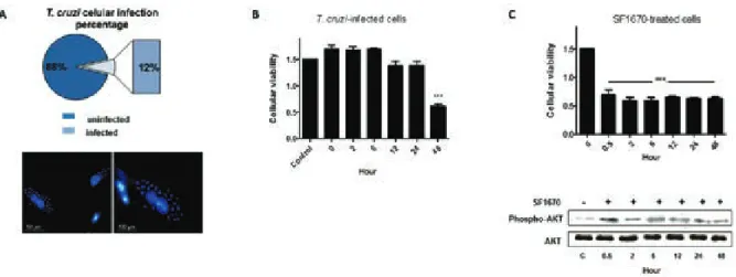
T. cruzi invasion and survival in host cells are criti-cal steps in the establishment of infection. Based on these observations, we first investigated the efficiency of the parasite infectivity in our in vitro model. After 2 h of parasite-host cell interaction, approximately 12% of cells were infected. The infection was also analysed 48 h p.i. via fluorescence microscopy (Fig. 1A). Next, cellular
viability of infected cells was examined at precise time-points after parasitic interaction (0 h, 2 h, 6 h, 12 h, 24 h, and 48 h), and uninfected cells were used as control in the assays. A 58.4% reduction in cellular viability at 48 h p.i. was observed (Fig. 1B). In addition, uninfected cells were treated with PTEN specific inhibitor (SF1670) and a reduction in cellular viability by ~50% (Fig. 1C) was observed, even after 30 min of PTEN activity inhibition, suggesting that the protein negatively modulates H9c2 cellular viability. The additional WB analyses reinforced the inhibitory effect of the drug on PTEN activity, which indirectly stimulates the phosphorylation of AKT pro-tein (Fig. 1C). The inhibition of PTEN activity favours the accumulation of PIP3 in cell membrane, which binds to AKT in its pleckstrin homology domain (Haugh et al. 2000, Schmid et al. 2004, Li et al. 2011). Only the AKT molecules on the plasma membrane can be phosphory-lated by protein kinases and become activated (Li et al. 2011). The analyses demonstrate that in the presence of SF1670, the phosphorylated AKT level considerably in-creased when compared to the untreated cells.
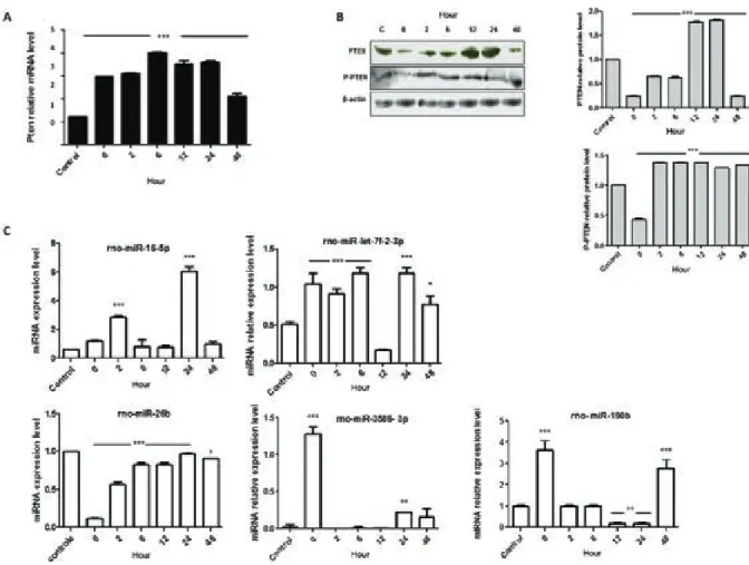
To investigate how PTEN could be possibly con-nected with the changes in cellular viability, mRNA expression and WB analyses were performed. In this acute cell culture model of infection, Pten mRNA sig-nificantly increased its expression level after cellular infection, reaching approximately 300% higher levels after 6 h of parasite infection (Fig. 2A). However, this mRNA expression pattern in T. cruzi-infected cells did not match with the PTEN protein expression. The WB analyses demonstrated that the PTEN protein levels decreased immediately after parasitic entry (0 h time-point) and increased considerably at 12 h and 24 h p.i., 77% and 81.09%, respectively, higher levels than the
Fig. 1: biological effects of the Trypanosoma cruzi strain Berenice-62 infection in H9c2 cellular survival. Cells were grown and infected with the parasite and the initial 48 h after parasitic infection were investigated. A: infected cells were analysed via fluorescence microscopy; the nuclear
material was labelled with DAPI (Bar = 50 μm and 100 μm). The representative rates of infected and uninfected cells are demonstrated in the
control. Moreover, despite the reduced functional activ-ity in cellular system, the phosphorylated form of PTEN was investigated (Fig. 2B). A reduced level of the phos-phorylated protein was also observed at 0 h p.i. (~57% lower than the control level) (Fig. 2B) and then its level increased its level to an average rate of 35% in the suc-ceeding time-points. However, the absolute quantifica-tion of PTEN forms (data not shown) demonstrated a reduced phosphorylated form of the protein when com-pared to the nonphosphorylated form.
Next, to describe the fine-tuning that controls gene expression, five miRNAs that possibly modulate PTEN expression were investigated (Fig. 2C). The results sug-gested a potential connection between the miRNA-190b and PTEN protein expression pattern, considering a nega-tive inverse correlation between the above two molecules. While the mRNA levels of Pten were found at higher lev-els in infected cells, the rno-miR-190b oscillated. Imme-diately after parasite entry into host cells, the miR-190b increased ~360%, returning to near basal expression level
in the next two investigated time-points (2 h and 6 h). Next, the expression pattern of miR-190b at 12 h and 24 h time-point was almost abolished, which is an inversed correlation to the PTEN protein expression pattern at such time-points. At 48 h time-point, the miR-190b presented a higher expression level that was also an inversed correla-tion to the expression pattern of PTEN protein.
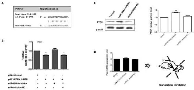
To confirm the mechanistic correlation between the miRNA-190b and PTEN, the potential target site at the 3’-UTR of Pten mRNA (514-528 bp) and the miRNA-190b were cloned and used in cellular transfection. The potential target site at the 3’-UTR of Pten mRNA and the miRNA-190b expression pattern is demonstrated in Fig. 3A. Next, luciferase assay followed by WB and qPCR analyses were performed. For luciferase assays (Fig. 3B), transfected pGL3-Control cells produced basal luciferase activity and the plasmid was used to maintain an equiva-lent amount of DNA inside the cells. On the other hand, the presence of pGL3-PTEN in H9c2 cells demonstrated the modulatory effect of the 3’-UTR sequence of PTEN
Fig. 2: modulatory effect of Trypanosoma cruzi in cellular metabolism of H9c2-infected cells. A: quantitative polymerase chain reaction
analy-ses of Pten mRNA; B: Western blots analyanaly-ses and quantification of PTEN, phospho-PTEN (P-PTEN), and the reference protein β-actin; C:
in the luciferase production. A reduction of ~22% lucifer-ase production was observed when compared with the ex-pression level of the protein by the control vector (pGL3-Control). In addition, the presence of the miR-190b inhibitor or its negative control reinforces the existence of a physical interaction between this miRNA and the Pten 3’-UTR. When cells were co-transfected with the pGL3-PTEN along with the specific inhibitor, the expression level of luciferase returned to near the basal level after 24 h transfection. The physical binding between the miR-inhibitor and miR-190b molecules decreased the small interfering RNA activity on PTEN-3’-UTR mRNA and facilitated the production of luciferase (Fig. 3). In addi-tion, the co-transfection assays performed with the miR-inhibitor negative control did not modulate the pattern of luciferase production in pGL3-PTEN-transfected cells.
Next, the effects of miRNA-190b inhibitor and its negative control were tested in WB and qPCR assays. The results demonstrated that the presence of specific miRNA-190b inhibitor increased the protein level by 40%, and the miRNA inhibitor negative control did not change the PTEN protein level when compared to the nontransfected culture in the 24 h investigation (Fig. 3C). The qPCR analyses reinforced the observation that the miRNA-190b blocks the Pten mRNA transla-tion (Fig. 3C), considering the presence of very small changes in Pten mRNA pattern of expression under the investigated conditions.
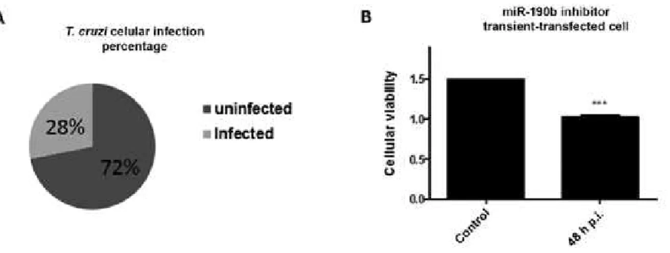
Finally, to evaluate the physiological effect of the miRNA-190b inhibitor in T. cruzi infection of H9c2 cells, transient transfected-cells were infected with the
parasite and infection rate and cellular viability were also measured. Fig. 4 presented the results and the pres-ence of miR inhibitor facilitated entry of the parasite, which raised the percentage of infected cells (Fig. 4A). An average infection rate of ~28% was observed in the three independent assays performed. Moreover, cellular viability measured in MTT assays also increased in the miRNA-190b inhibitor transient transfected-cells, when they were infected with the parasite. At 48 h time-point (Fig. 4B) p.i., the viability rate reduced ~31.8% when compared to the uninfected and transient transfected-cells with the miR inhibitor. The combined results re-inforced the negative functional activity of the miRNA-190b in T. cruzi-infected cells survival.
DISCUSSION
Several studies have demonstrated that T. cruzi induc-es multiple rinduc-esponsinduc-es in the heart that are necinduc-essary for the successful establishment of infection (Burleigh 2005, Yoshida 2006, Mott et al. 2009); however, the mechanis-tic understanding of this pathological process remains the focus of scientific investigations. In this study, we inves-tigated PTEN in T. cruzi Be-62-infected H9c2 cells, con-sidering its direct connection in controlling the AKT/pKB molecular pathway, which modulates cellular growth, hy-pertrophy, and even death of infected cells.
To understand the modulatory effect of the parasite in cellular survival during the onset of infection, cellu-lar viability was measured and considerable reduction in it was observed at 48 h time-point (Fig. 1B). PTEN was addressed as one important element correlated with
Fig. 3: molecular interplay between PTEN and miRNA-190b. A: schematic representation of the 3’-UTR sequence of Pten mRNA and the potential target region of the rno-miRNA-190b; B: relative luciferase activity in transfected cells with pGL3-Control and pGL3-PTEN-3’-UTR construct along with miRNA inhibitors. The results were plotted on representative graphs. The empty expression vector (pGL3-Control) was used to maintain an equivalent amount of DNA inside the cells and to express a basal level of luciferase; C: Western blots analyses and quantification of PTEN and the
Fig. 4: biological effects of the Trypanosoma cruzi strain Berenice-62 infection in H9c2 miRNA-190b inhibitor transient transfected. Cells were grown, transient transfected and infected with the parasite. A: infected cells were counted and the percentage rates of infected and uninfected cells are demonstrated in the circle; B: cellular viability assays of miRNA-190b inhibitor transient transfected-cells infected or uninfected with the T. cruzi after 48 h post-infection (p.i.) were represented in the graph. The presented values are the average from three independent experi-ments and the error bars represent the standard deviation of the mean. One-way analysis of variance testing showed significant differences between the control and cell samples and the significance level was set at p < 0.05 (***).
the control of cellular viability. Despite the low parasite infection rates of H9c2 cells (Fig. 1A), it has been well described that T. cruzi infection induces global host cell response in cardiomyocytes(Petersen & Burleigh 2003, Manque et al. 2011), due to the cellular mediators pro-duced by infected cells. In our cellular model, PTEN protein level was measured, and the increased amount of PTEN at 12 h and 24 h post-interaction (Fig. 2B) seems to correlate with a down-regulated expression of miR-190b (Fig. 2C), instead of the Pten mRNA expression level (Fig. 2A), which is maintained at higher level all along the investigated time-points. In addition, the high relative levels of P-PTEN in the assay, when compared to the phosphorylated protein in uninfected culture, re-inforce the observation that this molecular pathway is strictly regulated to assure homeostasis. It was well de-scribed that, despite the reduced functional activity in the cellular system, the phosphorylated form of PTEN is more stable (Vazquez et al. 2000), which could be more convenient in a stressed biological system, where many players act to avoid apoptosis.
In fact, the complexity of PTEN activity and func-tion in cellular systems has many details. In this sce-nario, miRNAs are small molecules that are able to modulate gene expression at the post-transcriptional level by targeting mRNA for degradation or by repress-ing the translation of mRNAs (Nilsen 2007). Based on these observations, we checked the expression level of five miRNAs and the miR-190b seems to be involved in PTEN regulation (Fig. 2C).
Despite the asynchronous expression of this miRNA and Pten mRNA, the combined results suggest that this molecule could be a relevant player in the down regu-lation of PTEN protein at 0 h of parasitic infection. To validate this observation, the putative binding site of the
miR-190b at the 3’-UTR sequence of PTEN from R. nor-vegicus was cloned into the pGL3 vector and transfected into H9c2 cells along with the corresponding miRNA inhibitor (miR-190b). The physical interaction between the molecules (mRNA and miRNA) was verified. The pGL3 vector contains the SV40 promoter and enhancer sequences, which results in the strong expression of lu-ciferase in mammalian cells, and it is useful for moni-toring RNA degradation, or translation blockage based on the reduction in luciferase activity; the finding was corroborated by WB analysis using miRNAs inhibitors. The analyses of PTEN protein after 48 h transfection with miR-inhibitors (specific and negative control) mod-ulated the PTEN protein level, but did not affect signifi-cantly the Pten mRNA level.
In conclusion, it is known that parasite infection mod-ulates cellular behaviour to ensure T. cruzi survival. How-ever, in the investigated early stage of T. cruzi Be-62-in-fected cells, the reduced expression level of PTEN, by the modulatory post-transcriptional process of miRNA-190b on the messenger RNA, results in decreased cellular vi-ability rates, which negatively contributes to the estab-lishment of the infection. To reinforce such observations, our in vitro functional assay demonstrated increased rates of cellular infection and viability in H9c2 transient transfected cells with the rno-miR-190b inhibitor, when compared to the nontransfected cells. This finding cor-roborates the physiological negative function of the miR-190b in contributing to the survival of T. cruzi-infected cells. Probably, in vivo the parasite is able to evade this initial decrease in cellular viability and survives. Fur-ther assays will answer this question.
ACKNOWLEDGEMENTS
REFERENCES
Aoki MP, Guinazu NL, Pellegrini AV, Gotoh T, Masih DT, Gea S 2004. Cruzipain, a major Trypanosoma cruzi antigen, promotes arginase-2 expression and survival of neonatal mouse cardiomy-ocytes. Am J Physiol Cell Physiol286: C206-C212.
Burleigh BA 2005. Host cell signaling and Trypanosoma cruzi inva-sion: do all roads lead to lysosomes? Sci STKE2005: pe36.
Burleigh BA, Soldati D 2008. Molecular mechanisms of parasite in-vasion, Subcellular Biochemistry 47, Springer, London, 257 pp.
Burleigh BA, Woolsey M 2002. Cell signalling and Trypanosoma cruzi invasion. Cell Microbiol 4: 701-711.
Calvet CM, Melo TG, Garzoni LR, Oliveira Jr FO, Silva-Neto DT, Meire-lles MNSL, Pereira MCS 2012. Current understanding of the Try-panosoma cruzi-cardiomyocyte interaction. Front Immunol3: 327.
Chuenkova MV, Furnari FB, Cavenee WK, Pereira MA 2001. Try-panosoma cruzi trans-sialidase: a potent and specific survival factor for human Schwann cells by means of phosphatidylinositol 3-kinase/Akt signaling. Proc Natl Acad Sci USA 98: 9936-9941.
Corral RS, Guerrero NA, Cuervo H, Gironès N, Fresno M 2013. Try-panosoma cruzi infection and endothelin-1 cooperatively activat-ed phatogenic inflammatory pathways in cardiomyocytes. PLOS Negl Trop Dis7: e2034.
Crackower MA, Oudit GY, Kozieradzki I, Sarao R, Sun H, Sasaki T, Hirsch E, Suzuki A, Shioi T, Irie-Sasaki J, Sah R, Cheng HY, Ry-bin VO, Lembo G, Fratta L, Oliveira-dos-Santos AJ, Benovic JL, Kahn CR, Izumo S, Steinberg SF, Wymann MP, Backx PH, Pen-ninger JM 2002. Regulation of myocardial contractility and cell size by distinct PI3K-PTEN signaling pathways. Cell110: 737-749.
Haugh JM, Codazzi F, Teruel M, Meyer T 2000. Spatial sensing in fibro-blasts mediated by 3 phosphoinositides. J Cell Biol 151: 1269-1280.
Li Y, Prasad AA, Jia Y, Roy SG,Loison F, Mondal S, Kocjan P, Silber-stein LE, Ding S, Luo HR 2011. Pretreatment with phosphatase and tensin homolog deleted on chromosome 10 (PTEN) inhibi-tor SF1670 augments the efficacy of granulocyte transfusion in a clinically relevant mouse model. Blood117: 6702-6713.
Maehama T, Dixon JE 1998. The tumor suppressor, PTEN/MMAC1, dephosphorylates the lipid second messenger, phosphatidylinosi-tol 3,4,5-trisphosphate. J Biol Chem 273: 13375-13378.
Manque PA, Probst C, Pereira MCS, Rampazzo RCP, Ozaki LS, Pa-voni DP, Silva-Neto DT, Carvalho MR, Xu P, Serrano MG, Alves JMP, Meirelles MNSL, Goldenberg S, Krieger MA, Buck GA 2011. Trypanosoma cruzi infection induces a global host cell re-sponse in cardiomyocytes. Infect Immun79: 1855-1862.
Mosmann T 1983. Rapid colorimetric assay for cellular growth and survival: application to proliferation and cytotoxicity assays. J Immunol Methods65: 55-63.
Mott A, Lenormand G, Costales J, Fredberg JJ, Burleigh B 2009. Modulation of host cell mechanics by Trypanosoma cruzi. J Cell Physiol218: 315-322.
Nilsen TW 2007. Mechanisms of microRNA-mediated gene regula-tion in animal cells. Trends Genet23: 243-249.
Oudit GY, Penninger JM 2009. Cardiac regulation by phosphoinosit-ide 3-kinases and PTEN. Cardiovasc Res82: 250-260.
Petersen CA, Burleigh BA 2003. Role for interleukin-1β in Trypano-soma cruzi-induced cardiomyocyte hypertrophy. Infect Immun 71: 4441-4447.
Sambrook J, Fritsch EF, Maniatis T 2000. Molecular cloning: a labo-ratory manual, 3rd ed., Cold Spring Harbor, New York, 2100 pp.
Schmid AC, Byrne RD, Vilar R, Woscholski R 2004. Bisperoxovanadi-um compounds are potent PTEN inhibitors. FEBS Lett 566: 35-38.
Vazquez F, Ramaswamy S, Nakamura N, Seller W 2000. Phosphory-lation of the PTEN tail regulates protein stability and function.
Mol Cell Biol 20: 5010-5018.
WHO - World Health Organization 2013. Chagas disease (American trypanosomiasis). Available from: who.int/mediacentre/fact-sheets/fs340/en/.
Yoshida N 2006. Molecular basis of mammalian cell invasion by Try-panosoma cruzi. AnAcad Bras Cienc78: 87-111.