Squamous odontogenic tumor:
report of a case of unusual involvement
Tumor odontogênico escamoso: relato de um caso de envolvimento incomum
Priscilla S. C. Lúcio1; Marcelo Augusto O. Sales2; Gustavo P. Godoy3; Rivadávio F. B. Amorim4
1. Universidade Estadual da Paraíba (UEPB). 2. Universidade Federal da Paraíba (UFPB). 3. Universidade Federal de Pernambuco (UFPE). 4. Harvard Medical School.
First submission on 01/08/15; last submission on 25/08/15; accepted for publication on 28/08/15; published on 20/12/15
ABSTRACT
The squamous odontogenic tumor (SOT) is deined as a very rare benign neoplasm, locally iniltrative and can extend to neighboring structures. This study aimed to report a case of SOT unusual involvement. A female patient, 56-year-old, smoker showed mild swelling in the chin region in the portion between the left canine and right irst premolar. Axial, coronal and sagittal images showed expansive hypodense lesion in the mandibular symphysis and parasymphysis. Partial removal of the lesion was performed, which led to the diagnosis of SOT. The patient presented no recurrence during 4 years and 2 months follow-up.
Key words: squamous odontogenic tumor; mandible; neoplasms.
INTRODUCTION
Squamous odontogenic tumor (SOT) was described for the irst time in 1975 by Pullon et al.(1), who reported six cases
of an oral lesion showing radiolucent area of bone destruction associated with the roots of adjacent teeth. However, it was only in 1992 that this lesion was described and classiied as a distinct pathological entity by the World Health Organization (WHO).
Deined as a very rare benign neoplasm, SOT is locally iniltrative and can extend to neighboring structures such as the maxillary sinuses and nasal cavity. As observed during normal odontogenesis, SOT also arises from variable inductive interactions between the odontogenic epithelium and ectomesenchyme, and is the result of neoplastic transformation of epithelial cell rests of Malassez or rests of Serres, and its embryological origin derived from the dental lamina(2-4).
Clinically, SOT appears as an asymptomatic swelling in the alveolar process and the affected teeth can exhibit discrete mobility and mild discomfort to percussion. In most cases, the tumor is accidentally discovered on routine dental radiographs, which reveal a generally well-deined, radiolucent, unilocular, triangular-shaped image between or along the roots of adjacent teeth, mimicking severe periodontal bone loss(4, 5).
Treatment of SOT consists of the surgical removal of the whole lesion, curettage and extraction of the affected teeth, if present. On the other hand, more extensive lesions iniltrating neighboring structures require more radical interventions such as en bloc resection(2, 4-6).
The objective of the present study was to describe the clinical, radiographic and histopathologic features of an uncommon case of SOT involving the mandible.
CASE REPORT
A black female patient 56-year-old, smoker was seen at the Oral Maxillofacial Service complaining of maladjusted lower complete denture. Extraoral physical examination revealed mild swelling in the chin region which caused discrete facial asymmetry. The skin covering the region had normal color. Intraoral physical examination showed normal mucosa without signs of ulceration and the lesion was hard on palpation. The swelling and buccal cortical expansion occurred in the anterior region of the mandible, particularly in the portion between the left canine and right irst premolar.
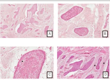
Axial, coronal and sagittal images (voxel size: 0.25 mm) were obtained from the mandibular arch. Analysis of the scans showed a
hyper dense image in the region of tooth 47 compatible with bone sclerosis, and an expansive hypodense lesion in the mandibular symphysis and parasymphysis, extending in the laterolateral direction until teeth 33 and 43 (Figure 1A). The image of the
lesion was multilocular and there were no calciications inside the lesion. Buccal cortical expansion and areas of bone destruction were also observed (Figure1B to 1F). The remaining structures
had a normal radiographic appearance. The lesion measured 16.75 mm (anteroposterior), 37.25 mm (laterolateral) and 20.51 mm (superior-inferior) in its major axes.
In view of this clinical presentation, the initial management adopted consisted of partial removal of the lesion (incisional biopsy), which led to the diagnosis of SOT. Thus, total resection of the tumor and curettage were performed, followed by the placement and adaptation of a reconstruction plate for mandibular reconstruction.
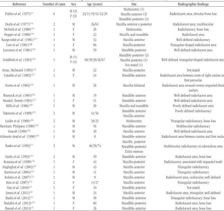
Microscopic analysis revealed the presence of epithelial islands of variables size and shape, which consisted of well-deined squamous epithelium with benign features (Figure 2A and B).
The epithelial islands were immersed in a stroma of cellular and
ibrous connective tissues and exhibited a peripheral layer of cuboidal cells without evidence of a palisade-like arrangement or nuclear polarization (Figure 2C). Intraepithelial microcystic
degeneration (Figure 2C – arrow) and circular areas of ibrous condensation (Figure 2D – asterisk) were identiied around
some epithelial islands, suggesting a reaction of the connective tissue stroma to epithelial proliferation. On the basis of these histopathologic features, the deinitive diagnosis was SOT.
The patient presented no postoperative complications and surgery provided good outcomes, with no recurrence during four years and two months follow-up.
FIGURE 1 − Axial, coronal, sagittal and 3D images
Expansive hypodense lesion in the mandibular symphysis and parasymphysis (A); multilocular image, buccal cortical expansion and areas of bone destruction (B, E and F) – posterior region (C) and anterior region (D).
FIGURE 2 − Epithelial islands of well-defined squamous epithelium with benign features
A and B) cellular stroma, peripheral layer of cuboidal cells; C) intraepithelial micro cystic degeneration (arrow); D) circular areas of fibrous condensation (asterisk) (HE, 100× and 400×).
HE: hematoxylin and eosin.
DISCUSSION
Odontogenic tumors arise from epithelial tissue, mesenchymal tissue, or both, which are involved in teeth development. The etiopathogenesis of these tumors and their causes or triggers are not well understood. Clinically, most of these tumors are asymptomatic, but tooth mobility, expansion and bone loss can be observed(2, 5, 6).
Squamous odontogenic tumor is a very rare benign tumor that arises from remnants of the dental lamina and occurs equally in the mandible and maxilla. Tumors located in the anterior region of the mandible are uncommon. Despite the bone involvement and benign nature of SOT, this tumor can iniltrate adjacent soft tissues, causing major esthetic and functional impairment. In addition, SOT can appear as a single tumor or affect multiple sites at the same anatomic location (multicentric)(2-6).
A
C
E
F
C
A B
D
B
TABLE − Cases published of SOT between1975-2015 years (PubMed Database)
Reference Number of cases Sex Age (years) Site Radiographic findings
Pullon et al. (1975)(1) 6 M (3)
F (3) 23/11/19/31/42/29
Multicentric (1) Maxilla-anterior (3) Mandible-posterior (2)
Radiolucent area; alveolar bone loss
Doyle et al. (1977)(14) 2 M 26/65 Maxilla-anterior e posterior Radiolucent area; multilocular
McNeill et al. (1980)(15) 1 F 26 Multicentric Radiolucency; bone loss
Hopper et al. (1980)(16) 1 F 22 Maxilla and mandible Radiolucent area
Kangvonkit et al. (1981)(17) 1 M 32 Maxilla-anterior Well-deined radiolucency Carr et al. (1981)(18) 1 F 66 Maxilla-posterior Triangular-shaped radiolucent area Leventon et al. (1981)(19) 1 M 59 Mandible-posterior Well-deined radiolucent area
Goldblatt et al. (1982)(20) 5 M (1)
F (4) 60/29/30/26/67
Mandible-posterior (3) Maxilla-posterior (1)
Not stated (1)
Well-deined; triangular-shaped radiolucent area
Swan, McDaniel (1983)(21) 1 M 32 Maxilla-posterior Not stated
Cataldo et al. (1983)(22) 1 F 24 Mandible-anterior Radiolucent area between roots of right canine and
irst premolar
Norris et al. (1984)(23) 1 M 26 Maxilla-bilateral Radiolucent area around crowns impacted third molars
Warnock et al. (1985)(24) 1 M 19 Mandible-anterior Well-deined radiolucent area
Monteil, Teresti (1985)(25) 1 F 51 Mandible-anterior Well-deined radiolucent area Mills et al. (1986)(26) 1 M 26 Maxilla and mandible Poorly deined radiolucent area
Tatemoto et al. (1989)(27) 2 M 41/56 Mandible-anterior
Maxilla-anterior
Poorly deined radiolucency
Leider et al. (1989)(28) 2 M 29/25 Multicentric Triangular radiolucency; bone loss
Reichart, Philipsen (1990)(29) 1 M 56 Mandible-anterior Multilocular radiolucency
Yaacob (1990)(30) 1 M 39 Maxilla-anterior Well-deined radiolucent area
Schwartz-Arad et al. (1990)(10) 1 M 8 Mandible-anterior Radiolucent area between canine and irst molar
Baden et al. (1993)(7) 3 M 46/39/74 Maxilla- posterior
Mandible-posterior Extra-osseous
Multilocular radiolucency in edentulous area
Saxby et al. (1993)(31) 2 M 59 Mandible-anterior Radiolucent area; bone loss
Kusama et al. (1998)(32) 1 F 42 Maxilla-posterior Radiolucency associated with impacted tooth Haghighat et al. (2002)(8) 1 M 43 Maxilla-anterior Triangular radiolucency
Barrios et al. (2004)(11) 1 M 11 Maxilla-anterior Triangular radiolucency
Ruhim et al. (2007)(13) 1 M 9 Maxilla-anterior Radiolucent area, unilocular, well-deined
Kim et al. (2007)(12) 2 F 15/27 Maxilla-anterior Triangular radiolucency Siar et al. (2010)(3) 1 F 10 Mandible-anterior Not stated
Jones et al. (2011)(6) 1 M 24 Maxilla-anterior Radiolucent area, triangular, well-deined Badni et al. (2012)(5) 1 M 58 Mandible-anterior Triangular radiolucency; bone loss Malathi et al. (2012)(33) 1 F 60 Mandible-posterior Radiolucent area; bone loss
Bansal et al. (2013)(2) 1 F 26 Mandible-anterior Radiolucent area; bone loss
SOT: squamous odontogenic tumor; M: male; F: female; anterior: anterior to the distal surface of the canine tooth; posterior: posterior to the distal surface of the canine tooth; multicentric: multiple sites at the same anatomical location.
Characterized for the irst time in 1975, SOT become largely known by the dissemination of a series of six cases described by
Pullon et al.(1). Until that time, some harmless lesions had been
diagnosed and treated in a mutilating and aggressive manner
since they were believed to be ameloblastomas or squamous cell carcinomas based on their histopathologic features. Since this irst report, diagnostic criteria and a surgical approach have been established and some of these criteria are used until today(2, 4).
Forty-seven cases of SOT have so far been reported in the English literature (PubMed Database). Adults are affected more frequently,
but the cases reported comprise a wide age-range (8-74 years), with a mean age of 36.1 years. There is no gender preference for SOT, the male to female ratio is 1:1.6. Squamous odontogenic tumors tend to arise in the canine-premolar area of the maxilla and in the molar region of the mandible, but it can occur along the jaws (Table). However, SOTs located in the maxilla show a more aggressive behavior due to the natural porous and medullary anatomy of this bone(2, 6-8). In
Most cases of SOT arise and develop in the periodontium of the permanent dentition. Pullon et al.(1) reported a case associated with the
deciduous dentition and cases involving edentulous areas(1, 7-10). The
present case is the 48th case of SOT reported in the English literature.
The location of SOT in an edentulous region is uncommon, since the tumor is more frequent in areas with teeth. In addition, involvement of the mandibular anterior region and the multilocular radiographic appearance are also uncommon indings(2).
Although not pathognomonic, most SOTs are detected during routine radiographic examination as a radiolucent, unilocular, triangular-shaped image associated with the roots of adjacent teeth. The tumor may be misdiagnosed from the time when the lesion is referred to as a severe periodontal defect. Some rare cases exhibit a multilocular radiolucency, circumscription of the tumor with a cortical border or, in more aggressive cases, lack of deinition of tumor margins. In addition to advanced periodontal bone loss, Langerhans cell disease, lateral periodontal cyst, keratocystic odontogenic tumor, central odontogenic ibroma and other odontogenic tumors should be included in the radiographic differential diagnosis(2-6, 11-13). Although computed
tomography permits to delimit the tumor, the present case exhibited an uncommon radiographic feature, i.e., the presence of a multilocular radiolucent image.
Microscopically, SOT is characterized by multiple proliferations of squamous epithelium surrounded by a moderately cellular ibrous-collagenous connective tissue stroma. The epithelial islands, which can be smoothly contoured and demarcated from
the stroma and vary in size and shape, occasionally exhibit foci of cystic degeneration and central calciication. The harmless appearance of the pavement epithelium characterizes the lesion, since ameloblastic nuclear polarization and a palisade-like arrangement of peripheral columnar cells are absent in SOTs. Furthermore, the squamous odontogenic tumor-like proliferations seen in the walls of inlammatory odontogenic cysts are not considered a manifestation of SOT(2-6, 11-13). Therefore,
the histopathological differential diagnoses for SOT include the acanthomatous and desmoplastic ameloblastoma and gingival squamous cell carcinoma; however, SOT shows no microscopic evidence of malignancy(2, 3, 5).
Treatment of SOT consists of conservative surgical removal (local excision), enucleation and/or complete curettage. However, more aggressive intervention may be necessary in the case of tumors located in the maxilla due to the aggressive potential of the lesion at this site. Recurrence is rarely reported in the literature and is attributed to incomplete removal of the initial tumor. In these cases, the extraction of adjacent teeth involved in the tumor is indicated(2, 4-6). Only one case of recurrence was observed among
the six cases evaluated by Pullon et al.(1).
The present results demonstrate the need for a comprehensive diagnostic evaluation considering all clinical, radiographic and especially histopathologic features in order to guarantee the success of treatment indicated in each case, promoting quality of life for patients with SOT. In addition, the present case highlights the importance of individual analysis of each case.
RESUMO
O tumor odontogênico escamoso (TOE) é definido como uma neoplasia benigna rara, localmente infiltrativa, podendo estender-se a estruturas vizinhas. Objetivou-se relatar um caso de TOE de acometimento incomum. Paciente do sexo feminino, 56 anos de idade, fumante, demonstrou leve aumento de volume na região do mento na porção compreendida entre o canino esquerdo e o primeiro pré-molar direito. Imagens axial, coronal e sagital demonstraram lesão expansiva hipodensa na sínfise e na linha média mandibular. Fez-se a remoção parcial da lesão, o que levou ao diagnóstico de TOE. A paciente não apresentou recidiva durante quatro anos e dois meses de acompanhamento.
Unitermos: tumor odontogênico escamoso; mandíbula; neoplasias.
REFERENCES
1. Pullon PA, Shafer WG, Elzay RP, Kerr DA, Corio RL. Squamous odontogenic tumor. Report of six cases of a previously undescribed lesion. Oral Surg Oral Med Oral Pathol. 1975; 40(5): 616-30.
2. Bansal S, Joshi SK. Squamous odontogenic tumor with unusual localization and appearance: a rare case report. Case Rep Med. 2013; 2013: 407967.
3. Siar CH, Nakano K, Ng KH, Tomida M, Nagatsuka H, Kawakami T. Squamous odontogenic tumor of the mandible: a case report demonstrating immunoexpression of Notch1, 3, 4, Jagged1 and Delta1. Eur J Med Res. 2010; 15(4): 180-4.
5. Badni M, Nagaraja A, Kamath V. Squamous odontogenic tumor: a case report and review of literature. J Oral Maxillofac Pathol. 2012; 16(1): 113-7.
6. Jones BE, Sarathy AP, Ramos MB, Foss RD. Squamous odontogenic tumor. Head Neck Pathol. 2011; 5(1): 17-9.
7. Baden E, Doyle J, Mesa M, Fabie M, Lederman D, Eichen M. Squamous odontogenic tumor. Report of three cases including the irst extraosseous case. Oral Surg Oral Med Oral Pathol. 1993; 75(6): 733-8.
8. Haghighat K, Kalmar JR, Mariotti AJ. Squamous odontogenic tumor: diagnosis and management. J Periodontol. 2002; 73(6): 653-6. 9. Philipsen HP, Reichart PA. Squamous odontogenic tumor (SOT): a benign neoplasm of the periodontium. A review of 36 reported cases. J Clin Periodontol. 1996; 23(10): 922-6.
10. Schwartz-Arad D, Lustmann J, Ulmansky M. Squamous odontogenic tumor. Review of the literature and case report. Int J Oral Maxillofac Surg. 1990; 19(6): 327-30.
11. Barrios TJ, Sudol JC, Cleveland DB. Squamous odontogenic tumor associated with an erupting maxillary canine: case report. J Oral Maxillofac Surg. 2004; 62(6): 742-4.
12. Kim K, Mintz SM, Stevens J. Squamous odontogenic tumor causing erosion of the lingual cortical plate in the mandible: a report of 2 cases. J Oral Maxillofac Surg. 2007; 65(6): 1227-31.
13. Ruhin B, Raoul G, Kolb F, et al. Aggressive maxillary squamous odontogenic tumour in a child: histological dilemma and adaptative surgical behaviour. Int J Oral Maxillofac Surg. 2007; 36(9): 864-6. 14. Doyle JL, Grodjesk JE, Dolinsky HB, Rafel SS. Squamous odontogenic tumor: report of three cases. J Oral Surg. 1977; 35(12): 994-6.
15. McNeill J, Price HM, Stoker NG. Squamous odontogenic tumor: report of case with long-term history. J Oral Surg. 1980; 38(6): 466-71. 16. Hopper TL, Sadeghi EM, Pricco DF. Squamous odontogenic tumor. Report of a case with multiple lesions. Oral Surg Oral Med Oral Pathol. 1980; 50(5): 404-10.
17. Kangvonkit P, Sirichitra V, Hansasuta C. [Squamous odontogenic tumor (report of a case and review of the literature)]. J Dent Assoc Thai. 1981; 31(1): 25-33.
18. Carr RF, Carlton DM Jr, Marks RB. Squamous odontogenic tumor: report of case. J Oral Surg. 1981; 39(4): 297-8.
19. Leventon GS, Happonen RP, Newland JR. Squamous odontogenic tumor. Am J Surg Pathol. 1981; 5(7): 671-7.
20. Goldblatt LI, Brannon RB, Ellis GL. Squamous odontogenic tumor. Report of ive cases and review of the literature. Oral Surg Oral Med Oral Pathol. 1982; 54(2): 187-96.
21. Swan RH, McDaniel RK. Squamous odontogenic proliferation with probable origin from the rests of Malassez (early squamous odontogenic tumor?). J Periodontol. 1983; 54(8): 493-6.
22. Cataldo E, Less WC, Giunta JL. Squamous odontogenic tumor. A lesion of the periodontium. J Periodontol. 1983; 54(12): 731-5.
23. Norris LH, Baghaei-Rad M, Maloney PL, Simpson G, Guinta J. Bilateral maxillary squamous odontogenic tumors and the malignant transformation of a mandibular radiolucent lesion. J Oral Maxillofac Surg. 1984; 42(12): 827-34.
24. Warnock GR, Pierce GL, Correll RW, Baker DA. Triangular-shaped radiolucent area between roots of the mandibular right canine and irst premolar. J Am Dent Assoc. 1985; 110(6): 945-6.
25. Monteil RA, Terestri P. Squamous odontogenic tumor related to an unerupted lower canine. J Oral Maxillofac Surg. 1985; 43(11): 888-95. 26. Mills WP, Davila MA, Beuttenmuller EA, Koudelka BM. Squamous odontogenic tumor. Report of a case with lesions in three quadrants. Oral Surg Oral Med Oral Pathol. 1986; 61(6): 557-63.
27. Tatemoto Y, Okada Y, Mori M. Squamous odontogenic tumor: immunohistochemical identiication of keratins. Oral Surg Oral Med Oral Pathol. 1989; 67(1): 63-7.
28. Leider AS, Jonker LA, Cook HE. Multicentric familial squamous odontogenic tumor. Oral Surg Oral Med Oral Pathol. 1989; 68(2): 175-81. 29. Reichart PA, Philipsen HP. Squamous odontogenic tumor. J Oral Pathol Med. 1990; 19(5): 226-8.
30. Yaacob HB. Squamous odontogenic tumor. J Nihon Univ Sch Dent. 1990; 32(3): 187-91.
31. Saxby MS, Rippin JW, Sheron JE. Case report: squamous odontogenic tumor of the gingiva. J Periodontol. 1993; 64(12): 1250-2.
32. Kusama K, Kawashima A, Nagai H, et al. Squamous odontogenic tumor of the maxilla: report of a case. J Oral Sci. 1998; 40(3): 119-22. 33. Malathi N, Radhika T, Thamizh CH, Nandakumar N. Peripheral squamous odontogenic tumor. Indian J Dent Res. 2012; 23(2): 286-8.
MAILING ADDRESS
Priscilla Suassuna Carneiro Lúcio