Hypothermia Protects and Prolongs the
Tolerance Time of Retinal Ganglion Cells
against Ischemia
Maximilian Schultheiss1, Sven Schnichels1*, Thoralf Hermann2, Jose Hurst1,
Marita Feldkaemper3, Blanca Arango-Gonzalez3, Marius Ueffing3, Karl U. Bartz-Schmidt1, Guenther Zeck2, Martin S. Spitzer1
1Centre for Ophthalmology, University Eye Hospital Tübingen, Tübingen, Germany,2Natural and Medical Sciences Institute at the University Tübingen, Reutlingen, Germany,3Institute for Ophthalmic Research, University of Tübingen, Tübingen, Germany
Abstract
Purpose
Hypothermia has been shown to be neuroprotective in the therapy of ischemic stroke in the brain. To date no studies exist on the level of the inner retina and it is unclear if hypothermia would prolong the ischemic tolerance time of retinal ganglion cells, which are decisive in many ischemic retinopathies.
Methods
Bovine eyes were enucleated and stored either at 21°C or 37°C for 100 or 340 minutes, respectively. Afterwards the globes were dissected, the retina was prepared and either the spontaneous ganglion cell responses were measured or the retina was incubated as an organotypic culture for additional 24 hours. After incubation the retina was either processed for histology (H&E and DAPI staining) or real-time PCR (Thy-1 expression) was performed.
Results
Hypothermia prolonged ganglion cell survival up to 340 minutes under ischemic conditions. In contrast to eyes kept at 37°C the eyes stored at 21°C still showed spontaneous ganglion cell spiking (56.8% versus 0%), a 5.8 fold higher Thy-1 mRNA expression (not significant, but a trend) and a preserved retinal structure after 340 minutes of ischemia.
Conclusion
Hypothermia protects retinal ganglion cells against ischemia and prolongs their ischemic tolerance time.
OPEN ACCESS
Citation:Schultheiss M, Schnichels S, Hermann T, Hurst J, Feldkaemper M, Arango-Gonzalez B, et al. (2016) Hypothermia Protects and Prolongs the Tolerance Time of Retinal Ganglion Cells against Ischemia. PLoS ONE 11(2): e0148616. doi:10.1371/ journal.pone.0148616
Editor:Alexandre Hiroaki Kihara, Universidade Federal do ABC, BRAZIL
Received:May 21, 2015
Accepted:January 21, 2016
Published:February 5, 2016
Copyright:© 2016 Schultheiss et al. This is an open access article distributed under the terms of the Creative Commons Attribution License, which permits unrestricted use, distribution, and reproduction in any medium, provided the original author and source are credited.
Data Availability Statement:All relevant data are within the paper.
Funding:The authors have no support or funding to report.
Introduction
Central retinal artery occlusion leads to irreversible vision loss within hours (h)[1]. Up to date, no causative treatment exists for this ophthalmologic urgency. Past trials using intra-arterial and intravenous thrombolysis could not confirm their efficacy[2,3], although previously pub-lished retrospective studies showed encouraging results[4,5]. Nevertheless, it has been
observed, that thrombolysis finally leads to reperfusion of the retinal circuit, but apparently the timespan in clinical practice until reperfusion of the retinal circuit occurs, seems to be too long. Therefore, like in ischemic stroke, the crucial factor for the functional result after thrombolysis is the timespan of ischemia[6]. By reducing this timespan to a minimum, thrombolysis as ther-apy could probably also work in central retinal artery occlusion. In order to increase the poten-tial therapeutic window, we are seeking for substances or procedures, which prolong the tolerance time of the retina to an ischemic insult.
Hypothermia, understood as temperature below 37°C, is supposed to be neuroprotective in cerebral stroke and to increase the tissue tolerance to ischemic insults[7]. At the moment a
large efficacy trial in ischemic stroke—the European Stroke Research Network for
Hypother-mia (EuroHYP)-1 trial—is recruiting up to 1500 patients in order to determine if hypothermia
can improve the functional outcome (further information athttp://www.eurohyp1.eu/).
Hypo-thermia can prolong the tolerance time to an ischemic insult presumably in every tissue. How-ever, no neuroprotective treatments at the retina have been established so far in the clinics. Comparable to cerebral stroke, hypothermia could protect the retinal ganglion cells during a central retinal artery occlusion. To consider hypothermia as possible effective treatment for acute retinal ischemia, its application should preserve the retina long enough allowing that a curative therapy like fibrinolysis could be performed.
Hypothermia as neuroprotective mechanism at the retina has been already evaluated and proved to be protective[8,9,10]. Earlier investigations always focused on a single time point and thus, important clinical questions remain open[8,9,10]. It is therefore unclear if hypothermia opens a realistic therapeutic time window for causative therapies like thrombolysis. Addition-ally, no studies exist on the level of retinal ganglion cells, which are the cells of the retina most vulnerable to ischemic insults of the inner retina[11,12].
We therefore conducted a study, in which the effects of hypothermia on retinal function and ganglion cell survival under ischemic conditions were investigated. The shortest ischemic period was 100 minutes (min), because according to a study of Hayreh et al. beyond 97 min significant irreversible damage of the inner retina occurs[13]. Furthermore Hayreh et al. showed, that after 240 min of ischemia no functional recovery can be observed after reperfusion anymore[13]. The aim of our study was to investigate, whether hypother-mia extends the retinal tolerance time against ischehypother-mia. Therefore, we added additional 100 min to the reported 240 min. According to the published data, the retinal ganglion cells should be dead or dying by then. If hypothermia would preserve the retinal function up to that time point, hypothermia would prolong the retinal tolerance time against ischemia and could possibly open a clinically realistic therapeutic window for a causative therapy (e.g. lysis).
Materials and Methods
Preparation of retinal biopsies
Bovine eyes were obtained from the local abattoir (Gärtringen; Schlachthof e.G., Germany). At this abattoir, animals are butchered for food production. No additional animals were killed for this study. The permission to use this otherwise discarded tissue was obtained from the local regulatory authorities (Az.34-9181.30 from the Landratsamt Böblingen, Germany issued in
2006). Ten min after the cow`s death, eyes were enucleated by the abattoir’s veterinarian. The
globes were stored either at 21°C or 37°C for 100 or 340 min respectively. After arriving at the lab the globes were cleaned of additional tissue down to the sclera. Then the eyes were disin-fected in 10% iodine diluted in 0.9% NaCl (Braun, Germany) for 5 min. After disinfection the globes were washed twice for 2 min using 0.9% NaCl. Thereafter the eyes were dissected and retinal biopsies were prepared under sterile conditions.
Dissection was performed after 100 and 340 min of anoxic ischemia. The preparation of the retina was performed in Dulbecco`s modified Eagle`s medium (Invitrogen-Gibco; Rockville, MD), which was not saturated with oxygen.
Retinal biopsies were taken at a distance of 8 mm from the optic nerve. The biopsy locations were superior of the optic nerve for real-time PCR, temporal for histology and inferior for the retinal ganglion cell recordings. By using defined biopsy-locations the amount of RGCs in the analyzed retinal region was constant for each assay. Additionally one bovine eye could serve as source for different assays. Retinal biopsies were placed on an insert with a permeable mem-brane (Millipore AB, Solna, Sweden; PIHA03050) facing the vitreous side upwards. Under the permeable membrane, R16 serum free culture medium (Invitrogen Life Technologies, Paisley, UK; 07490743A) was used for cultivation. Retinal biopsies for real-time PCR and histology
were cultivated at 37°C in an environment containing 5% CO2for 24 h, the retinal biopsies for
measuring spontaneous ganglion cell responses were measured directly after preparation. After cultivation the samples were frozen using liquid nitrogen. For the real-time PCR
sam-ples were stored at -80°C. For Histology unfixed samsam-ples were embedded in Tissue-Tek1O.C.
T.™(Sakura, Nehterlands), frozen and stored at -20°C.
We did not randomize our eyes during the retina preparation and the MEA-recordings. However, neither, the person who performed the Thy-1 mRNA nor the person who performed the MEA-recordings was aware to which treatment group the respective samples belonged.
Recording of ganglion cell activity in bovine retina
The retinal biopsies for the 10 min control group were processed directly at the abattoir. The biopsies for the other groups were prepared as described above.
The recordings of RGC-activity was performed like previously described[14]. Briefly, retinal tissue was mounted, ganglion cell-side down, on a microelectrode array (MEA) comprising 60 TiN electrodes (Multichannelsystems MCS GmbH, Reutlingen). The electrodes of this MEA
are arranged in an 8 x 8 grid with 200μm spacing between the electrode centres. Spontaneous
activity of the ganglion cells was recorded for 10 min after an initial adaptation period of 10
min. During the recording the retina was continuously superfused with carbogenated Ames’
medium (Sigma Aldrich) at 35–37°C. Data were amplified, filtered (0.3–3 kHz) and saved for
offline analysis. Ganglion cell activity, the timing of extracellular spike waveform, is detected
from raw recordings if they exceed six times the root mean square (rms)noise on the
corre-sponding electrode. Correlated timestamps among different electrodes are eliminated to avoid duplicates. Only electrodes with a mean average spike rate of 1 Hz (1 spike/sec) are counted as
electrodes for each preparation was evaluated. This number is an estimate of the average activ-ity within the investigated retinal portion.
Real-time PCR
Tissue preparation and cultivation was performed like described above. Only for the 10 min of ischemia control group the eye balls were stored and processed on ice, in order to avoid mRNA degradation on the way to the lab.
From the retinas the mRNA was isolated and cDNA was synthesized and purified using MultiMACS cDNA Synthesis Kit according to the manufactures protocol (MultiMACS mRNA Isolation Kit (8x12), Miltenyi Biotec #130-092-520, Germany)).
Aliquots of cDNA (5 ng) were analyzed in duplicate reactions by qPCR using 1μM
gene-spe-cific primers and Sso Advanced Universal SYBR Green Supermix (BIORAD; California—USA)
in a total volume of 20μl. PCRs were carried out in a CFX96 and analyzed with CFX Manager
Software, version 3.0 (BIORAD). Relative expression levels were calculated as previously described[15] with ß-actin as a reference gene. Briefly explained, the relative expression ratio of a target gene is calculated based on the amplification efficiency and threshold cycle in compari-son to a reference gene. The following gene-specific primers were (designed with Primer 3 plus software (http://www.bioinformatics.nl/cgi-bin/primer3plus/primer3plus.cgi)) used: Thy1 (F: 5´-ctcggcaccatgaaccct-3´, R: 5´-agacgaaggctctggttc-3´), ß-Actin (F: 5´-ctcttccagccttccttc-3´; R: 5´-gggcagtgatctctttct-3´).
Histology
Frozen retinas were cut on a cryostat (12μm sections). Staining with haematoxylin and eosin
(H&E) as well as with 40,6-Diamidin-2-phenylindol (DAPI) was performed to view the retinal
structure. For DAPI staining tissue was first fixed with cold methanol. Sections were blocked in 10% BSA and stained with DAPI. H&E staining was performed on unfixed tissue to visualize the retinal structure. Photographs were taken with an Axiovert 135 fluorescent microscope (Zeiss, Göttingen, Germany) which used the AxioVision 4.6 software (Zeiss, Göttingen, Germany).
Statistics
Statistical analysis was performed using JMP1(SAS Institute Inc., Cary, NC, USA). To test for
differences between the groups the Wilcoxon-Kruskal-Wallis test was used for the MEA-data. A sided t-test was used for the real-time PCR data. Additionally we performed for the two-sided t-test a Bonferroni adjustment as multiple testing was performed. Differences were
con-sidered to be significant at p<0.05 and after Bonferroni adjustment at p<0.025.
Results
Spontaneous retinal ganglion cell responses
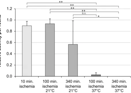
After 100 min of ischemia the number of spiking retinal ganglion cells was not altered in the eyes stored at 21°C when compared to the freshly prepared eyes which were enucleated within 10 min after death (Fig 1). In contrast after 100 min of ischemia and storage at 37°C the sponta-neous retinal ganglion cell responses were statistically significant reduced (Fig 1). Additionally, only in the eyes which were stored at 21°C, but not in the eyes stored at 37°C, spontaneous reti-nal ganglion cells spiking was observed even after 340 min of ischemia (Fig 1).
data had a non-Gaussian distribution) of the electrodes recorded spikes after 100 min of ische-mia. The percentage of electrodes recording ganglion cell activity dropped to 56.7% ± 40.9% after 340 min of ischemia (Fig 1). In contrast, in the eyes stored at 37°C only 2.8% ± 4.1% of the electrodes detected retinal ganglion cell activity after 100 min of ischemia. After 340 min of ischemia at 37°C no ganglion cell spikes at all were detected. The attachment of the retina on the micro-electrode array was carefully monitored by analyzing the mean noise level[16], to prevent misinterpretation of the results caused by inhomogeneous interfacing and thus record-ing of smaller areas.
Thy-1-Expression
Thy-1 expression levels were normalized toβ-actin. The arbitrary units were set as 1 for the
samples with 10 min of ischemia and no cultivation for 24 h. These eyes served as control group, as it was not possible to receive the eyes faster than ten min after death.
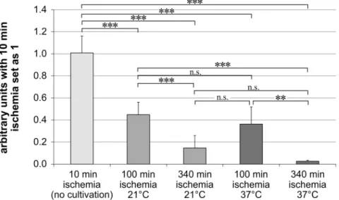
Longer ischemia times resulted in a lower Thy-1 expression (Fig 2). In our control-group with 10 min of ischemia and no cultivation for 24 h, we observed the highest Thy-1 expression (1.01 ± 0.15). This group was followed by the groups with 100 min of ischemia (100 min ische-mia at 21°C: 0.45 ± 0.11; 100 min ischeische-mia at 37°C: 0.31 ± 0.08) and the lowest expression was seen in the groups with 340 min of ischemia (340 min ischemia at 21°C: 0.14 ± 0.11; 340 min ischemia at 37°C: 0.03 ± 0.01).
The eyes stored at 21°C showed after 100 and 340 min of ischemia significantly higher
Thy-1 mRNA levels than the eyes stored at 37°C in the two-sided t-test (p<0.05). However after
Bonferroni adjustment it was only a trend at both temperatures (p>0.025). Nevertheless after
100 min and 340 min of ischemia the Thy-1 expression was 1.5 respectively 5.8 fold higher when the eyes were stored at 21°C (Fig 2).
Fig 1. Spontaneous retinal ganglion cell responses are preserved under hypothermic conditions.Bar graph represents the relative amount of electrodes measuring spontaneous retinal ganglion cell activity per retina. Spontaneous action potentials of retinal ganglion cells were measured simultaneously by the 60 electrodes of a Multi-Electrode-Array (MEA). Data are depicted as mean±SD with*p<0.05 and**p<0.01; n = 6 retinas.
Histology
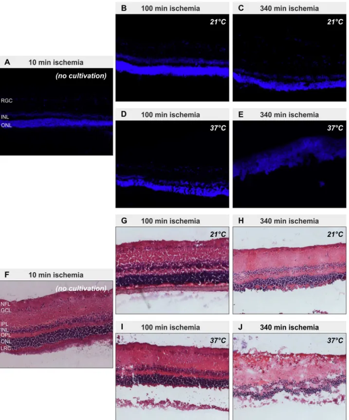
Representative pictures of cryosections revealed preservation of the retinal structure in all eyes except for those stored at 37°C for 340 min (Fig 3). In these eyes the retinal architecture was severely disturbed (Fig 3E and 3J). The inner and outer nuclear layer could be hardly distin-guished from each other, because the outer plexiform layer had nearly disappeared. Further-more, the inner plexiform layer was thickened and some of the cells of the retinal ganglion cell
layer were“dislocated”into the nerve fiber layer (Fig 3J). The severely disturbed retinal
archi-tecture goes in line with the observation during tissue preparation, that only in this group the retina was like a viscous fluid which could not be grasped anymore by forceps. All other groups still had the typical consistency of a retina.
Discussion
The results of the presented study suggest that hypothermia can prolong the tolerance time of retinal ganglion cells against ischemia.
The analysis of the spontaneous retinal ganglion cell spikes (action potentials) showed that under hypothermic conditions the retinal ganglion cells can survive an acute ischemic insult of 340 min. With real-time PCR we could detect 5.8 fold more living retinal ganglion cells in the tissue when the eyes exposed to 340 min of ischemia were kept under hypothermic conditions
during the ischemic period—even after subsequent 24h of cultivation. This conclusion can be
drawn, because Thy-1 mRNA is exclusively produced by the retinal ganglion cells in the retina and the amount of Thy-1 mRNA expression can be used as equivalent to the number of living retinal ganglion cells[17]. In contrast to ischemia at body temperature hypothermia preserved the retinal architecture for many h as observable in the stained retinal cryosections (Fig 3C). Additionally in the group with 340 min of ischemia at 37°C the retina was like a thick, sticky fluid and no tangible tissue anymore. The results of the real-time PCR and the measurements of the spontaneous retinal ganglion cell responses are supported by the histological findings.
Fig 2. Thy-1 mRNA expression is higher under hypothermic conditions.Bar graph represents the Thy-1 mRNA expression levels in arbitrary units after normalizing them to the samples with 10 min of ischemia. Thy-1 mRNA expression were measured by real-time PCR. Data are depicted as mean±SD, with**p<0.01 and ***p<0.001; n = 5–6 retinas.
Fig 3. Retinal structure is preserved at 21°C storage.Representative pictures of cryosections of the five different groups (10 min ischemia (no cultivation) (A, F); 100 min ischemia at 21°C (B, G); 340 min ischemia at 21°C (C, H); 100 min ischemia at 37°C (D, I); 340 min ischemia at 340°C (E, J)) after staining with DAPI (A-E) or haematoxylin and eosin (F-J) to visualize nuclei and retinal structure are shown. Except for the probes with 340 min of ischemia stored at 37°C, which show a degenerated retina, all other sections show a preserved retinal structure. Layers of the retina: nerve fiber layer (NFL), ganglion cell layer (GCL), inner plexiform layer (IPL), inner nuclear layer (INL), outer plexiform layer (OPL), outer nuclear layer (ONL), layer of rods and cones (LRC)
Interestingly the results of the RGC recordings (analyzed directly after preparation) and the Thy-1 mRNA (analyzed after 24 h of cultivation) differed especially in the 100 min ischemia at 37°C-group. This group showed a nearly fully depressed RGC-activity but a substantially retained expression of Thy-1 mRNA. The reason for this could be that the ischemic insult at the beginning leaves RGCs structurally intact but functionally inactive. The same effect occurs with neurons in the penumbra of an ischemic stroke[18,19]. This could explain, why the neu-rons were electrophysiological silent directly after the ischemic insult, but still somehow alive, because the Thy-1 mRNA expression was maintained even after 24h of cultivation.
By extending the retinal ganglion cell tolerance time to ischemia, hypothermia could open a therapeutic window for a causal therapy of acute retinal ischemic diseases such as central reti-nal artery occlusion. Nevertheless, hypothermia cannot ameliorate the impact of ischemia completely. The deleterious effect of ischemia is only slowed down and not stopped by hypothermia.
Already other studies showed that hypothermia is neuroprotective against ischemia [8,9,10]. Nevertheless, we are the first who analyzed the neuroprotective effect of hypothermia at the level of retinal ganglion cells. These retinal ganglion cells are the decisive cells for the
visual outcome after an ischemic retinal insult in the clinical setting and therefore should be–
in our opinion- the center of investigations.
Recently Salido et al. reported that global or local hypothermic preconditioning (32 and 33°C for 20 min) protects the rat retina from ischemic damage[20] on the level of retinal gan-glion cells. However, preconditioning with hypothermia is difficult to achieve in real life as nobody can know in advance when an acute ischemic event will take place. Thus, inducing hypothermia as soon as possible after the onset of acute ischemia, by a cooled irrigation solu-tion during vitrectomy for example, seems to be more promising. Vitrectomy can be performed within 15 min after diagnosis in local anesthesia.
Importantly hypothermia not only reduces the metabolism and energy consumption, but also protects cells against glutamate excitotoxicity, which is a nearly ubiquitous existing damag-ing mechanism in most acute and chronic ischemic retinal diseases[21,22]. Additionally hypo-thermia reduces the activity of neuronal-damaging neuroinflammatory cascades[23].
Our study was carried out on enucleated bovine eyes. Therefore, our results have to be con-firmed in an in vivo model, before conclusions can be drawn for a clinical setting. Nevertheless in our model the retinal survival time could be extended to 340 min of ischemia when the ret-ina was stored at 21°C during the ischemic period.
In conclusion, hypothermia protects retinal ganglion cells against an ischemic insult and prolongs their ischemic tolerance time. Hypothermia therefore could open a therapeutic win-dow to initiate causal treatments of acute ischemic retinal diseases such as central retinal artery occlusion. Nevertheless it still has to be investigated which temperature of the irrigation solu-tion results in the best neuroprotective effect.
Acknowledgments
The work has been presented in parts at: ARVO 2013, Fort Lauderdale, Florida, USA.
Author Contributions
References
1. Beatty S, Au Eong KG (2000) Local intra-arterial fibrinolysis for acute occlusion of the central retinal artery: a meta-analysis of the published data. Br J Ophthalmol 84: 914–916. PMID:10906103
2. Schumacher M, Schmidt D, Jurklies B, Gall C, Wanke I, Schmoor C, et al. (2010) Central retinal artery occlusion: local intra-arterial fibrinolysis versus conservative treatment, a multicenter randomized trial. Ophthalmology 117: 1367–1375 e1361. doi:10.1016/j.ophtha.2010.03.061PMID:20609991
3. Chen CS, Lee AW, Campbell B, Lee T, Paine M, Fraser C, et al. (2011) Efficacy of intravenous tissue-type plasminogen activator in central retinal artery occlusion: report from a randomized, controlled trial. Stroke 42: 2229–2234. doi:10.1161/STROKEAHA.111.613653PMID:21757667
4. Aldrich EM, Lee AW, Chen CS, Gottesman RF, Bahouth MN, Gailloud P, et al. (2008) Local intraarterial fibrinolysis administered in aliquots for the treatment of central retinal artery occlusion: the Johns Hopkins Hospital experience. Stroke 39: 1746–1750. doi:10.1161/STROKEAHA.107.505404PMID:18420951
5. Hattenbach LO, Kuhli-Hattenbach C, Scharrer I, Baatz H (2008) Intravenous thrombolysis with low-dose recombinant tissue plasminogen activator in central retinal artery occlusion. Am J Ophthalmol 146: 700–706. doi:10.1016/j.ajo.2008.06.016PMID:18718570
6. Gumbinger C, Reuter B, Stock C, Sauer T, Wietholter H, Bruder I, et al. (2014) Time to treatment with recombinant tissue plasminogen activator and outcome of stroke in clinical practice: retrospective anal-ysis of hospital quality assurance data with comparison with results from randomised clinical trials. BMJ 348: g3429. doi:10.1136/bmj.g3429PMID:24879819
7. Wu TC, Grotta JC (2013) Hypothermia for acute ischaemic stroke. Lancet Neurol 12: 275–284. doi:10. 1016/S1474-4422(13)70013-9PMID:23415567
8. Tamai K, Toumoto E, Majima A (1997) Local hypothermia protects the retina from ischaemic injury in vitrectomy. Br J Ophthalmol 81: 789–794. PMID:9422935
9. Quinones-Hinojosa A, Malek JY, Ames A 3rd, Ogilvy CS, Maynard KI (2003) Metabolic effects of hypo-thermia and its neuroprotective effects on the recovery of metabolic and electrophysiological function in the ischemic retina in vitro. Neurosurgery 52: 1178–1186; discussion 1186–1177. PMID:12699563 10. Wang X, Niwa M, Hara A, Matsuno H, Kawase K, Kozawa O, et al. (2002) Neuronal degradation in
mouse retina after a transient ischemia and protective effect of hypothermia. Neurol Res 24: 730–735. PMID:12392214
11. Osborne NN, Casson RJ, Wood JP, Chidlow G, Graham M, Melena J (2004) Retinal ischemia: mecha-nisms of damage and potential therapeutic strategies. Prog Retin Eye Res 23: 91–147. PMID:14766318 12. Stevens WD, Fortin T, Pappas BA (2002) Retinal and optic nerve degeneration after chronic carotid
ligation: time course and role of light exposure. Stroke 33: 1107–1112. PMID:11935068
13. Hayreh SS, Zimmerman MB, Kimura A, Sanon A (2004) Central retinal artery occlusion. Retinal survival time. Exp Eye Res 78: 723–736. PMID:15106952
14. Stutzki H, Leibig C, Andreadaki A, Fischer D, Zeck G (2014) Inflammatory stimulation preserves physi-ological properties of retinal ganglion cells after optic nerve injury. Front Cell Neurosci 8: 38. doi:10. 3389/fncel.2014.00038PMID:24574973
15. Pfaffl MW (2001) A new mathematical model for relative quantification in real-time RT-PCR. Nucleic Acids Res 29: e45. PMID:11328886
16. Zeitler R, Fromherz P, Zeck G (2011) Extracellular voltage noise probes the interface between retina and silicon chip. Applied Physics Letters 99: 263702.
17. Nash MS, Osborne NN (1999) Assessment of Thy-1 mRNA levels as an index of retinal ganglion cell damage. Invest Ophthalmol Vis Sci 40: 1293–1298. PMID:10235569
18. Murphy TH, Corbett D (2009) Plasticity during stroke recovery: from synapse to behaviour. Nat Rev Neurosci 10: 861–872. doi:10.1038/nrn2735PMID:19888284
19. Astrup J, Symon L, Branston NM, Lassen NA (1977) Cortical evoked potential and extracellular K+ and H+ at critical levels of brain ischemia. Stroke 8: 51–57. PMID:13521
20. Salido EM, Dorfman D, Bordone M, Chianelli M, Gonzalez Fleitas MF, Rosenstein RE (2013) Global and ocular hypothermic preconditioning protect the rat retina from ischemic damage. PLoS One 8: e61656. doi:10.1371/journal.pone.0061656PMID:23626711
21. Brandt SK, Weatherly ME, Ware L, Linn DM, Linn CL (2010) Calcium preconditioning triggers neuropro-tection in retinal ganglion cells. Neuroscience.
22. Baltmr A, Duggan J, Nizari S, Salt TE, Cordeiro MF (2010) Neuroprotection in glaucoma—Is there a future role? Exp Eye Res 91: 554–566. doi:10.1016/j.exer.2010.08.009PMID:20800593