RESEARCH ARTICLE
Regulation of Budding Yeast CENP-A levels
Prevents Misincorporation at Promoter
Nucleosomes and Transcriptional Defects
Erica M. Hildebrand1,2, Sue Biggins1*
1Howard Hughes Medical Institute, Division of Basic Sciences, Fred Hutchinson Cancer Research Center, Seattle, Washington, United States of America,2Molecular and Cellular Biology Program, University of Washington, Seattle, Washington, United States of America
Abstract
The exclusive localization of the histone H3 variant CENP-A to centromeres is essential for accurate chromosome segregation. Ubiquitin-mediated proteolysis helps to ensure that CENP-A does not mislocalize to euchromatin, which can lead to genomic instability. Consis-tent with this, overexpression of the budding yeast CENP-ACse4is lethal in cells lacking Psh1, the E3 ubiquitin ligase that targets CENP-ACse4for degradation. To identify additional mechanisms that prevent CENP-ACse4misincorporation and lethality, we analyzed the genome-wide mislocalization pattern of overexpressed CENP-ACse4in the presence and absence of Psh1 by chromatin immunoprecipitation followed by high throughput sequenc-ing. We found that ectopic CENP-ACse4is enriched at promoters that contain histone H2A. ZHtz1nucleosomes, but that H2A.ZHtz1is not required for CENP-ACse4mislocalization. Instead, the INO80 complex, which removes H2A.ZHtz1from nucleosomes, promotes the ectopic deposition of CENP-ACse4. Transcriptional profiling revealed gene expression changes in thepsh1Δcells overexpressing CENP-ACse4. The down-regulated genes are enriched for CENP-ACse4mislocalization to promoters, while the up-regulated genes corre-late with those that are also transcriptionally up-regucorre-lated in anhtz1Δstrain. Together, these data show that regulating centromeric nucleosome localization is not only critical for maintaining centromere function, but also for ensuring accurate promoter function and tran-scriptional regulation.
Author Summary
Chromosomes carry the genetic material in cells. When cells divide, each daughter cell must inherit a single copy of each chromosome. The centromere is the locus on each chro-mosome that ensures the equal distribution of chrochro-mosomes during cell division. One essential protein involved in this task is CENP-ACse4, which normally localizes exclusively to centromeres. Here, we investigated where CENP-ACse4spreads in the genome when parts of its regulatory machinery are removed. We found that CENP-ACse4becomes OPEN ACCESS
Citation:Hildebrand EM, Biggins S (2016) Regulation of Budding Yeast CENP-A levels Prevents Misincorporation at Promoter Nucleosomes and Transcriptional Defects. PLoS Genet 12(3): e1005930. doi:10.1371/journal.pgen.1005930
Editor:Beth A. Sullivan, Duke University, UNITED STATES
Received:June 17, 2015
Accepted:February 22, 2016
Published:March 16, 2016
Copyright:© 2016 Hildebrand, Biggins. This is an open access article distributed under the terms of the Creative Commons Attribution License, which permits unrestricted use, distribution, and reproduction in any medium, provided the original author and source are credited.
Data Availability Statement:All relevant data are within the paper and its Supporting Information files or are available from the NCBI GEO repository (accession number GSE69696,http://www.ncbi.nlm. nih.gov/geo/query/acc.cgi?acc=GSE69696).
mislocalized to promoters, the region upstream of each gene that controls the activity of the gene. Consistent with this, the mislocalization of CENP-ACse4to promoters leads to problems with gene activity. Our work shows that mislocalization of centromeric proteins can have effects beyond chromosome segregation defects, such as interfering with gene expression on chromosome arms.
Introduction
The eukaryotic genome is packaged into chromatin, which consists of 147 bp repeating units of DNA wrapped around histone proteins to form nucleosomes [1]. Chromatin is important not only for packaging and protecting DNA, but also for regulating access of genes and other DNA elements to nuclear proteins involved in processes such as transcription, replication, and chro-mosome segregation. Most nucleosomes are composed of the canonical histone proteins, H2A, H2B, H3, and H4 [2]. However, the behavior and functions of nucleosomes can be altered both by chemically modifying canonical histones through post-translational modifications and by exchanging canonical histones for histone variants that alter nucleosome composition [2]. For example, H2A.Z is a variant of histone H2A and is found at promoter nucleosomes genome-wide where it regulates transcription [2–4]. In contrast, the conserved CENP-A variant (also called CenH3) replaces H3 in nucleosomes exclusively at the centromere where it regulates chromosome segregation [5–7]. Because changes in nucleosome composition can have a major impact on the underlying functions of the genome, it is critical to understand the mechanisms that control the localization of histone modifications and variants.
The genomic incorporation of the budding yeast H2A.ZHtz1(SGD ID: S000005372) histone variant is regulated by the SWR1 (SWR-C) and INO80 (INO80-C) chromatin remodeling complexes [8]. H2A.ZHtz1localizes to intergenic regions, specifically near transcription start sites (TSS) at the +1 and -1 nucleosomes surrounding nucleosome-depleted regions (NDRs) [3,4,9–12]. In budding yeast, H2A.ZHtz1nucleosomes are correlated with high nucleosome turnover [13], which is proposed to assist transcriptional initiation or rapid changes between transcriptional states [14–16]. SWR-C incorporates H2A.ZHtz1into nucleosomes by exchang-ing H2A/H2B dimers for H2A.ZHtz1/H2B dimers [17–19]. In contrast, the mechanism of H2A. ZHtz1removal from nucleosomes by INO80-C is less well understood because it has two reported activities that both lead to H2A.ZHtz1exchange, either by swapping H2A.ZHtz1/H2B dimers for H2A/H2B dimers [20] or by promoting turnover of the entire nucleosome [8,19].
The localization of the CENP-A variant is regulated by the histone chaperone HJURP (Scm3 in budding yeast), which is targeted specifically to centromeres [21–25]. Centromeric sequence and size are highly variable throughout eukaryotes and can be specified by either an underlying sequence or through epigenetic inheritance [26,27]. Despite the diversity of centro-meres, CENP-A is a conserved hallmark of all centromeres. The presence of CENP-A directs the formation of the kinetochore, a large protein complex that mediates attachments between the microtubules of the mitotic spindle and the chromosome during cell division [26,28,29]. CENP-A mislocalization to euchromatin through overexpression or tethering can lead to ectopic kinetochore formation and genomic instability [30–32]. However, CENP-A mislocali-zation has not been reported to disrupt other genomic processes [33,34].
Multiple mechanisms ensure that CENP-A does not mislocalize to euchromatin. A number of chromatin remodelers and histone chaperones are reported to help maintain centromeric chromatin or prevent CENP-A mislocalization, including Fun30, RSC, INO80-C, CAF-1, HIR, FACT, and RbAp48 and SWI/SNF [35–41]. In addition, ubiquitin-mediated proteolysis
funding/pgm_summ.jsp?pims_id=6201). SB is an Investigator of the Howard Hughes Medical Institute (https://www.hhmi.org). The funders had no role in study design, data collection and analysis, decision to publish, or preparation of the manuscript.
prevents ectopic CENP-A localization by controlling total CENP-A protein levels [42–45]. In budding yeast, proteolysis of CENP-ACse4(SGD ID: S000001532) is mediated by an E3 ubiqui-tin ligase called Psh1 (SGD ID: S000005415) [46,47]. When CENP-ACse4is overexpressed in the absence of Psh1-mediated proteolysis [42,46–48], cells accumulate high levels of CEN-P-ACse4in euchromatin. This also results in lethality, although the underlying cause has not been determined [42,46,47].
Similar to CENP-A, H2A.Z also contributes to chromosome segregation. In human cells, H2A.Z is found at pericentromeric regions, where it is incorporated at the inner centromere between the CENP-A nucleosome domains, and helps to establish centromeric heterochroma-tin [49,50]. Similarly, H2A.ZHtz1is also a component of pericentromeric chromatin in budding yeast, where it localizes to nucleosomes flanking the CENP-ACse4nucleosome and is important for chromosome segregation through unknown mechanisms [4,51,52]. However, it is unclear whether there is a connection between the localization of the histone variants. In human cells, overexpressed CENP-A was found to mislocalize to regions enriched for H2A.Z, although no physical interaction was detected between these two histone variants [33]. In contrast, studies inS.pombehave shown that CENP-ACnp1tends to mislocalize to ectopic regions that are depleted of H2A.ZHtz1[53].
We set out to determine whether there are features of euchromatin that normally prevent budding yeast CENP-A misincorporation as well as to identify the functional consequences of CENP-A mislocalization to euchromatin. The identification of euchromatic sites that strongly misincorporate CENP-A may also shed light on the underlying cause of the lethality. To address these questions, we performed the first genome-wide analysis of CENP-A overexpres-sion in the absence of ubiquitin-mediated degradation. We found that overexpressed CEN-P-ACse4mislocalizes to promoters that are enriched for NDRs flanked by H2A.ZHtz1, and this mislocalization is dramatically enhanced in cells that cannot degrade CENP-ACse4. This locali-zation pattern appears to be due in part to co-opting of INO80-C to incorporate excess CEN-P-ACse4into promoter nucleosomes that normally contain H2A.ZHtz1. Consistent with this, there was a significant correlation between transcripts that were misregulated in cells lacking H2A.ZHtz1and those with high levels of CENP-ACse4mislocalization. We also found that a sub-set of promoters that misincorporate CENP-ACse4have decreased transcription, which may be the underlying cause of lethality. Together, these data suggest that it is essential that cells regu-late CENP-ACse4localization not only to ensure proper chromosome segregation, but also to protect cells from promoter nucleosome disruption and transcriptional misregulation.
Results
Excess CENP-A
Cse4mislocalizes to intergenic regions of the genome
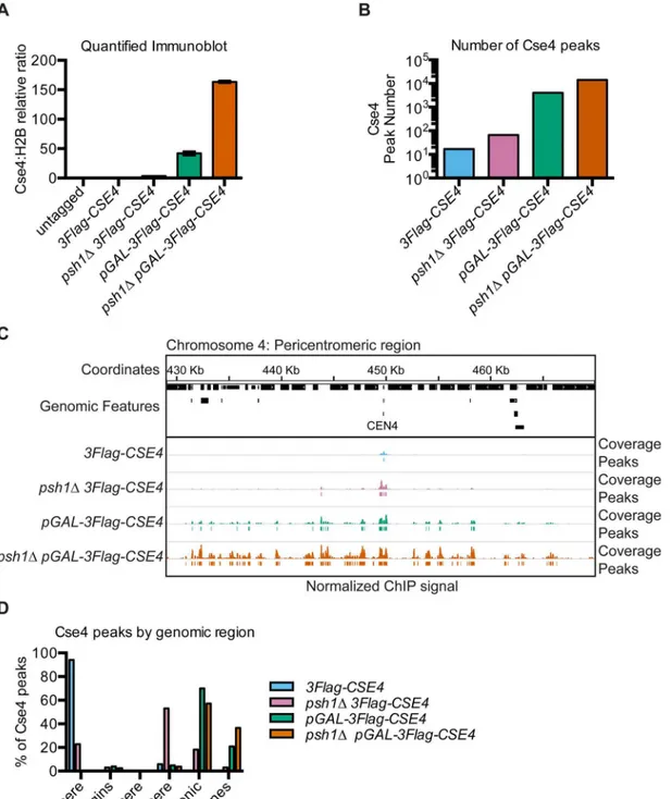
To identify the precise genomic sites of CENP-ACse4mislocalization in budding yeast, we per-formed ChIP-seq on endogenous and overexpressed CENP-ACse4in the presence and absence of Psh1-mediated proteolysis. All strains contained a fully functional ectopic3Flag-CSE4gene integrated at theURA3locus under the endogenous promoter and were deleted for the endoge-nousCSE4gene. Cells overexpressing CENP-ACse4contained an additional copy under the con-trol of theGALpromoter (pGAL-3Flag-CSE4). As seen previously, CENP-ACse4overexpression inhibited the growth of WT cells and resulted in lethality inpsh1Δcells (S1A Fig) [46,47]. The growth inhibition correlated with the total amount of chromatin-bound CENP-ACse4protein (Fig 1A,S1B Fig).
Fig 1. Intergenic regions are the major sites of overexpressed CENP-ACse4mislocalization.(A) Quantification of CENP-ACse4levels in MNase-digested chromatin in untagged (SBY3, black),3Flag-CSE4(SBY10419, blue),psh1Δ3Flag-CSE4(SBY10484, pink),pGAL-3Flag-CSE4(SBY10425, green) and psh1ΔpGAL-3Flag-Cse4(SBY10483, orange) strains. The ratio of CENP-ACse4:H2B in the chromatin in each strain was quantified relative to the
CENP-ACse4:H2B ratio from the3Flag-CSE4(SBY10419) strain. Quantification is based on two biological replicates. Error bars are +/- 1 standard error of the mean (SEM) of the two biological replicates. (B) The total number of CENP-ACse4peaks called in the indicated strains:3Flag-CSE4(SBY10419, blue),psh1
Δ
3Flag-CSE4(SBY10484, pink),pGAL-3Flag-CSE4(SBY10425, green) andpsh1ΔpGAL-3Flag-CSE4(SBY10483, orange). (C) A representative region of the CENP-ACse4ChIP-seq coverage on Chromosome 4 between 429,000 base pairs (bp) and 470,000 bp is shown. The CENP-ACse4ChIP-seq coverage for the strains in (B) is normalized to the coverage at the centromeres after subtracting the input. Peaks are shown as lines below each coverage signal (the cutoff is the average minimum coverage at the centromere in the3Flag-CSE4strain). The scale of the normalized coverage is from 0–20,000 for all strains.
MNase-treated chromatin by immunoprecipitation of 3Flag-Cse4. The amount of CENP-ACse4 recovered in the ChIP samples reflected the starting levels in the chromatin (S1C Fig). The input samples (MNase-digested chromatin) and ChIP samples (3Flag-Cse4-bound chromatin after immunoprecipitation) were made into paired-end sequencing libraries using a modified Solexa library preparation protocol that captures DNA particles down to ~25 bp (S1D Fig) [54,
55]. Paired-end sequencing resulted in greater than 1.5 million reads/sample, with an average read length ranging from 147–164 bp (S1 Table). The mononucleosome-sized sequencing reads from the input and ChIP samples for each strain were mapped to theS.cerevisiae refer-ence genome versionSacCer3[56].
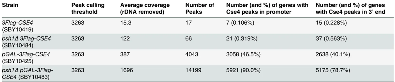
The peaks of CENP-ACse4enrichment genome-wide correlated with the levels of chromatin-bound CENP-ACse4(Fig 1B,Table 1). Seventeen peaks were identified for the3Flag-CSE4strain, representing the sixteen centromeres as well as a peak 150 bp fromCEN9. A small amount of CENP-ACse4mislocalization was seen starting in thepsh1Δstrain with 66 peaks, and was further increased in cells overexpressing CENP-ACse4with 4043 peaks. The greatest enrichment in the euchromatin was detected in thepsh1Δcells overexpressing CENP-ACse4with 14,199 peaks. An example of the coverage data and corresponding peaks for a representative region around Cen-tromere 4 shows a single cenCen-tromere peak for the WT strain and additional peaks around the centromere in the other strains (Fig 1C). The increased CENP-ACse4mislocalization in sur-rounding euchromatin is especially apparent in thepGAL-3Flag-CSE4andpsh1Δ pGAL-3Flag-CSE4strains that have the highest levels of CENP-ACse4. We independently confirmed the CEN-P-ACse4enrichment atCEN4and at other representative peaks by ChIP-qPCR (S2A Fig). Our initial analysis also identified a CENP-ACse4peak at the rDNA locus in all strains. This did not show significant enrichment in the3Flag-CSE4strain by ChIP-qPCR but did in the cells with overexpressed CENP-ACse4, similar to previously reported data [46,57] (S2A Fig). However, due to the difficulty in analyzing this repetitive region by standard mapping algorithms, ChIP coverage of this region was excluded from further computational analyses.
To determine if mislocalized CENP-ACse4favors certain genomic regions, we analyzed the percentage of CENP-ACse4peaks in various functional regions of the genome, including cen-tromeres, pericencen-tromeres, telomeres, replication origins, genes, and intergenic regions (Fig 1D). We defined pericentromeres as 20 Kilobases (Kb) flanking each centromere, consistent with the 20–50 Kb size of cohesin enrichment around each centromere in budding yeast [58,
59]. As expected, the majority of CENP-ACse4peaks in WT cells were at centromeres, with an increase in pericentromeric peaks in thepsh1Δmutant. However, the majority of peaks in the strains overexpressing CENP-ACse4were in the intergenic regions, with a smaller percentage within genes. As intergenic regions make up less than 30% of the entire genome, these data indicate a strong enrichment of CENP-ACse4in intergenic regions in cells overexpressing CENP-ACse4.
We next asked whether the intergenic enrichment correlates with features known to be asso-ciated with centromeres. ChIP-seq of mildly overexpressed CENP-ACse4previously identified 23 centromere-like regions (CLRs) on chromosome arms that are enriched for mislocalized CENP-ACse4and other kinetochore proteins [60]. These CLRs share characteristics with cen-tromeric sequences such as having a high AT% and conferring stability to plasmid DNA. As expected, most of the CLRs have CENP-ACse4peaks in thepsh1ΔpGAL-3Flag-CSE4strain (S2B Fig). However, CENP-ACse4was overexpressed to much higher levels in our study (150-fold compared to 3-fold), so the CLRs are a small fraction of the total peaks. Consistent with this, there was also enrichment in low confidence negative control regions (LCNCRs), indicating there is no preference for CLR localization. We also analyzed the AT content of the DNA bound by mislocalized CENP-ACse4, as this is a defining characteristic of centromeric DNA in budding yeast. As expected, CENP-ACse4peaks were highly enriched for AT
nucleotides in the WT strain. However there was only a moderate increase in AT% in the psh1Δstrain compared to the input nucleosomes, and almost no AT bias in the strains with overexpressed CENP-ACse4(S2C–S2F Fig). Together, these data indicate that the mislocaliza-tion of CENP-ACse4is due to a more widespread effect than just centromere-like
characteristics.
Mislocalized CENP-A
Cse4is enriched in promoters but is not correlated
with basal transcription levels
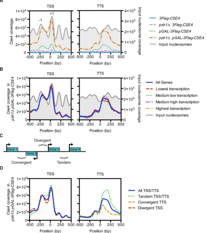
We next asked whether the intergenic enrichment of overexpressed CENP-ACse4was specific to either promoters (defined as 500 bp upstream of the transcription start site (TSS)) or tran-scription terminators (defined as 500 bp downstream of the trantran-scription termination site (TTS)) by calculating the number of peaks in these regions (Table 1). CENP-ACse4was enriched in both regions when overexpressed, so we more precisely analyzed the pattern by plotting the average coverage in 10 bp windows for regions 500 bp upstream and downstream of all TSS or TTS (the TSS or TTS is plotted at position 0 based on previously reported RNA-seq transcription start positions [61]). In thepsh1Δcells overexpressing CENP-ACse4, there was enrichment -200 bp from the TSS and directly over the TSS, which correspond to the -1 and +1 nucleosomes respectively (Fig 2A). At the TTS, CENP-ACse4was enriched in the nucleosome just after the termination site, and was shifted slightly into the NDR compared to the WT nucleosomes. Although the level of CENP-ACse4enrichment in the other three strains was much lower overall, the trend is similar in the cells with increased CENP-ACse4. This pattern is reminiscent of the pattern of CENP-ACse4mislocalization upon deletion ofCAC1andHIR1, which leads to ectopic CENP-ACse4enrichment at promoters in the presence of Psh1 [38].
We next asked whether the accumulation of CENP-ACse4in promoters and terminators is associated with the basal level of transcription in WT cells. We plotted CENP-ACse4 enrich-ment at the TSS and TTS of genes binned into quartiles by the published transcription levels in a WT strain, ranked from lowest transcription to highest transcription [62]. However, there was no correlation between CENP-ACse4enrichment and the different transcription levels (Fig 2B,S3 Fig). Therefore, the CENP-ACse4localization to promoters in thepsh1Δ pGAL-3Flag-CSE4strain was not an artifact of increased chromatin accessibility in areas of high transcrip-tion, such as was found in the previously reported CENP-ACse4ChIP-seq for slightly overex-pressed or hypomorphic CENP-ACse4[63]. We also analyzed whether CENP-ACse4
mislocalization correlated with the direction of transcription of the surrounding genes, since this has been shown for cohesin localization, which is specifically enriched in convergent inter-genic regions outside of the pericentromere [58,64]. We classified the intergenic regions as tan-dem (between two genes transcribed in the same direction), convergent (between two genes
Table 1. CENP-ACse4peak information.
Strain Peak calling
threshold
Average coverage (rDNA removed)
Number of Peaks
Number (and %) of genes with Cse4 peaks in promoter
Number (and %) of genes with Cse4 peaks in 3’end
3Flag-CSE4 (SBY10419)
3263 15.3 17 7 (0.106%) 15 (0.228%)
psh1Δ3Flag-CSE4 (SBY10484)
3263 122 66 21 (0.319%) 37 (0.563%)
pGAL-3Flag-CSE4 (SBY10425)
3263 387 4043 3058 (46.5%) 2638 (40.1%)
psh1Δ pGAL-3Flag-CSE4(SBY10483)
3263 1696 14199 5921 (90.0%) 5175 (78.7%)
Fig 2. Overexpressed CENP-ACse4mislocalizes to promoters.(A) Mean CENP-ACse4ChIP coverage 500 bp upstream and downstream of all
transcription start sites (TSS) or transcription termination sites (TTS), for3Flag-CSE4(SBY10419, blue),psh1Δ3Flag-CSE4(SBY10484, pink), pGAL-3Flag-Cse4(SBY10425, green) andpsh1ΔpGAL-3Flag-Cse4(SBY10483, orange). The input nucleosome positions are from the input MNase-seq data from the 3Flag-CSE4strain (SBY10419) and are plotted in grey. (B) Mean CENP-ACse4ChIP coverage for thepsh1
ΔpGAL-3Flag-CSE4(SBY10483) strain separated by transcription levels of the corresponding genes [62]. (C) Diagram of the classification of the different types of intergenic regions based on the direction of transcription of the flanking genes. (D) Mean CENP-ACse4ChIP coverage for thepsh1ΔpGAL-3Flag-CSE4strain (SBY10483) separated by the direction of transcription for the corresponding TSS or TTS.
doi:10.1371/journal.pgen.1005930.g002
transcribed towards each other), or divergent (between two genes transcribed away from each other) (Fig 2C). In promoters, CENP-ACse4was enriched at the tandem and divergent genes (Fig 2D,S4 Fig). At the terminators, CENP-ACse4was enriched at the tandem TTS and depleted at the convergent TTS. Because convergent regions lack promoters, these data are consistent with the enrichment of CENP-ACse4to promoter regions.
Mislocalized CENP-A
Cse4is found at H2A.Z
Htz1-enriched nucleosomes
flanking NDRs
Since CENP-ACse4mislocalization to promoters was not correlated with transcription levels,
we looked for another chromatin feature specific to promoters that might enhance CEN-P-ACse4incorporation. One characteristic of promoters that is less commonly found at the 3
’
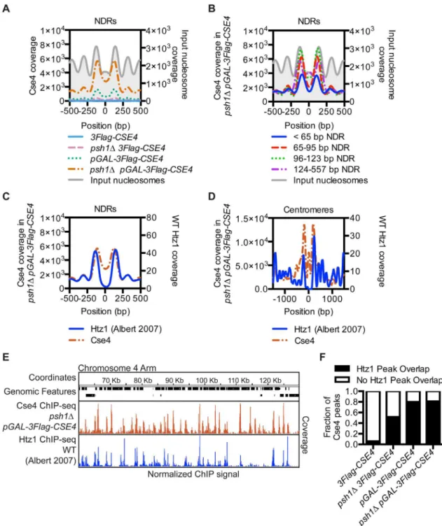
ends of genes is the NDR between the -1 and +1 nucleosomes at the TSS [65]. We therefore compared CENP-ACse4profiles centered on all NDRs and found a strong CENP-ACse4
enrich-ment in the nucleosomes flanking the NDRs in thepsh1ΔpGAL-3Flag-CSE4strain (Fig 3A). Because NDRs vary in length up to 557 bp, we asked whether there was a specific NDR length that correlated with CENP-ACse4mislocalization and found the highest enrichment in NDRs longer than 65 bp (Fig 3B). We obtained similar results when the analysis was centered on the TSS instead of the NDR (S5A–S5D Fig), consistent with the enrichment of CENP-ACse4in NDR containing promoters.
The localization of CENP-ACse4to the nucleosomes flanking the NDRs is similar to H2A. ZHtz1, the only other histone variant in budding yeast [4]. In addition, the SWR-C
chromatin-remodeling complex that incorporates H2A.ZHtz1preferentially binds to NDRs greater than 50 bp [66], similar to the length of NDRs that have the highest CENP-ACse4enrichment (greater
than 65 bp) (Fig 3B). We therefore investigated the relationship between previously reported H2A.ZHtz1localization [4] and the mislocalization of overexpressed CENP-ACse4inpsh1
Δ cells. There was a striking similarity in their enrichment at NDRs (Fig 3C), as well as a similar trend of co-enrichment in the nucleosomes flanking replication origins (S5E and S5F Fig) and centromeres (Fig 3D,S5G Fig). The CENP-ACse4coverage at the TSS was also similar to H2A. ZHtz1coverage, while at the TTS CENP-ACse4was shifted more into the 3’NDR than H2A. ZHtz1(S6A and S6B Fig). The histone variants exhibited a genome-wide trend to co-localize, as seen in a representative region of the arm of Chromosome 4 (Fig 3EandS6C Fig). There was a high coincidence of overlap between CENP-ACse4peaks in the experimental strains with H2A. ZHtz1peaks in WT cells, although they were not specifically enriched in any of the genomic
fea-tures correlated with CENP-ACse4mislocalization (Fig 3F,S6D and S6E Fig) Together, these data indicate a significant enrichment of misincorporated CENP-ACse4at sites where H2A.
ZHtz1nucleosomes are normally located genome-wide inpsh1Δcells overexpressing CENP-ACse4.
CENP-A
Cse4accumulation in chromatin does not depend on H2A.Z
Htz1chromatin-Fig 3. CENP-ACse4mislocalizes to regions that are enriched for the histone variant H2A.ZHtz1.(A) Mean CENP-ACse4
ChIP coverage for the 3Flag-CSE4(SBY10419, blue),psh1Δ3Flag-CSE4(SBY10484, pink),pGAL-3Flag-CSE4(SBY10425, green) andpsh1ΔpGAL-3Flag-CSE4(SBY10483, orange) strains centered on annotated NDRs [65]. (B) Mean CENP-ACse4ChIP coverage for thepsh1
ΔpGAL-3Flag-CSE4(SBY10483) strain centered on annotated NDRs, binned by NDR length [65]. (C) Mean CENP-ACse4ChIP coverage for thepsh1
ΔpGAL-3Flag-CSE4(SBY10483, orange) strain (left y axis) vs. mean H2A.ZHtz1ChIP coverage [4] (blue) (right y axis) at all NDRs. (D) Mean CENP-ACse4ChIP coverage for thepsh1ΔpGAL-3Flag-CSE4(SBY10483, orange) strain (left y axis) vs. mean normalized H2A.ZHtz1ChIP coverage [4] (blue) (right y axis) at all centromeres. (E) CENP-ACse4ChIP coverage for thepsh1
Δ
pGAL-3Flag-CSE4(SBY10483, orange) strain and WT H2A.ZHtz1coverage [4] (blue) on the chromosome 4 arm between 60,000 bp and 130,000 bp. The scale of the normalized coverage is from 0–20,000 for the CENP-ACse4ChIP-seq and from 0–301 for the H2A.ZHtz1ChIP-seq. (F) Overlap between
CENP-ACse4peaks in3Flag-CSE4(SBY10419),psh1Δ3Flag-CSE4(SBY10484),pGAL-3Flag-CSE4(SBY10425) orpsh1ΔpGAL-3Flag-CSE4 (SBY10483) compared to WT H2A.ZHtz1nucleosomes [4].
doi:10.1371/journal.pgen.1005930.g003
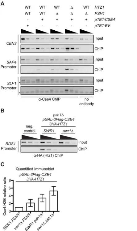
bound CENP-ACse4inswr1Δcells. We confirmed that H2A.ZHtz1was reduced at a previously reported promoter nucleosome locus by ChIP-PCR (Fig 4B)[17,68,69]. Similar to our findings with thehtz1Δmutant, bulk H2A.ZHtz1was not depleted in the chromatin fraction inswr1Δ, but CENP-ACse4chromatin levels were somewhat higher in theswr1Δpsh1Δcells compared to psh1Δ(Fig 4C,S7A and S7B Fig). In addition, there was no change in CENP-ACse4stability in swr1Δcells (S7C Fig). We also tested whether CENP-ACse4overexpression in thepsh1Δmutant affects H2A.ZHtz1promoter occupancy, but did not detect an effect at the loci analyzed (S7D Fig). However, given that H2A.ZHtz1is estimated to occupy only a small proportion of nucleo-somes at any given locus in the population, it may be difficult to detect a significant difference [11,19]. Together, our data suggest that although ectopic CENP-ACse4and WT H2A.ZHtz1 local-ize to similar sites, the H2A.ZHtz1incorporation machinery does not promote CENP-ACse4 mis-localization and may instead help to prevent CENP-ACse4promoter incorporation.
INO80-C contributes to CENP-A
Cse4mislocalization in
psh1
Δ
cells
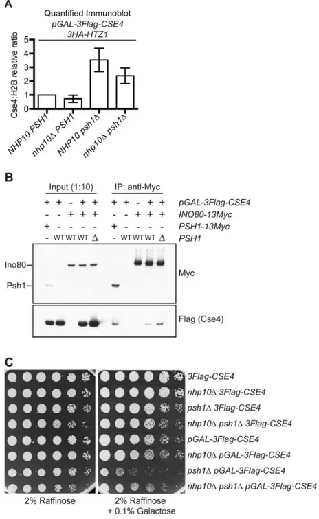
Since the ectopic localization of CENP-ACse4does not depend on H2A.ZHtz1incorporation, we asked whether chromatin remodelers that remove H2A.ZHtz1are involved. INO80-C has been reported to act preferentially on H2A.ZHtz1-containing +1 nucleosomes and to promote full nucleosome turnover [19,20]. We therefore hypothesized that CENP-ACse4might be incorpo-rated into chromatin when canonical H3 is removed by INO80-C-mediated nucleosome turn-over. Previous work showed that deletion of the ATPase Ino80 (SGD ID: S000003118) leads to a global alteration of H2A.ZHtz1localization patterns genome-wide without affecting the overall lev-els of H2A.ZHtz1incorporation in the genome [20,70]. However, this deletion mutant is not viable in the strain background we used in this study [71]. We therefore used a deletion ofNHP10(SGD ID: S000002160), a non-essential INO80-C subunit that facilitates binding to nucleosomes and DNA, but that does not affect catalytic activityin vitro[72–74]. To analyze CENP-ACse4levels, we performed chromatin fractionation in WT andnhp10Δcells overexpressing CENP-ACse4. Similar to previously reported work, we did not detect a change in total H2A.ZHtz1levels in the chromatin in thenhp10Δstrain (S8A and S8B Fig)[20,70]. However, CENP-ACse4chromatin levels were somewhat reduced whenNHP10was deleted (Fig 5AandS8B Fig), suggesting that INO80-C his-tone exchange activity contributes to CENP-ACse4misincorporation. To more directly test this possibility, we asked whether Ino80 associates with CENP-ACse4in vivo. CENP-ACse4 co-immu-noprecipitated with Ino80 (Fig 5B), and this interaction increased in the absence of Psh1. To determine how this affects cell viability, we also analyzed the growth ofnhp10Δmutant cells over-expressing CENP-ACse4. Although strong CENP-ACse4overexpression is lethal topsh1Δcells regardless of the presence ofNHP10(S8C Fig), a deletion ofNHP10improved the growth of psh1Δmutant cells that were moderately overexpressing CENP-ACse4(Fig 5C). We confirmed these effects were not due to altered levels or stability of CENP-ACse4innhp10Δmutant cells (S8D and S8E Fig). Together, these data suggest that at least some of the ectopic CENP-ACse4 deposition is likely coupled to the chromatin remodeling activity of INO80-C.
Mislocalized CENP-A
Cse4perturbs transcription in the absence of Psh1
Fig 4. CENP-ACse4mislocalization does not depend on H2A.ZHtz1incorporation.(A) ChIP was performed with anti-Cse4 antibody on negative control cells (SBY15924), as well aspTET-CSE4 (SBY15903),psh1ΔpTET-CSE4(SBY15904) andpsh1Δhtz1ΔpTET-CSE4(SBY15906) cells overexpressing CENP-ACse4for six hours. As a control, we also performed a ChIP experiment with no antibody inpsh1ΔpTET-CSE4cells. Input dilutions are 1:100, 1:300, 1:100 and ChIP dilutions are 1x, 1:3, 1:9. (B) ChIP-PCR of 3HA-Htz1 at theRDS1promoter. Strains used: negative (neg.) control (SBY3),psh1Δ
pGAL-3Flag-CSE4 3HA-HTZ1(SBY12833),psh1ΔpGAL-3Flag-CSE4,3HA-HTZ1 swr1Δ(SBY12924). Input dilutions: 1:100, 1:300, 1:900. ChIP dilutions: 1:3, 1:9, 1:21. (C) Relative CENP-ACse4levels were measured by quantifying the mean chromatin CENP-ACse4:H2B fold change vs.pGAL-3Flag-CSE4 HA-HTZ1+/- 1 SEM using quantitative immunoblots of chromatin fraction (n = 3), p = 0.0204 (paired t-test comparing the Cse4:H2B relative ratio inpsh1ΔpGAL-3Flag-CSE4toswr1Δpsh1ΔpGAL-3Flag-CSE4). Strains used werepGAL-3Flag-CSE4 3HA-HTZ1(SBY12832),swr1ΔpGAL-3Flag-CSE4 3HA-HTZ1 (SBY12956),psh1ΔpGAL-3Flag-CSE4 3HA-HTZ1(SBY12833) andswr1Δpsh1ΔpGAL-3Flag-CSE4 3HA-HTZ1(SBY12924).
doi:10.1371/journal.pgen.1005930.g004
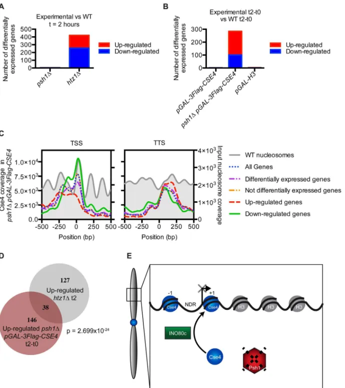
containing just aPSH1deletion or overexpressing CENP-ACse4or H3 had very little change in transcription (Fig 6A and 6B). However, a large number of genes were misregulated inpsh1Δ cells overexpressing CENP-ACse4, as well as inhtz1Δcells as previously described [75,76]. We confirmed that these gene expression changes were not due to an indirect effect of CENP-ACse4 mislocalization to the rDNA by measuring the rDNA copy number and rRNA transcript levels, which were not significantly different between the strains (S9A and S9B Fig). We also con-firmed that the differentially transcribed genes in thepsh1ΔpGAL-3Flag-CSE4strain are not a consequence of altered cell cycle progression [47,77] (S9C Fig).
To determine whether CENP-ACse4mislocalization to promoters correlates with transcrip-tional misregulation of downstream genes, we compared the promoters with CENP-ACse4 peaks to the genes showing altered transcription in thepsh1Δstrain overexpressing CEN-P-ACse4. While there was a significant overlap (p = 0.0009, hypergeometric distribution) between the down-regulated genes and those with promoter CENP-ACse4peaks (S9D Fig), the vast majority of genes with CENP-ACse4promoter peaks do not have changes in transcription. This is similar to the relationship between H2A.ZHtz1peaks and the genes that are differentially regulated inhtz1Δ[9], confirming that changes in the histone composition of promoters does not always lead to direct transcriptional effects. However, the downregulated genes have much higher CENP-ACse4coverage at the +1 nucleosome compared to other promoters, suggesting that both the amount and position of CENP-ACse4misincorporation may determine which downstream genes become misregulated (Fig 6C). Analysis of transcription factor binding sites enriched at promoters of the downregulated genes with CENP-ACse4promoter peaks identified Cse2 (SGDID: S000005293) as the most significantly enriched transcription factor (S2 File). Cse2 is a subunit of the RNA Polymerase II Mediator complex, and has also been shown to be required for chromosome segregation [79,80], leading to the possibility that the transcriptional defects are correlated with altered Cse2 function.
Given the relationship between CENP-ACse4and H2A.ZHtz1localization, we also asked whether there was a correlation between the transcriptional changes inpsh1Δ pGAL-3Flag-CSE4andhtz1Δmutant cells. Interestingly, there was a significant overlap between the genes that increased transcription in both strains (Fig 6D), and these were also enriched for CEN-P-ACse4in the NDR (S9E Fig). We analyzed the promoters of the affected genes for common transcription factors and found 24 that are enriched at the promoters of these genes (S2 File), so the underlying mechanism for the misregulation is not clearly associated with one factor. However, these data are consistent with the relationship between CENP-ACse4mislocalization and the INO80-C chromatin remodeling machinery that controls H2A.ZHtz1.
Discussion
In this study, we performed the first genome-wide localization of the centromeric histone vari-ant CENP-ACse4in the absence of Psh1-mediated proteolysis and found that it mislocalizes to intergenic regions when overexpressed. There was a significant correlation between the sites of CENP-ACse4mislocalization and nucleosomes that normally incorporate the H2A.ZHtz1 vari-ant. Consistent with this, we found that INO80-C, which acts on H2A.ZHtz1nucleosomes, also contributes to the ectopic localization of CENP-ACse4, identifying another mechanism that promotes CENP-ACse4mislocalization. We also found that the number of CENP-ACse4ectopic peaks is significantly enhanced and leads to transcriptional defects when Psh1 is absent, under-scoring the importance of proteolysis in maintaining genome stability through the exclusive localization of the centromeric histone variant.
Fig 5. INO80-C contributes to CENP-ACse4misincorporation.(A) Mean chromatin CENP-ACse4:H2B fold change vs.pGAL-3Flag-CSE4 HA-HTZ1strain +/- 1 SEM from quantitative immunoblots of the chromatin fraction. Strains used:pGAL-3Flag-CSE4 HA-HTZ1(SBY12832),nhp10ΔpGAL-3Flag-CSE4 3HA-HTZ1 (SBY12930),psh1ΔpGAL-3Flag-CSE4 3HA-HTZ1(SBY12833), andnhp10Δpsh1ΔpGAL-3Flag-CSE4 3HA-HTZ1(SBY12959). (n = 3). p = 0.0593 (paired t-test comparing the Cse4:H2B relative ratio inpsh1Δ
pGAL-3Flag-CSE4tonhp10Δpsh1ΔpGAL-3Flag-CSE4). (B) Co-Immunoprecipitation (co-IP) of
overexpressed 3Flag-Cse4 with either Psh1-13Myc or Ino80-13Myc from the following strains:PSH1-13Myc pGAL-3Flag-CSE4(SBY14482),pGAL-3Flag-CSE4(SBY9540),INO80-13Myc(SBY14527),INO80-13Myc pGAL-3Flag-CSE4(SBY14526),INO80-13Myc psh1ΔpGAL-3Flag-CSE4(SBY14515). (C) Five-fold serial dilutions of strains3Flag-CSE4(SBY10419),nhp10Δ3Flag-CSE4(SBY12958),psh1Δ3Flag-CSE4 (SBY10484),nhp10Δpsh1Δ3Flag-CSE4(SBY12928),pGAL-3Flag-CSE4(SBY10425),nhp10Δ pGAL-3Flag-CSE4(SBY12930),psh1ΔpGAL-3Flag-CSE4(SBY10484), andnhp10Δpsh1ΔpGAL-3Flag-CSE4 (SBY12959) were plated on indicated media.
doi:10.1371/journal.pgen.1005930.g005
overexpressed CENP-ACse4is much stronger in the absence of proteolysis. In human cells, CENP-A overexpression misincorporates at CTCF binding sites, which are associated with the histone variants H2A.Z and H3.3 and have high levels of histone turnover [33]. In budding yeast, the connection between histone turnover and CENP-ACse4mislocalization is less clear. High histone turnover and more open chromatin have been shown to be permissive for CEN-P-ACse4mislocalization [54,60]. This is consistent with our results, as promoters have a higher level of turnover than intragenic regions [13]. However, acaf1Δhir1Δdouble mutant that decreases histone turnover genome-wide still mislocalizes even endogenous levels of CEN-P-ACse4to promoters [38]. Therefore, histone turnover is not strictly required for CENP-ACse4 mislocalization and there must be additional mechanisms that promote the ectopic deposition of CENP-ACse4.
INO80-C promotes CENP-A
Cse4mislocalization
We identified a strong similarity between H2A.ZHtz1localization and CENP-ACse4 mislocaliza-tion in nucleosomes flanking NDRs, such as replicamislocaliza-tion origins, centromeres, and +1 nucleo-somes at promoters. We also found that INO80-C contributes to CENP-ACse4mislocalization. CENP-ACse4co-immunoprecipitates with INO80-C, and this interaction is increased in the psh1Δmutant where there are higher levels of CENP-ACse4. Consistent with this, annhp10Δ mutant reduced the ectopic localization and partially rescued the growth defect of thepsh1Δ mutant when CENP-ACse4was overexpressed. However,nhp10Δdoes not fully rescue the lethality or ectopic deposition, so additional chromatin remodelers or histone chaperones must also contribute to ectopic CENP-ACse4incorporation. In humans, the chaperone activity of DAXX is involved in CENP-A deposition in euchromatin [33], but there is no ortholog of this protein in budding yeast.
H2A.ZHtz1localization to nucleosomes flanking NDRs requires SWR-C binding, and SWR-C enrichment is increased with longer NDRsin vivo[66]. Similarly, we found that CEN-P-ACse4is enriched at longer NDRs. However, we determined that H2A.ZHtz1and SWR-C are not required for CENP-ACse4deposition. Our work is instead consistent with the possibility that the two yeast histone variants could have an antagonistic relationship, such that they are found at the same places in the genome, but never at the same time. This is reminiscent of the relationship between CENP-ACnp1and H2A.ZHtz1in fission yeast, where CENP-ACnp1forms neocentromeres in regions with low H2A.ZHtz1when the endogenous centromere is deleted [53]. However, we detect CENP-ACse4mislocalization at nucleosomes that normally have high H2A.ZHtz1enrichment. We speculate that this is due to different mechanisms leading to ectopic deposition. In fission yeast, the ectopic CENP-ACnp1localization to neocentromeres depended on the centromeric chaperone [53], while our data suggests a role for INO80-C in the ectopic deposition of highly expressed CENP-ACse4. Given that INO80-C acts in opposition to SWR-C to remove H2A.ZHtz1from nucleosomes, we propose that the full nucleosome turn-over activity of INO80-C leads to the removal of H3 and the incorporation of CENP-ACse4into promoter nucleosomes (Fig 6E). This model explains both the co-localization of the histone variants and the potentially antagonistic relationship between H2A.ZHtz1and CENP-ACse4in the chromatin.
Psh1 acts on CENP-A
Cse4throughout the euchromatin
Fig 6. CENP-ACse4mislocalization to promoters alters transcription.(A) Graph of the number of transcripts significantly increased or decreased compared to the WT strain by RNA-seq ofpsh1Δandhtz1Δcells at t = 2 hours. Differential expression analysis statistics were performed using edgeR [78]. (B) Graph of the number of transcripts significantly increased or decreased at t = 2 hours compared to t = 0 inpGAL-3Flag-CSE4,psh1ΔpGAL-3Flag-CSE4, andpGAL-H3that are not also changed at t = 2 hours compared to t = 0 in the WT strain. (C) TSS and TTS profiles of CENP-ACse4enrichment based on the measured transcriptional changes by RNA-seq in thepsh1ΔpGAL-3Flag-CSE4strain. (D) Proportional Venn diagram of genes up-regulated inpsh1Δ
pGAL-3Flag-CSE4at t = 2 hours and genes up-regulated inhtz1Δat t = 2 hours. p = 2.699x10⁻²⁴(p-value from a cumulative hypergeometric distribution test, which represents the probability of the number of genes overlapped or greater between the two strains.) (E) Model: At chromosome arms, INO80-C-mediated full nucleosome turnover may lead to CENP-ACse4deposition and H2A.ZHtz1removal. Psh1 blocks stable CENP-ACse4promoter incorporation by
ubiquitylating mislocalized CENP-ACse4and targeting it for degradation. Mislocalization of CENP-ACse4to promoters leads to misregulation of a subset of downstream genes, which could affect survival of these cells.
doi:10.1371/journal.pgen.1005930.g006
incorporated into genes even in the absence of Psh1, suggesting that additional mechanisms control its localization. We previously showed that the FACT complex, which was recently demonstrated to remove H2A.ZHtz1from genes, interacts with Psh1 to facilitate CENP-ACse4 degradation [47,70,81]. However, FACT does not interact with CENP-ACse4in the absence of Psh1 [47]. One possibility is that FACT could indirectly antagonize CENP-ACse4 mislocaliza-tion into genes by ensuring that H3 is quickly reincorporated into nucleosomes following tran-scription, similar to its role in fission yeast [40]. In the future, it will be important to
understand how intragenic regions are protected from CENP-ACse4deposition.
CENP-A
Cse4mislocalization causes defects in transcription
For the first time in any organism, we detected large-scale changes in transcription when CENP-A mislocalized to euchromatin. This only occurred in cells lacking Psh1, and the down-regulated genes had very high levels of CENP-ACse4in their promoters. This suggests that strong misincorporation of CENP-ACse4at a promoter may be required to cause
transcrip-tional defects, and may explain why this has not been previously observed. The levels of CEN-P-ACse4overexpression achieved in the absence of proteolysis are much higher than previous
studies that have analyzed CENP-ACse4mislocalization. It is not clear whether mislocalization of CENP-ACse4at a given promoter is sufficient to directly decrease transcription. We found a
significant enrichment of the Cse2 transcription factor in the promoters of the downregulated genes, leading to the intriguing possibility that CENP-ACse4incorporation alters Cse2 function
at a subset of genes to inhibit transcription. It is interesting to note that Cse2 and Cse4 were identified in the same genetic screen for mutants in chromosome segregation [5,79], and it will be important to further explore their relationship in the future.
We also identified genes that increased transcription when CENP-ACse4was mislocalized,
and these significantly overlap with those altered inhtz1Δmutant cells. This further confirms the potential antagonistic relationship between the yeast histone variants, and suggests that high levels of CENP-ACse4may lead to similar chromatin changes at a subset of promoters as cells lacking H2A.ZHtz1. The underlying mechanism for why only a fraction of promoters that
contain H2A.ZHtz1are transcriptionally up-regulated in its absence is not known. We speculate that a change in nucleosome positioning or stability occurs at these promoters that facilitates the access of transcriptional machinery. Consistent with this, we found that the up-regulated gene promoters have CENP-ACse4enrichment within rather than flanking the NDR and lack strong +1 enrichment.
We found that regulating the levels and localization of the centromeric histone variant is critical to prevent transcriptional misregulation in budding yeast. Although CENP-A mislocali-zation leads to the formation of ectopic kinetochores in other organisms, we have not been able to determine whether this occurs in budding yeast due to the difficulty of detecting ectopic kinetochores [47]. Our work suggests the possibility that transcriptional defects due to the mis-localization of CENP-ACse4in the absence of proteolysis may be the underlying cause of lethal-ity in these cells. These data highlight the need to accurately regulate the localization of the centromeric histone variant CENP-ACse4to both ensure genomic stability through its centro-meric functions, as well as to prevent the disruption of euchromatic functions.
Materials and Methods
Yeast strain construction and microbial techniques
induced for 2 hours with 2% galactose. Yeast strains were constructed using standard genetic techniques. Epitope-tagged proteins were constructed using either a PCR integration technique [84] or by the integration of plasmids after restriction digestion. Specific plasmids and yeast strains used in this study are described in theS2andS3Tables.
General protein techniques
Protein extracts to check total CENP-ACse4levels were prepared as described [85]. Immuno-blots using chemiluminescence were performed as previously described [85]. For all immuno-blots, the antibody dilutions were as follows: Mouse anti-Pgk1 monoclonal antibodies (Invitrogen Catalog # 459250) at a 1:10,000 dilution were used as a loading control. Mouse anti-Flag M2 monoclonal antibodies (Sigma-Aldrich Catalog # F3165) were used at a 1:3000 dilution, Mouse anti-HA 12CA5 monoclonal antibodies (Roche Catalog # 1-583-816) were used at a 1:10,000 dilution, and rabbit anti-H2B polyclonal antibodies (Active Motif Catalog # 39237) were used at a 1:3,000 dilution. Mouse anti-Myc 9E10 monoclonal antibodies were used at a 1:10,000 dilution (Covance Catalog # MMS-150R). Co-IP experiments were per-formed as previously described [81] for Psh1-Myc and Ino80-Myc strains using 5ul Protein G Dynabeads conjugated with 1.5ul anti-Myc (A-14, SC-789) and run on a gradient SDS-PAGE gel. Quantitative immunoblots were carried out according to [86] with the modification of using 4% non-fat milk in PBS as the blocking agent for the anti-Flag immunoblot. Briefly, IRDye anti-mouse and anti-rabbit secondary antibodies from LI-COR were used at a 1:15,000 dilution. The immunoblots were imaged on a LI-COR imaging system, and the protein levels were quantified using Image Studio Lite.
Chromatin fractionation assay
Chromatin fractionation assays were performed as described [81], followed by quantitative immunoblots. The mean and SEM of three independent experiments is reported. anti-PGK1 was used as a marker and loading control for the soluble fraction, and anti-H2B was used as a marker and loading control for the chromatin fraction. The Cse4:H2B and H2A.Z:H2B ratios were normalized to thepGAL-3Flag-CSE4strain. Note that the levels of H2A.ZHtz1and H2B are somewhat variable between strains. This may be due to differential susceptibility of the cell wall to zymolyase digestion during the chromatin fractionation procedure, which seems to vary between strains. To control for this, we used H2B to determine the level of total chromatin in each condition.
ChIP-seq
3Flag-Cse4-containing nucleosomes were isolated by ChIP of 3Flag-Cse4 using monoclonal anti-Flag M2 antibodies (Sigma-Aldrich Catalog # F3165). ChIPs were performed with Micro-coccal nuclease (MNase, Worthington Biochemical Corporation Catalog # LS004798)-treated chromatin as described [55] with the following addition. Before nuclei isolation, proteins were crosslinked to DNA with 1% formaldehyde for 15 minutes. Crosslinks were then reversed before DNA extraction by the addition of 1% SDS and an overnight incubation at 65°C [87]. DNA was extracted using phenol:chloroform extraction and ethanol precipitation, and was treated with RNAse and purified using a Qiagen Reaction Clean-up kit before library construc-tion. Paired-end sequencing libraries of both input DNA from MNase-digested chromatin and 3Flag-Cse4 ChIP DNA were prepared using a modified Solexa library preparation protocol that captures DNA particles down to ~25 bp [55]. Cluster generation, followed by 25 cycles of paired-end sequencing on an Illumina HiSeq 2000, was performed by the Fred Hutchinson Cancer Research Center Genomics Shared Resource facility, resulting in 24 bp paired end
reads. Base calling was performed using Illumina's Real Time Analysis software v1.13.48.0. Raw FASTQ sequence files were deposited in the NCBI GEO Series GSE69696.
Identification of CENP-A
Cse4-enriched loci from ChIP-seq data
Raw reads (passing Solexa quality test) were mapped to theS.cerevisiaereference genome ver-sion SacCer3 (SaccharomycesGenome Database (SGD)/UCSC) using the Burrows-Wheeler Aligner (BWA) [88]. The resulting Binary Sequence Alignment/Map (BAM) files were filtered for proper pairs with a mapping score>= 30 using samtools [89]. Mononucleosomes were
iden-tified as paired-end reads with insert sizes between 50 bp and 240 bp using R Bioconductor pack-ages GenomicRanges, rtracklayer, Rsamtools, nucleR, and the UCSC SacCer3 reference genome [56,90–92]. ChIP reads were compared to the input reads for each strain using the Dynamic Analysis of Nucleosome and Protein Occupancy by Sequencing, version 2 (DANPOS2) function Dpos with background subtraction [93], and the background-subtracted ChIP signal was nor-malized to the coverage at centromeric regions for each strain, which contains a CENP-ACse4 nucleosome throughout the cell cycle [87], and smoothed using the default DANPOS2 Dpos smoothing parameters [93]. The resulting normalized coverage data was visualized using the Integrated Genomics Viewer (IGV) [94,95]. Wiggle track format (WIG) files of the normalized coverage for each sample in 10 bp steps are available under NCBI GEO Series GSE69696.
To identify genomic loci enriched for CENP-ACse4, we analyzed the coverage relative to the centromere. Although CENP-ACse4is constitutively localized to the centromere [96], its cover-age at the centromere is under-represented relative to other genomic regions. This effect is likely due to the decreased solubility of the centromere to MNase digestion due to kinetochore protein binding, which makes it possible for other genomic regions to appear enriched above its occupancy at the centromere [54]. We called peaks of CENP-ACse4occupancy in each strain as any region where the CENP-ACse4enrichment was above the threshold of the minimum average coverage at any centromere in the3Flag-CSE4strain using R Bioconductor packages Genomic Ranges, rtracklayer, and the UCSC SacCer3 reference genome [56,90–92] and the DANPOS2 function Dtriple to call peaks without any further normalization or smoothing [93]. rDNA ChIP coverage was set to 0 before peak calling due to the high copy number of this region, and this locus was excluded from subsequent computational analyses. Input nucleo-some peaks were also called using DANPOS2 [93]. Browser Extensible Data (BED) files of the called peaks for each sample are available at NCBI GEO Series GSE69696.
Overlap of CENP-A
Cse4peaks with genomic regions
Genomic regions were annotated using the following strategy:SaccharomycesGenome Database (SGD) annotations of the SacCer3 genome were used to call regions of centromeres, pericentro-meres, telopericentro-meres, origins of replication, genes, and intergenic regions in that order, such that each base was assigned to only the first overlapping region type. To analyze the percentage of peaks from each strain in each genomic region, 1 bp regions at the center of each CENP-ACse4 peak were overlapped with each region so that each peak was counted only once using R Bio-conductor packages Genomic Ranges, rtracklayer, and UCSC SacCer3 [56,90–92]. The same analysis was performed with CENP-ACse4peaks that either did or did not overlap WT H2A. ZHtz1peaks.
Meta-analysis of CENP-A
Cse4and H2A.Z
Htz1enrichment at gene ends
and other genomic loci
function [65,93]. H2A.ZHtz1ChIP data is from [4]. H2A.ZHtz1coverage was calculated from the mapped reads with greater than 90% identity using the DANPOS2 function dpos with the default parameters [93], after lifting over the coordinates to the SacCer3 genome using R Bio-conductor packages Genomic Ranges, rtracklayer, and UCSC SacCer3 [56,90–92]. For the analysis of the transcription start sites (TSS) and transcription termination sites (TTS), the mean CENP-ACse4or H2A.ZHtz1coverage in 10 bp windows was calculated for 500 bp upstream and downstream of 3987 transcripts using custom gene files modified to use experi-mentally derived TSS data instead of open reading frame (ORF) start sites from Nagalakshmi et al, 2008 (GSE11209) [61]. For the analysis of specific groups of genes, the gene file was divided into the specified bins using R Bioconductor packages before using the DANPOS2 function. For NDRs, origins, and centromeres, DANPOS2 profile was run centered on the genomic features using bed files containing either each NDR [65], origin (from SacCer3 anno-tation) or centromere (from SacCer3 annoanno-tation). All plots were made using GraphPad Prism version 6.0 for OSX, GraphPad Software, La Jolla California USA,www.graphpad.com.
Comparison of CENP-A
Cse4and H2A.Z
Htz1nucleosome localization
Coverage data was visualized using IGV [94,95]. The fraction of overlap between CENP-ACse4 peaks for each strain and reported H2A.ZHtz1peaks (coarse grain nucleosome positions) from wild-type (WT) cells [4] was calculated using R Bioconductor packages GenomicRanges and rtracklayer [90–92].
ChIP-PCR with sonication for H2A.Z
Htz1and
pTet-CSE4
ChIP was performed from 50 ml formaldehyde-crosslinked cultures as described [97]. Chro-matin was fragmented by sonication to approximately 500 bp fragments. For HA-Htz1 ChIP, 3HA-Htz1 was immunoprecipitated using anti-HA (12CA5) antibodies (Roche). 3-fold serial dilutions of the Input (1:100, 1:300, 1:900) and ChIP (1:3, 1:9, 1:27) DNA were used for PCR reactions to detect the amount of DNA pulled down with 3HA-Htz1 in each strain [97] and were analyzed on 1.4% agarose gels. Primers for theRDS1promoter are from [69] and are listed inS4 Table. For CENP-ACse4ChIP, CENP-ACse4was immunoprecipitated using anti-Cse4 (235N) antibodies [6] from strains with CENP-ACse4expression induced from an induc-ible tetracycline repressed promoter [98] after a 6-hour washout of doxycycline (5ug/ml) in YC-URA media. 3-fold serial dilutions of the Input (1:100, 1:300, 1:900) and ChIP (1x, 1:3, 1:9) DNA were used for PCR reactions atCEN3[87], theSAP4promoter and theSLP1promoter.
RNA-seq
Total RNA was extracted from each sample using a hot acid phenol extraction protocol [99], followed by DNAse I treatment (Invitrogen Amplification Grade) phenol:chloroform extrac-tion, and ethanol precipitation. Two or three independent biological replicates of each geno-type were used. Total RNA integrity was checked using an Agilent 2200 TapeStation (Agilent Technologies, Inc., Santa Clara, CA) and quantified using a Trinean DropSense96 spectropho-tometer (Caliper Life Sciences, Hopkinton, MA). RNA-seq libraries were prepared from total RNA using the TruSeq RNA Sample Prep v2 Kit (Illumina, Inc., San Diego, CA, USA) and a Sciclone NGSx Workstation (PerkinElmer, Waltham, MA, USA). Library size distributions were validated using an Agilent 2200 TapeStation (Agilent Technologies, Santa Clara, CA, USA). Additional library QC, blending of pooled indexed libraries, and cluster optimization were performed using Life Technologies’Invitrogen Qubit1
2.0 Fluorometer (Life Technolo-gies-Invitrogen, Carlsbad, CA, USA).
RNA-seq libraries were pooled (18-plex) and clustered onto a flow cell lane. Sequencing was performed using an Illumina HiSeq 2500 in“rapid run”mode employing a single-read, 50 base read length (SR50) sequencing strategy. Image analysis and base calling was performed using Illumina's Real Time Analysis v1.18 software, followed by 'demultiplexing' of indexed reads and generation of FASTQ files, using Illumina's bcl2fastq Conversion Software v1.8.4 (http:// support.illumina.com/downloads/bcl2fastq_conversion_software_184.html). Reads of low quality were filtered prior to alignment to the reference genome (UCSC SacCer3 assembly) using TopHat v2.1.0[100]. Counts were generated from TopHat alignments for each gene using the Python package HTSeq v0.6.1[101]. Genes with low counts across all samples were removed, prior to identification of differentially expressed genes using the Bioconductor pack-age edgeR v3.12.0[78]. A false discovery rate (FDR) method was employed to correct for multi-ple testing[102]. Differential expression was defined as |log2(ratio) |0.585 (± 1.5-fold) with
the FDR set to 5%. Normalized differential expression data are available as excel files (S3 File), and raw data is available under NCBI GEO Series GSE69696.
Comparison of transcriptional changes with
htz1
Δ
The overlap between lists of genes with significantly changed transcription compared to WT yeast inhtz1Δat t = 2 hours, andpsh1ΔpGAL-3Flag-CSE4at t = 2 hours—t = 0 vs. WT t = 2 hours—t = 0 was found using the Whitehead Institute Compare Two Lists tool (http://jura.wi. mit.edu/bioc/tools/compare.php). The number of significantly up or down regulated tran-scripts overlapped between the genotypes was compared using the hypergeometric distribution (p-value is probability of getting more than the observed number of successes) using the total number of genes in the edgeR result files as the total population, using the GeneProf hypergeo-metric distribution calculator [103].
Supporting Information
S1 Fig. Characterization of ChIP-seq strains.(A) 5-fold serial dilutions of ChIP-seq strains (3Flag-CSE4(SBY10419),psh1Δ3Flag-CSE4(SBY10484),pGAL-3Flag-CSE4(SBY10425) and psh1ΔpGAL-3Flag-CSE4(SBY10483)) grown on the indicated media at the indicated tempera-tures. (B) Immunoblot of CENP-ACse4and H2B levels in MNase-treated. chromatin (input) in the following strains: untagged WT (SBY3),3Flag-CSE4(SBY10419),psh1Δ3Flag-CSE4 (SBY10484),pGAL-3Flag-CSE4(SBY10425) andpsh1ΔpGAL-3Flag-CSE4(SBY10483). (C) Immunoblot of samples from the ChIP-seq experiment showing 3Flag-Cse4 in the insoluble pellet (Pellet), the MNase-treated input chromatin (Input), the unbound material (after anti-FLAG IP) (Unbound), and the anti-anti-FLAG IP for the following strains:3Flag-CSE4
(SBY10419),pGAL-3Flag-CSE4(SBY10425),psh1Δ3Flag-CSE4(SBY10484), andpsh1Δ pGAL-3Flag-CSE4(SBY10483). Whole cell extract (WCE) from an untagged strain (SBY3) is also shown as a comparison. (D) Prepared Solexa libraries before sequencing for3Flag-CSE4 (SBY10419),pGAL-3Flag-CSE4(SBY10425),psh1ΔpGAL-3Flag-CSE4(SBY10483) andpsh1Δ 3Flag-CSE4(SBY10484), Both Input (IN) and IP are shown for each strain.
(TIF)
[60] that overlap CENP-ACse4ChIP-seq peaks in each strain. (C-F) AT% per CENP-ACse4 ChIP or input nucleosome peak for the3Flag-CSE4strain (SBY10419), thepsh1Δ3Flag-CSE4 strain (SBY10484), thepGAL-3Flag-CSE4strain (SBY10425), or thepsh1ΔpGAL-3Flag-CSE4 strain (SBY10483).
(TIF)
S3 Fig. Basal transcription levels are not correlated with CENP-ACse4mislocalization.
(A-C) TSS and TTS profiles of CENP-ACse4ChIP compared to input nucleosome ChIP for 3Flag-CSE4(SBY10419),psh1Δ3Flag-CSE4(SBY10484), orpGAL-3Flag-CSE4(SBY10425) strains. Genes are binned by basal transcription level [61].
(TIF)
S4 Fig. Transcription direction is correlated with CENP-ACse4mislocalization.(A-C) TSS and TTS profiles of CENP-ACse4ChIP for3Flag-CSE4(SBY10419),psh1Δ3Flag-CSE4 (SBY10484), orpGAL-3Flag-CSE4(SBY10425) strains. The 5’and 3’ends of genes are binned by direction of upstream or downstream transcription.
(TIF)
S5 Fig. CENP-ACse4is mislocalized to nucleosomes flanking NDRs, origins of replication, and centromeres.(A-D) Mean CENP-ACse4ChIP coverage 500 bp upstream and downstream of all transcription start sites (TSS), for3Flag-CSE4(SBY10419),psh1Δ3Flag-CSE4(SBY10484), pGAL-3Flag-CSE4(SBY10425), andpsh1ΔpGAL-3Flag-CSE4(SBY10483) strains separated by the presence of an annotated NDR [65] within the promoter. (E) Mean CENP-ACse4ChIP cov-erage for thepsh1ΔpGAL-3Flag-CSE4(SBY10483) strain (left y axis) vs. mean H2A.ZHtz1ChIP coverage [4] (right y axis) at all origins of replication. (F) Mean CENP-ACse4ChIP coverage for 3Flag-CSE4(SBY10419),psh1Δ3Flag-CSE4(SBY10484),pGAL-3Flag-CSE4(SBY10425),psh1Δ pGAL-3Flag-CSE4(SBY10483) strains at all origins. (G) Mean CENP-ACse4ChIP coverage for 3Flag-CSE4(SBY10419),psh1Δ3Flag-CSE4(SBY10484),pGAL-3Flag-CSE4(SBY10425), and psh1ΔpGAL-3Flag-CSE4(SBY10483) strains at all centromeres.
(TIF)
S6 Fig. CENP-ACse4localization to H2A.ZHtz1nucleosomes.(A) Mean CENP-ACse4(from psh1ΔpGAL-3Flag-CSE4(SBY10483)) and H2A.ZHtz1(from WT strain [4]) ChIP coverage 500 bp flanking all TSS. (B) Mean CENP-ACse4(frompsh1ΔpGAL-3Flag-CSE4SBY10483) and H2A.ZHtz1(from WT strain [4]) ChIP coverage 500 bp flanking all TTS. (C) CEN-P-ACse4ChIP coverage for the3Flag-CSE4(SBY10419, blue),psh1Δ3Flag-CSE4(SBY10484, pink),pGAL-3Flag-CSE4(SBY10425, green),psh1ΔpGAL-3Flag-CSE4(SBY10483, orange) strain and WT H2A.ZHtz1coverage [4] (blue) on the chromosome 4 arm between 60,000 bp and 130,000 bp. (D) Boxplot showing H2A.ZHtz1score distributions for each H2A.ZHtz1 nucleosome with (black) or without (blue) overlapping CENP-ACse4peaks in the3Flag-CSE4 (SBY10419),psh1ΔpGAL-3Flag-CSE4(SBY10484),pGAL-3Flag-CSE4(SBY10425) orpsh1Δ pGAL-3Flag-CSE4(SBY10483) strains. (E) The percentage of CENP-ACse4peak centers sepa-rated by overlap with H2A.ZHtz1peaks in each type of genomic region is graphed for the psh1ΔpGAL-3Flag-CSE4strain (SBY10483). SeeFig 1Bfor the percentages of the total CEN-P-ACse4peaks in each genomic region.
(TIF)
S7 Fig. The role of H2A.ZHtz1and SWR-C in CENP-ACse4mislocalization.(A) Means +/- 1 SEM from quantitative immunoblots of chromatin fractionations measuring H2A.ZHtz1:H2B fold change vs. thepGAL-3Flag-CSE4strain. Strains used:pGAL-3Flag-CSE4 3HA-HTZ1 (SBY12832),swr1ΔpGAL-3Flag-CSE4 3HA-HTZ1(SBY12956),psh1ΔpGAL-3Flag-CSE4
3HA-HTZ1(SBY12833), andswr1Δpsh1ΔpGAL-3Flag-CSE4 3HA-HTZ1(SBY12924). n = 3. (B) Immunoblot of a chromatin fractionation experiment from strains as in (A). Pgk1 is a marker of the soluble fraction and H2B is a marker of the chromatin fraction. (C) Stability assay ofpGAL-3Flag-CSE4in WT (SBY12332), vs.swr1Δ(SBY12333) background. (-) samples were taken before adding 2% galactose to inducepGAL-3Flag-CSE4overexpression. 0–60 min-ute timepoints were taken after adding 2% glucose and cyclohexamide (50ug/ml) to inhibit transcription and translation ofpGAL-3Flag-CSE4. Pgk1 is shown as a loading control. (D) HA-Htz1 ChIP in untagged WT (SBY3),3HA-HTZ1 3Flag-CSE4(SBY12918),psh1Δ 3HA-HTZ1 3Flag-CSE4(SBY13998),3HA-HTZ1 pGAL-3Flag-CSE4(SBY12832),psh1Δ 3HA-HTZ1 pGAL-3Flag-CSE4(SBY12833) at theSAP4andRDS1promoters and within the ARE1gene.
(TIF)
S8 Fig. The role of INO80-C in CENP-ACse4localization.(A) Mean +/- 1 SEM from quanti-tative immunoblots of chromatin fractionations measuring H2A.ZHtz1:H2B fold change vs. pGAL-3Flag-CSE4strain. Strains used:pGAL-3Flag-CSE4 HA-HTZ1(SBY12832),nhp10Δ pGAL-3Flag-CSE4 3HA-HTZ1(SBY12930),psh1ΔpGAL-3Flag-CSE4 3HA-HTZ1(SBY12833), andnhp10Δpsh1ΔpGAL-3Flag-CSE4 3HA-HTZ1(SBY12959). n = 3. (B) Immunoblot of chro-matin fractionation from the strains in (A). Pgk1 is a marker of the soluble fraction and H2B is a marker of the chromatin fraction. (C) 5-fold serial dilutions of WT (SBY3939),nhp10Δ (SBY11577),psh1Δ(SBY8336),psh1Δnhp10Δ(SBY12346),pGAL-3Flag-CSE4(SBY12349), nhp10ΔpGAL-3Flag-CSE4(SBY12317),psh1ΔpGAL-3Flag-CSE4(SBY12350),nhp10Δpsh1Δ pGAL-3Flag-CSE4(SBY12348) strains on indicated media. (D) Immunoblot of CENP-ACse4 levels when induced with low levels of galactose (0.1%) inpsh1ΔpGAL-3Flag-CSE4 3HA-HTZ1 (SBY12833) andnhp10Δpsh1ΔpGAL-3Flag-CSE4 3HA-HTZ1(SBY12959) cells. Pgk1 is shown as a loading control. (E) Stability assay ofpGAL-3Flag-CSE4in wild-type (SBY12349) vs.nhp10Δ(SBY12317) background. (-) samples were taken before adding 2% galactose to inducepGAL-3Flag-CSE4overexpression. 0–60 minute timepoints were taken after adding 2% glucose and cyclohexamide (50ug/ml) to stop transcription and translation of pGAL-3Flag-CSE4. Pgk1 is shown as a loading control.
(TIF)
S9 Fig. Comparison of CENP-ACse4promoter mislocalization and changes in gene expres-sion.(A) rDNA copy number ratio comparing the rDNA toUTH1, a single copy gene. Graph shows the mean ratio +/- 1 SEM for 3 biological replicates. Strains used were:3Flag-CSE4 (SBY10419),psh1Δ3Flag-CSE4(SBY10484),pGAL-3Flag-CSE4(SBY10425),psh1Δ pGAL-3Flag-CSE4(SBY10425). (B) rRNA expression analysis compared toACT1at t0 and t2 for strains used in the RNA-seq experiment (seeS5 Table). Graph shows mean ratio +/- 1 standard deviation. (n = 2–3) (C) Cell cycle distribution of cell cycle regulated genes differentially expressed (DE) at t2 inpsh1ΔpGAL-3Flag-CSE4. (D) Proportional Venn diagram of genes upregulated inpsh1ΔpGAL-3Flag-CSE4at t = 2 hours and genes with CENP-ACse4peaks in their promoters in thepsh1ΔpGAL-3Flag-CSE4strain. p = 0.009 from a cumulative hypergeo-metric distribution test. (E) TSS and TTS profiles of CENP-ACse4ChIP coverage in thepsh1Δ pGAL-3Flag-CSE4(SBY10483) strain at all genes (blue), upregulated differentially expressed (DE) genes inpsh1ΔpGAL-3Flag-CSE4only (red), upregulated DE genes inhtz1Δonly (green), or upregulated DE genes in bothpsh1ΔpGAL-3Flag-CSE4andhtz1Δ(purple). (TIF)
S1 Table. ChIP-seq information.
S2 Table. Yeast strains used in this study.
(PDF)
S3 Table. Plasmids used in this study.
(PDF)
S4 Table. Oligonucleotides used in this study.
(PDF)
S5 Table. RNA-seq information.
(PDF)
S1 File. RNA-seq differential gene expression data.
(XLSX)
S2 File. Transcription factor enrichment analysis.
(XLSX)
S3 File. Supplemental methods.
(PDF)
Acknowledgments
We thank the Henikoff, Tsukiyama, and Wu labs for primer sequences and Jerry Davison, Jorja Henikoff, Christine Codomo, Adam Yadon, Jairo Rodriguez, Kristina Krassovsky, Sheila Teves, and Florian Steiner for assistance with ChIP-seq library preparation and analysis. We are also grateful to Eric Alcid, Jeff Delrow, Ryan Basom, Alyssa Dawson, and the FHCRC Genomic Resource for RNA purification assistance, RNA-seq library preparation, Illumina sequencing, and RNA-seq analysis, and Adrienne Barber for technical assistance. We also thank members of the Biggins, Tsukiyama, and Henikoff labs for helpful discussions and gen-erous gifts of strains, and Toshio Tsukiyama, Geert Kops and the Biggins lab for critical com-ments on the manuscript.
Author Contributions
Conceived and designed the experiments: EMH SB. Performed the experiments: EMH. Ana-lyzed the data: EMH SB. Contributed reagents/materials/analysis tools: EMH. Wrote the paper: EMH SB.
References
1. Luger K, Mader AW, Richmond RK, Sargent DF, Richmond TJ. Crystal structure of the nucleosome core particle at 2.8 A resolution. Nature. 1997; 389(6648):251–60. PMID:9305837.
2. Venkatesh S, Workman JL. Histone exchange, chromatin structure and the regulation of transcription. Nat Rev Mol Cell Biol. 2015 Mar; 16(3):178–89. PMID:25650798. doi:10.1038/nrm3941
3. Sarma K, Reinberg D. Histone variants meet their match. Nature Reviews Molecular Cell Biology. 2005 Mar; 6(2):139–49. PMID:15688000
4. Albert I, Mavrich TN, Tomsho LP, Qi J, Zanton SJ, Schuster SC, et al. Translational and rotational set-tings of H2A.Z nucleosomes across theSaccharomyces cerevisiaegenome. Nature. 2007 Apr 29; 446(7135):572–6. PMID:17392789
5. Stoler S, Keith KC, Curnick KE, Fitzgerald-Hayes M. A mutation inCSE4, an essential gene encoding a novel chromatin-associated protein in yeast, causes chromosome nondisjunction and cell cycle arrest at mitosis. Genes Dev. 1995; 9:573–86. PMID:7698647
6. Furuyama S, Biggins S. Centromere identity is specified by a single centromeric nucleosome in bud-ding yeast. Proc Natl Acad Sci U S A. 2007 Sep 11; 104(37):14706–11. PMID:17804787. PMCID:
1976213. Epub 2007/09/07.
7. Yamagishi Y, Sakuno T, Goto Y, Watanabe Y. Kinetochore composition and its function: lessons from yeasts. FEMS microbiology reviews. 2014 Mar; 38(2):185–200. PMID:24666101.
8. Gerhold CB, Gasser SM. INO80 and SWR complexes: relating structure to function in chromatin remodeling. Trends in Cell Biology. 2014 Nov 01; 24(11):619–31. doi:10.1016/j.tcb.2014.06.004
PMID:25088669
9. Raisner RM, Hartley PD, Meneghini MD, Bao MZ, Liu CL, Schreiber SL, et al. Histone variant H2A.Z marks the 5' ends of both active and inactive genes in euchromatin. Cell. 2005 Oct 21; 123(2):233–48.
PMID:16239142.
10. Li B, Pattenden SG, Lee D, Gutiérrez J, Chen J, Seidel C, et al. Preferential occupancy of histone vari-ant H2AZ at inactive promoters influences local histone modifications and chromatin remodeling. Proc Natl Acad Sci U S A. 2005 Dec 20; 102(51):18385–90. PMID:16344463. PMCID: PMC1317944.
11. Luk E, Ranjan A, FitzGerald PC, Mizuguchi G, Huang Y, Wei D, et al. Stepwise histone replacement by SWR1 requires dual activation with histone H2A.Z and canonical nucleosome. Cell. 2010 Nov 24; 143(5):725–36. PMID:21111233. doi:10.1016/j.cell.2010.10.019
12. Henikoff S. Epigenetic profiling of histone variants. 2009. In: Epigenomics [Internet]. Netherlands: Springer. 1. [101–18]. Available from:http://www.springer.com/gp/book/9781402091865.
13. Dion MF, Kaplan T, Kim M, Buratowski S, Friedman N, Rando OJ. Dynamics of replication-indepen-dent histone turnover in budding yeast. Science. 2007 Apr 09; 315(5817):1405–8. PMID:17347438
14. Wang AY, Aristizabal MJ, Ryan C, Krogan NJ, Kobor MS. Key functional regions in the histone variant H2A.Z C-terminal docking domain. Mol Cell Biol. 2011 Sep; 31(18):3871–84. PMID:21791612.
PMCID: 3165728. doi:10.1128/MCB.05182-11
15. Wood TJ, Thistlethwaite A, Harris MR, Lovell SC, Millar CB. Mutations in non-acid patch residues dis-rupt H2A.Z's association with chromatin through multiple mechanisms. PloS one. 2013 Oct 01; 8(10): e76394. PMID:24098487
16. Weber CM, Ramachandran S, Henikoff S. Nucleosomes are context-specific, H2A.Z-modulated barri-ers to RNA polymerase. Mol Cell. 2014 Mar 6; 53(5):819–30. PMID:24606920. doi:10.1016/j.molcel. 2014.02.014
17. Krogan NJ, Keogh MC, Datta N, Sawa C, Ryan OW, Ding H, et al. A Snf2 family ATPase complex required for recruitment of the histone H2A variant Htz1. Mol Cell. 2003 Dec; 12(6):1565–76. PMID: 14690608.
18. Watanabe S, Radman-Livaja M, Rando OJ, Peterson CL. A histone acetylation switch regulates H2A. Z deposition by the SWR-C remodeling enzyme. Science. 2013 May 11; 340(6129):195–9. doi:10. 1126/science.1229758PMID:23580526
19. Yen K, Vinayachandran V, Pugh BF. SWR-C and INO80 chromatin remodelers recognize nucleo-some-free regions near +1 nucleosomes. Cell. 2013 Sep 12; 154(6):1246–56. PMID:24034248.
PMCID: 4090706.
20. Papamichos-Chronakis M, Watanabe S, Rando OJ, Peterson CL. Global regulation of H2A.Z localiza-tion by the INO80 chromatin-remodeling enzyme is essential for genome integrity. Cell. 2011 Jan 21; 144(2):200–13. PMID:21241891. PMCID: 3035940. doi:10.1016/j.cell.2010.12.021
21. Stoler S, Rogers K, Weitze S, Morey L, Fitzgerald-Hayes M, Baker RE. Scm3, an essential Saccharo-myces cerevisiaecentromere protein required for G2/M progression and Cse4 localization. Proc Natl Acad Sci U S A. 2007 Jul 19; 104(25):10571–6. PMID:17548816. PMCID: PMC1885823.
22. Camahort R, Li B, Florens L, Swanson SK, Washburn MP, Gerton JL. Scm3 is essential to recruit the histone H3 variant Cse4 to centromeres and to maintain a functional kinetochore. Mol Cell. 2007 Jun 22; 26(6):853–65. PMID:17569568. Epub 2007/06/16.
23. Mizuguchi G, Xiao H, Wisniewski J, Smith MM, Wu C. Nonhistone Scm3 and histones CenH3-H4 assemble the core of centromere-specific nucleosomes. Cell. 2007 Jun 15; 129(6):1153–64. PMID: 17574026. Epub 2007/06/19.
24. Foltz DR, Jansen LE, Bailey AO, Yates JR 3rd, Bassett EA, Wood S, et al. Centromere-specific assembly of CENP-A nucleosomes is mediated by HJURP. Cell. 2009 May 1; 137(3):472–84. PMID: 19410544. PMCID: 2747366. Epub 2009/05/05. doi:10.1016/j.cell.2009.02.039
25. Dunleavy EM, Roche D, Tagami H, Lacoste N, Ray-Gallet D, Nakamura Y, et al. HJURP is a cell-cycle-dependent maintenance and deposition factor of CENP-A at centromeres. Cell. 2009 May 1; 137(3):485–97. PMID:19410545. Epub 2009/05/05. doi:10.1016/j.cell.2009.02.040