Bond
strengths,
degree
of
conversion
of
the
cement
and
molecular
structure
of
the
adhesive–dentine
joint
in
fibre
post
restorations
Renata
Marques
de
Melo
a,*,
Marco
Antonio
Bottino
a,
Roberto
Kawakami
Harrop
Galva˜
o
b,
Winston
O.
Soboyejo
caDepartmentofDentalMaterialsandProsthodontics,Sa˜oPauloStateUniversity,Sa˜oJose´ dosCampos,Sa˜oPaulo,Brazil
bElectronicEngineeringDivision,AerospaceInstituteofTechnology,Sa˜oJose´ dosCampos,Sa˜oPaulo,Brazil
cDepartmentofAerospaceEngineering,PrincetonUniversity,Princeton,NJ,USA
1.
Introduction
Whilsttheuseofpostsofvaryingdesignareofdemonstrated successinrestoringendodonticallytreatedteeth,the intro-duction of polymer-like post materials, that can bond to dentine, has special application for restoring functionality
whenthetoothhasbeenseverelydamagedbycaries,trauma, acongenitaldisorderorinternalresorption.1–3Prefabricated
fibrepostsmadeofatransparentepoxy-resinmatrixcanbe used with chemical-, light- and dual-cure luting agents, providing a low-modulus restoration that is analogous to the soft dentine layer and may help to prevent cracks propagatingintothedentine.4,5
a
r
t
i
c
l
e
i
n
f
o
Articlehistory:
Received4August2011 Receivedinrevisedform 28December2011 Accepted2January2012
Keywords:
Bondingagents Rootcanaldentine Fibreposts
Degreeofconversion Ramanspectroscopy
a
b
s
t
r
a
c
t
Objectives: Becausefibrepostrestorationsareinfluencedbymultiplefactorssuchasthe typesofbondingmaterials,thedentineregionandthetimeundermoistexposure,this studysoughttodeterminethebondstrengthofendodonticrestorationsanditsrelationto thedegreeofconversionofthecementlayerandthemolecularstructureofthe dentine-bondedjoints.
Methods: Theperformanceof2etch-and-rinse(All-Bond2andOne-StepPlus)and2 self-etch(ClearfilSEBondandXenoIII)adhesivesatpostspacesregions,after7dor4m,was evaluated.FRCPostecPluspostswerecementedtotherootcanalwithadual-cureresin cement(Duo-Link).Transversesectionsofthetoothweresubjectedtopush-outtesting,to degree-of-conversion measurements and to hybrid layer evaluation through m-Raman
spectroscopy.
Results: Coronalbondingwashigherthancervicalandmiddlebonding.Thehybridlayer wasthickerfortheetch-and-rinsesystems,withthicknessesdecreasingtowardsthemiddle region.Thedegreeofconversionmeasuredforthe3-stepetch-and-rinsegroupafter4m wassignificantlyhigherthanthatfortheself-etchinggroups.
Conclusions:Althoughnottotallystableattheadhesive–dentineinterface,the3-step etch-and-rinse adhesivein the coronal dentine providedthe bestbond strength, degreeof conversionofthecementandhybridlayerthicknessinpostrestorations,inboth short-andlong-termanalyses.
# 2012ElsevierLtd.Allrightsreserved.
*Correspondingauthor.Tel.:+551239479032.
E-mailaddress:[email protected](R.MarquesdeMelo).
Available
online
at
www.sciencedirect.com
journalhomepage:www.intl.elsevierhealth.com/journals/jden
0300-5712/$–seefrontmatter# 2012ElsevierLtd.Allrightsreserved.
Thelowlightirradiance6–8andthedifficultyofmoisture
control9 at the deepest levels of the root preparation are
amongsttheissuesthatcomplicatethecementcureinpost restorations. Moreover, ultrastructural observations have confirmedthepresenceofasuperficialinteraction between theacidicresinmonomersoftheoxygeninhibitionlayerof certainadhesivesandtheresincementinitiators,decreasing thecementcuring.8Theacidicmonomersarehydrophilicand
can also create water channels across the adhesive layer, disturbingcementpolymerisationandhydrolysingthe adhe-sive–dentineinterface.9–11
More recently, Raman spectroscopy has been used to assessthedegreeofconversionofmethacrylateresinsinthe initialstagesofcure.12Hardnesstestsareindirectformsof
measuringthedepthofcureofcementsinpostspaces,6,13but
theyareineffectiveformeasurementsbeyond10mm,6where
Ramanspectroscopyseemstoindicatethatasharpcontact indenter will not be needed. If the factors contributing to failureattheadhesive/dentineinterfacearetobeeffectively isolatedandunderstood, anunderstandingofthechemical structureofthesesurfacesisrequired.Ramanspectroscopy withlateral spatialresolutionlessthan 2mmhasalsobeen
usedtostudytheadhesivedentineinterfaceatamolecular level,14andtothecurrentauthors’knowledge,therehasnot
been an investigation into the chemical differences ofthe interfaceinadhesiveendodonticrestorations.
Asfor bondstrength tests, theydo notallowfor direct identificationofthehybridlayercomponents,aswellasthe degradingcomponentsthatwillappearovertime. Neverthe-less, strength tests, namely, the push-out test, along with morphologicevaluationshavebeenlargelyusedforthestudy ofbondedendodonticrestorations.15–19
Since the primary function of the post is to provide retention,it is important to address separately the events thataffecttheadhesionoffibrepostrestorationsandtoknow howtheyacttogetherforoverallperformance.Accordingly, thepurposeofthiscurrentworkwastostudythemechanical propertiesofbondedendodonticrestorations,consideringthe degreeofconversionofthecementlayerandthemolecular structureoftheadhesive/hybridlayerdentineinterface.Our hypothesiswasthatthebondstrength,thecementcureand thehybridlayerformationwouldbesuperiorforthe etch-and-rinsesystemsinthecrownregion.Totestthishypothesis,we usedpush-outbondstrengthtestsandm-Raman
spectrosco-py,asafunctionofsomeofthemostimportantvariablesthat cancompromisebondingaccordingtopreviousliterature:the typeofbondingagent,18thetoothregion19and thetimein
aqueousstorage.20,21
2.
Materials
and
methods
Thecurrent study was conducted on 80 human maxillary incisorsandcaninesobtainedfromtheHumanToothBankof the Department of Odontology at Taubate´ University at Taubate´,Brazil.Thetoothbankprocedureincludescleaning the teeth with periodontal curettes and storing them in distilledwater( 48C).Selectionofspecimenswasbasedon teeth with straight root canals and without caries or root resorption.Teethofsimilarlengthwerealsogivenpreference.
Theteethwererandomlydividedinto8groups(n=10):(a) Group 1—treated with the 3-step total dentine etching adhesive system,All-Bond 2(BISCO,Schaumburg,IL,USA); (b) Group 2—treated with the 2-step total etch dentine adhesive system, One-Step Plus (BISCO, Schaumburg, IL, USA); (c) Group 3—treated with the 2-step self-etching adhesive system, Clearfil SE Bond (Kuraray Medical Inc., Kurashiki,Okayama,Japan);and(d)Group4—treatedwiththe 1-stepself-etchadhesivesystem,XenoIII(DeTreyDentsply, Konstanz,Germany).Groups1–4weretreatedandevaluated after7dofstorageinartificialsalivaat378C.Groups5,6,7and 8 were respectively treated with the same adhesives and analysed after 4months ofstorage inartificial saliva. The chemicalcompositionoftheadhesivesispresentedinTable1. Thecrownofeachtoothwasremoved4mmcoronaltothe cement–enameljunction(CEJ),perpendiculartothelongaxis ofthetoothonthebuccalaspect,bymeansofawater-cooled diamondsawatlowspeed(Saw,SouthBayTechnology,San Clemente,CA,USA).Thepulpwasthenremovedwithano. 15Kfile(DentsplyMaillefer;Ballaigues,Switzerland).Theroot canalwaswidenedupto4mmshortoftheapexwithno.15, 20,25,and30files(DentsplyMaillefer;Ballaigues,Switzerland) followedbyano.2Largobur(DentsplyMaillefer;Ballaigues, Switzerland).Ateachchangeofinstrument,therootcanalwas thoroughly irrigated with 0.5% NaOCl, and suction was performed. To receive the posts, the roots were prepared with a no. 3 bur of the FRC Postec post system (Ivoclar Vivadent,Schaan,Liechtenstein).Eachrootwaspositionedin thecentreofasiliconemould(3mm3mm3mm),andthe surrounding space was filled with clear, chemically cured acrylicresin(Jet,ArtigosOdontolo´gicosCla´ssico,Sa˜oPaulo,SP, Brazil).Toallowthetoothlongaxistobeasperpendicularas possibletotheground,embeddingwasperformedwiththeno. 3burofthepostsysteminsidetherootcanal,withitsupper partconnectedtoasurveyor(BioArtEquipamentos Odonto-lo´gicos;Sa˜oCarlos,SP,Brazil).Afterthestorageperiod,allof these procedures allowed the specimens to be cut into transverse segments where the adhesive interface format wasapproximatelythatofthefrustumofacone.
Beforecementation,theexternallateralwallsoftheteeth receivedacoatofblacknailvarnishtoallowforpassageof lightonlythroughthemostcoronalportion,sincetherootis clinicallycoveredbyperiodontaltissues.Thematerialswere applied followingthemanufacturers’ instructions(Table1). Twolayersofthebondingresinofeachadhesivewereapplied todentineandlight-curedfor10s.Thepostswerelutedwith Duo-Linkcement,preparedbymixingequalpartsofbaseand catalystfor10suntilahomogeneous colourwas achieved. Thecementwastheninsertedintotherootcanalusingano. 40 Lentulobur(DentsplyMaillefer;Ballaigues, Switzerland), andthepostwasplacedintoposition.Withtheintenttoseal the coronal entrance, until the storage period passed the excesscementwasleftontop.Eachtoothwaslight-curedfor 40s (Optilux 501-SDS Kerr, Danbury, CT, USA) at a light intensityof650mW/cm2.Theembeddedteethwereattached
excess cement could lead to overestimation of the bond strengthvalues.Overall,6sections,measuringnearly1.5mm inthickness,wereprepared,with2fromeachstudyregion (coronal;cervical/middleregionsoftheroot).
Threesegments(1perregion)ofeachtoothwererandomly selectedforthepush-outtest.Thesegmentwaspositionedona metallicdevicewithacentralopeninglargerthantherootcanal diameter.Themostcoronalportionwasalwaysplacedfacing downwardinrelationtotheloadtip(apical-coronalload).The tip,ametalliccylinderwithadiameterof0.85mmattheend, waspressedontothepostcentreinanattempttoavoidtouching the dentine. The test was performed in a servo-hydraulic machine(Instron8872,Instron,Canton,MA,USA)atacrosshead speedof1mm/minwithaloadcellof50kgf.Itshouldbenoted thatthecalculationoftheinterfaceareawasperformedwiththe formulaforcalculatingthelateralareaofarightconefrustum. Theradius(r)wasobtained bymeasurementoftheinternal diametersofthebasescorrespondingtotheinternaldiameterof therootcanalwallsinthesegment.Theloadforfracturewas attainedinkgf,andthebondstrengthwascalculatedinMPa.
The3remainingsegmentswere usedtodeterminethe degreeofconversionofthecementaswellasfortheanalysis oftheadhesivepenetration.Beforethespectraweretaken, the segmentswere smoothedfor 1min with4000-grit SiC paperandswabbedwith5%NaOCltoremovetheslurry.An argonionlaser514.5nmservedastheexcitationsource.To prevent chemical reactions or dehydration during the
experiments,thelaseroutputpowerwascontrolledtoless than5mW.Them-Ramansystemconsistedofholographic
optics,asingle1800groove/mmgrating,0.5-m spectrome-ter andaliquidnitrogen cooledCCD detector (11003 330 pixels).Thespectrometerwasregularlycalibratedaccording to the neon emission spectrum. The precision of the measuredfrequencywasbetterthan2cm 1.Thespectrum
wasobtained fromthefrequencyrangeof1200–1900cm 1
with 3scans. Therateofunreactedcarbon–carbon double bonds(%C C)wasdeterminedfromtheratioofabsorbance intensitiesofaliphaticC C(peakheightat1637cm 1)against
an internal standard before and after the specimen was cured. The aromatic carbon–carbon bond (peak height at 1608cm 1)absorbancewasusedasaninternalstandard.The
degreeofconversionwasdeterminedbysubtractingthe% C Cfrom100%.Theanalyseswerecarriedoutin5sectionsof every region, values averagedandmeanvaluesfitted toa combinationofLorentzianandGaussianmodesusingOrigin software(OriginLab,Northampton,MA,USA).
Linearspectraweretakenat1–1.5mmintervalsstartingatthe
cementlayerandgoingalonglinesacrossthedentine–adhesive interfaceatrandomsiteswithintheintertubulardentineofeach sectionregion.Thescanningwasdoneonceateachsection, correspondingto1regionfromatotalof3teethpergroup.
The Raman spectra of the bonding agents occurred at 1720cm 1(carbonyl),1609cm 1(phenylC
C),1454cm 1(C–H
deformation),1185(dimetil-gem),and1111–1118cm 1(C–O–C).
Table1–Chemicalcompositions,batchnumbersandbondingproceduresoftheadhesivesystemstested.
Adhesive Component Batch# Composition Applicationprotocol
All-Bond2(3-stepetch andrinse)
Etchant(UniEtch) 32%Phosphoricacid Etchadentinefor15s.
Rinseaanddryb. PrimerA 0600004826 Acetone,ethanol,Na-N-tolylglycine
glycidylmethacrylate
Dispenseandmixequal amountsofPrimerAand B.Applycandairdry thoroughly. PrimerB 0600003705 Acetone,ethanol,Biphenyl
dimethacrylate
Pre-BondResin 0600004127 BisphenolAdiglycidylmethacrylate Triethyleneglycoldimethacrylate Benzoylperoxide
Mixequalnumbersofdrops ofPre-bondTMandDentine/
EnamelBondingResin. Applycandlight-curefor20s. Dentine/Enamel
BondingResin
0600004127 BisphenolAdiglycidylmethacrylate Urethanedimethacrylate,HEMA
One-stepPlus(2-step etchandrinse)
Etchant(Uni-Etch) – 32%Phosphoricacid Etchadentinefor15s.
Rinseaanddryb. Adhesive 0500005247 Biphenyldimethacrylate,HEMA,
acetone,glass
Applycandlight-cure.
ClearfilSEBond(2-step self-etch)
Self-etchingprimer 00727A HEMA,hydrophilicdimethacrylate,10-MDP, toluidine,camphorquinone,water
ApplycPrimer.Leave undisturbedfor20s. Adhesive 01044A BisGMA,Silanatedsilica,HEMA,
hydrophilicdimethacrylate10-MDP, toluidine,camphorquinone
ApplycBond.Light-cure.
XenoIII(1-stepself-etch; two-componentsystem)
LiquidA 0605000261 HEMA,purifiedwater,ethanol, 2,6-Di-tert-butyl-phydroxytoluene, nanofiller
MixliquidsAandB.Applyc andleaveundisturbedfor20s. Airthin.Light-cure.
LiquidB 0605000261 Pyro-EMA,PEM-F,UDMA,BHT, Camphorquinone,EPD
a Etchingandwater-rinsingwereperformedwithlong-tippedsyringes.
b Dryingwasperformedwiththreeabsorbentpaperpoints(#60).
The major features related to dentine collagen were 1238– 1245cm 1(Amide III,NH
2deformation, random coil), 1273–
1280cm 1(AmideIII,NH
2deformation,alpha-helix),1453cm 1
(C–H deformation), and 1660–1667cm 1 (Amide I, carbonyl
stretching).Themineralcontentwasidentifiedbythefeatures 961cm 1(phosphate)and1072cm 1(carbonate).
Thetreatmentsofslopingandcurvedbackgroundswere performedusingthewavelettransformtechnique,mainlydue tothelowsignal-to-noiseratiointhesamplespectra.Forthis
purpose, the spectra were decomposed using the db4
Daubechieswaveletwith8decompositionlevels.22Baseline
correction was achieved by replacing the approximation coefficients at the last decomposition level with zero. In addition,‘denoising’wascarriedoutbymeansoftheuniversal methodforthresholdselection.23
Initial raw spectra for representative samples of each bonding agent and region were also collected from an integratedfullyautomatedconfocalRamanimagingsystem (LabRamARAMIS,HoribaJobinYvon Inc.,Edison, NJ,USA). These spectra served as a reference against which all subsequentspectrawerecomparedbecausetheformerwere less influenced by background noise, receiving no further treatment.
Non-testedspecimensweredemineralisedwith phospho-ric acid for 5min and deproteinised with NaOCl 5% and examinedfortagformation.Thefracturemodesofthetested specimenswerealsoexamined.Allspecimenswere desiccat-edfor24handgold-sputteredundervacuum(10 6Torr).The
analysesweredoneinaPhilipsXL30SEM(Eindhoven,The Netherlands)undervacuum(210 5mbar).
Minitab15.1(MinitabInc.,StateCollege,PA,USA),Statistica (StatSoft, Tulsa, OK, USA) and Statistix 8.0 (Analytical Software, Tallahassee, FL, USA) were used for statistical analysis. Statistical assumptions were considered in the conducting ofanormality test, andthe modelsfollowed a normaldistribution.Inadditiontothedescriptivestatistics, parametricinferentialstatisticswereconductedwith repeat-ed-measuresANOVA(RMANOVA),Tukey’sadjustment test andpairedStudent’sttest.Pearson’scorrelationwasusedto studythedegreeoflinearrelationshipbetweenbondstrength and degree of conversion. p values less than 0.05 were consideredtobestatisticallysignificantinalltests.
3.
Results
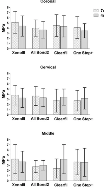
The bond strength means and standard deviations are displayed in Fig. 1. Only the ‘‘region’’ had a significant influenceonthebondstrength(3-wayRMANOVA,p<0.05). Statistically significantdifferenceswerefoundbetweenthe coronalregion(4.21.9MPa)andthecervical(3.32.2MPa) andmiddle(3.41.8MPa) regions,which werenot statisti-callydifferentfromoneanother.
The main effects, as well as the interaction between ‘‘adhesive’’ and ‘‘time’’,had a significant influence on the degreeofconversionofthecements. Homogeneousgroups wereestablishedwithTukey’stest,showingthatthedegreeof conversion ofAll-Bond2after4mwas significantlyhigher thanthatofClearfilSEBondafter7dandXenoIIIafterboth times(Table2).
The separate effect of the region was studied with the paired Student t test followed by Bonferroni’s correction (p<0.05).Thedataforallregionsweresignificantlydifferent (p=0.017),beingcoronal>cervical>middle.
Fig.1–Bondstrengthobtainedwiththeadhesivesystems asafunctionoftoothregionandstoragetime.
Table2–Meansa(%)ofthedegreeofconversionasa
functionoftheadhesivesystemandtime.(Numbersin parenthesesarestandarddeviations.).
Adhesive Time
7d 4m
All-Bond2 52(13.9AB) 72(12.1A)
XenoIII 26(23.0C) 46(19.0BC)
ClearfilSEBond 38(20.0BC) 51(7.6AB)
One-stepPlus 53(10.0AB) 59(2.5AB)
Weak correlation coefficients (Pearson, r<0.5) with no statistically significant differences (p<0.05) resulted inno lineardependencebetweenthebondstrengthandthedegree ofconversiondata.
TheAll-Bond2specimensshowedapatternof deminer-alisationandadhesivepenetrationthatdecreasedtowardsthe middleregion.Attimes,thebondingagentdidnotpenetrate the whole extension of the demineralised dentine of the deepestlevel(Fig.2AandB).
Generally,the adhesivepenetration decreasedalongthe rootcanalforallsystems.ClearfilSEBondpresentedonlya superficial interaction with dentine (ca. 1–2mm), which
remained roughly the same throughout the regions. The mildest interaction with dentine resulted in increased amounts of bonding agent coming into contact with the cementlayer,formingacombinedlayerofincreasedpacking ofC–O–Cdensityat1111cm 1.
After4m,theAll-Bond2bandsappearedlessintense,and anenlargementofthefeaturesbetween1600and1700cm 1at
about 5mm of coronal and cervical regions was observed.
Similarly,One-StepPluspresentedweakersignalsafter4m, withevidenceforcollagenexposition.
ClearfilSEBondwasthemoststableadhesiveintermsof the intensity of its bands as well as the extension of its penetrationintothedentine.AsforXenoIII,theelutionofthe bondingagentledtocollagenexpositionafter4m.
The tag formation characteristics according to bonding agentaredisplayedinFig.3A,B,CandD.
At 4m, somesections ofOne-Step Plus presentedsigns suggestiveofdegradation(notshown).
The fracture analysis was based on the main features observed under SEM of representative specimens. Overall, after7dofstorage,allthebondedinterfaceswereextruded
(cement debonded from tooth and post debonded from
cement),includingthepostfibres(Fig.3EandF).At4m,the hybridlayer/cementinterfacewasdisruptedwithhardlyany harmtothepost-cementinterfaceandthepostfibres(Fig.3G andH).
4.
Discussion
Theliteratureaboutfibrepostrestorationsisvast,mainlydue to the various issues surrounding them. In this study, we aimedtoinvestigatehowsomeofthoseissuesarerelatedand whicharepreponderantforretention.Thefindings suggest thatthedentine–adhesivejointisdefinitelytheweakestlinkof suchrestorations.20
Thebondstrengthdifferencesinthecrownandcervical/ middle parts of the post space suggest that the middle hybridisation of the post space is negligible for overall retention.Thebondstrengthsuperiorityofthecoronalregion inthepresentstudyisinaccordancewithreportsfromother studiesconcerningadhesiveendodonticrestorations.13,19,24–26
Inthatregard,severalfactorscontributetoimproved reten-tioninthecrown,fromtheefficiencyofrootcanalbrushesfor spreadingthe adhesive, to moisturecontrol and structural differencessuchastubulinumbers.9,27
Thereasonwedidnotusearootfillingmaterialwasthat removalofthefillingiscritical.Thus,canalobturationwasnot
performedbecauseitcouldleadtogreatervariabilityinthe bondstrengthresultsandnotshowtheadhesionbetweenthe adhesive and the dentine, which was the objective of the presentstudy.Plus,afillinglikegutta-perchacomplicatesthe Ramanreadingsbyincreasingthesamplefluorescence.
Thedegradationprocessesdependmostlyonthediffusion ratethatcanbeacceleratedbyworkingwithtinyspecimens. Fewer than 90d of storage were necessary to degrade microtensile specimens.28 The current findings showedno
differencesinthebondstrengthsofthe7-dayand4-month specimens,whichmayhaveoccurredduetothestorageofthe intact roots asopposed tothin sections.This methodwas preferredbecausethedeepestpartsofthepostspacearenotin directcontactwithsaliva.
Although no statisticaldifferencesregardingthe typeof bondingagentandthetimeforanalysiswereobserved,the SEMmicrographsshoweddifferencesintagformation,hybrid layerappearanceandfracturecharacteristics.Thetag forma-tion looked more intricate, with the presence of lateral branches,27fortheetch-and-rinsesystemsthanforthe
self-etching systems. Previous investigators found that, under push-out testing, failures occurred predominantly at the cement/dentine interface, which was corroborated by the presentresults.9,24,25However,animportantobservationwas
thatthefractureanalysisshowedthatthehybridlayerbecame morevulnerableafter4-monthstorageinwater,asthe7-day specimens generallyshowed disruptionofallbonded inter-facesandextrusionofthepostfibres.
Inadditiontothedifferencesinhybridlayerappearances showedby SEM,the hybridlayer qualitythroughm-Raman
spectroscopyvarieddependingonthebondingagent.These findingsdidnotseemtohaveinfluencedthebondstrengths, reinforcingtheideathattherelationshipamongstthebonding agentwettability,thehybridlayerandthebondstrengthisnot obvious.29 After 7d, the hybrid layer seemed intact, with
thicknessesthatdecreasedtowardsthemiddleregionofthe post space. The amount of bonding agent that infiltrated dentinewashigherfortheetch-and-rinsesystems.Afterthe 4-monthstorage,adecreaseofBisGMAbandsinallgroups, followedbyanincreaseofcollagenbandsinthefirstmicrons, suggestedcollagenexpositionasaresultofpolymer hydroly-sis/monomer elution. The collagen features were mainly AmideIdisorderedcollagenwithbroadersmallpeaksat1626, 1650 and 1680cm 1 (normalsharp collagenbands occur at
1663–1667cm 1).
Theextrinsicandintrinsicwater30inunprotectedcollagen
zones causes fibril loosening due to the loss of collagen helicity.31 This usuallyhappens with the use ofsimplified
bondingagentsbecauseoftheirhydrophilicnature.9Wateris
also responsible for activating MMPs, which are enzymes presentinthecollagenmatrixthatmaydegradecollagenin thelongterm.32Therefore,inthepresentstudy,the
micro-graphs and spectraof4-monthOne-step Plus and XenoIII presentedsignsofdegradation,whichwasplausiblytriggered byhydrophiliccomponentssuchasHEMA.33,34The
degrada-tionwasalsoextendedtothecementclosetodentine,where crackswereformedprobablyduetowatersorption.35
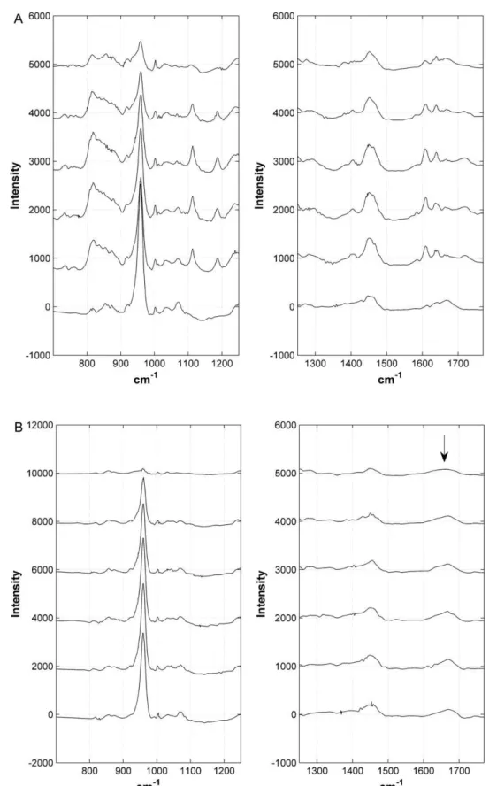
Fig.2–ThedifferencesinthepenetrationofAll-Bond2after7darediscerniblebetweenthecoronalregion(A)andthe middlepartoftherootcanalpreparation(B).Theheightsofthebandsat1610(aromaticring)and1640cmS1(vinyl)are
similar(A).Incontrastwiththesharpbandsat1663cmS1(AmideI)atthecoronallevel,broadbandsofdisorderedcollagen
agents.Overthemonths,however,theintensityofthebands andtheextensionoftheadhesivepenetrationdidnotchange asintheothermaterials,whichmayhavebeeninfluencedby thepresenceof10-MDP,whichbondschemicallytodentine throughionicbindingtocalcium.36,37Although not
statisti-cally significant, in corroboration withthe Raman findings regarding the stability of Clearfil SE Bond there was an increase in the bond strength means for the cervical and
middleregionsofthe4-monthgroups.The1-stepself-etching agentwasslightlymoreaggressiveinmineralremoval,butits 4-month spectra were quite difficult to read due to the heterogeneityofthedegradedhybridlayer.
Thelackofpenetrationinthewholeextensionof deminer-aliseddentinewasdetectedinbothetch-and-rinseand self-etchmodalities.Clinically,thismightbetranslatedintoapoor sealingthatthreatenstheprognosisofendodonticallytreated
teeth.26Moreover,non-encapsulatedcollagenissusceptibleto
degradation.38
Cementcuring was influenced by all 3factors: bonding agent,rootcanalregionandtime.Thecurrentresultsagree withmoststudies thatmeasured thedegree ofconversion indirectlythrough hardnesstesting, showing adecrease of valuesin the depths ofthe post space.5,13,25,39 Theunique
aspectofthisstudywasusingm-Ramanspectroscopy,which
doesnotrequirecontact,makingthemeasurementpossible evenifthecementisnothardenough.
In the current study,in which only the bondingagent varied,theimpactoftheadverseacid–basereactiononthe degreeofconversionofthecementwasobserved.XenoIIIled tothelowestdegreeofconversion,mainlyintheregionwhere thecuringlightwaslessintense.After4m,despitethelower conversiontendencyinthe deepest regions,the degreeof conversion seemed to have increased. Nevertheless, the differencescaused by the bondingagent in the degree of conversion of the cement did not correlate to the bond
strength, and the same was seen in a study of post
restorationswherethe hardnessandthebondstrength of thecementweremeasured.40Ontheonehand,greaterdegree
ofconversioninthepolymermatrixcorrelatedwithhigher contractionstresses.41Asaresult,thestressthatappearsas
tensileforcesattheinterfacemayformstress-relievinggaps, diminishing the bond strength. On the other hand, the residualmonomerspresentat7dmaywellhavebeenleached to the medium,decreasing the number of carbon double bondsandnotincreasingstiffnessoftherestoration.That meanstheincreaseinthedegreeofconversionwaspossibly apparentwiththe residualmonomerelutedafter4m.42In
thisregard, one could say that at 7d the reaction is still occurring,thusexplainingthelowinitialvaluesofthe self-etch groups (Table 2). However, the monomer elution rationaleismore likely,because thedegree ofconversion increasewasnotaccompaniedbybondstrength improve-ments.Thehighstandarddeviationsareprobablyaresultof thediscrepanciesofconversioninthethreeregionsofthe self-etchgroups.
5.
Conclusions
Hitherto, it seems that conventional 3-step etch-and-rinse bondedtocoronaldentineprovidessuperiorbondstrength, degreeofconversionofthecementandhybridlayerquality, althoughthesealingabilityhasstilltobequestionedinfuture studies. This conforms to our anticipated hypothesis. But theseresultsarevalidonlyforthematerialsandprocedures presented herein. The great variety of marketed bonding agents,fibrepostandcements,plusthenecessitytobuildupa coreandtoreproducemechanicalfatigueforbettersimilarity tothat achievedin the clinicsaresomeofthe issues that futureworkwillhavetoaddress.
Acknowledgements
Thisresearch was supportedby the BrazilianGovernment (FAPESP 2005/59205-4; CAPES BEX 0031-07-0; and CNPq
researchfellowship)andbytheNationalScienceFoundation (DMR0231418).TheauthorsaregratefultoProf.ThomasDuffy andFumingJiang(TheDepartmentofGeosciences,Princeton University) for the use of their laboratory and for their invaluable assistance with the Raman measurements. The authorsarealsogratefultoProf.PauletteSpencerandQiang Ye(TheUniversityofKansas)forhelpinconductingsomeof theRamanevaluationsandforusefultechnicaldiscussions. AppreciationisalsoextendedtoProf.IvanBalducciforhelpin thestatisticalanalysis.
R
e
f
e
r
e
n
c
e
s
1. NewmanMP,YamanP,DennisonJ,RafterM,BillyE. Fractureresistanceofendodonticallytreatedteethrestored withcompositeposts.JournalofProstheticDentistry
2003;89:360–7.
2. Go´mez-PoloM,Llido´ B,RiveroA,delRı´oJ,Celemı´nA.A 10-yearretrospectivestudyofthesurvivalrateofteeth restoredwithmetalprefabricatedpostsversuscast metalpostsandcores.JournalofDentistry2010;38: 916–20.
3. Hatta.ShinyaA,VallittuPK,ShinyaA,LassilaLVJ.High volumeindividualfibrepostversuslowvolumefibrepost: thefractureloadoftherestoredtooth.JournalofDentistry
2011;39:65–71.
4. PegorettiA,FambriL,ZappiniG,BianchettiM.Finite elementanalysisofaglassfibrereinforcedcomposite endodonticpost.Biomaterials2002;23:2667–82.
5. TahaNA,PalamaraJE,MesserHH.Fracturestrengthand fracturepatternsofrootfilledteethrestoredwithdirect resinrestorations.JournalofDentistry2011;39:527–35. 6. YouldasO,Alac¸amT.Microhardnessofcompositesin
simulatedrootcanalscuredwithlighttransmittingposts andglass-fiber.JournalofEndodontics2005;31:104–6. 7. HoY,LaiY,ChouI,YangS,LeeS.Effectsoflightattenuation
byfibrepostsonpolymerizationofadual-curedresin cementandmicroleakageofpost-restoredteeth.Journalof Dentistry2011;39:309–15.
8. KimYK,KimSK,KimKH,KwonTY.Degreeofconversionof dual-curedresincementlight-curedthroughthreefibre postswithinhumanrootcanals:anexvivostudy.
InternationalEndodonticJournal2009;42:667–74.
9. ChersoniS,AcquavivaGL,PratiC,FerrariM,GrandiniS, PashleyDH,etal.Invivofluidmovementthroughdentin adhesivesinendodonticallytreatedteeth.JournalofDental Research2005;84:223–7.
10.CheongC,KingNM,PashleyDH,FerrariM,ToledanoM,Tay FR.Incompatibilityofself-etchadhesiveswithchemical/ dual-curedcomposites:two-stepvsone-stepsystems.
OperativeDentistry2003;28:747–55.
11.WangY,SpencerP.Physiochemicalinteractionsatthe interfacesbetweenself-etchadhesivesystemsanddentine.
JournalofDentistry2004;32:567–79.
12.PianelliC,DevauxS,BebelmanS,LeLoupG.The micro-Ramanspectroscopy,ausefultooltodeterminethedegree ofconversionoflight-activatedcompositeresins.Journalof BiomedicalMaterialsResearch1999;48:675–81.
13.FoxtonRM,NakajimaM,TagamiJ,MiuraH.Adhesionto rootcanaldentineusingoneandtwo-stepadhesiveswith dual-curecompositecorematerials.JournalofOral Rehabilitation2005;32:97–105.
15.WakefieldCW,DraughnRA,SneedWD,DavisTN.Shear bondstrengthsofsixbondingsystemsusingpush-out methodofinvitrotesting.OperativeDentistry1998;23:69–76. 16.BoschianPestL,CavalliG,BertaniP,GaglianiM.Adhesive
post-endodonticrestorationswithfiberposts:push-out testsandSEMobservations.DentalMaterials2002;18: 596–602.
17.GoracciC,TavaresAU,FabianelliA,MonticelliF,RaffaelliO, CardosoPC,etal.Theadhesionbetweenfiberpostsandroot canalwalls:comparisonbetweenmicrotensileand push-outbondstrengthmeasurements.EuropeanJournalofOral Sciences2004;112:353–61.
18.KurtzJS,Perdiga˜oJ,GeraldeliS,HodgesJS,BowlesRW.Bond strengthsoftooth-coloredposts.Effectofsealer,dentin adhesiveandrootregion.AmericanJournalofDentistry
2003;16:31A–6A.
19.ValandroLF,BaldissaraP,GalhanoGA,MeloRM,Mallmann A,ScottIR,etal.Effectofmechanicalcyclingonthe push-outbondstrengthoffiberpostsadhesivelybondedto humanrootdentin.OperativeDentistry2007;32:579–88. 20.SpencerP,YeQ,ParkJ,ToppEM,MisraA,MarangosO,etal.
Adhesive/dentininterface:theweaklinkinthecomposite.
AnnalsofBiomedicalEngineering2010;38:1989–2003. 21.StewardsonDA,ShortallAC,MarquisPM.Theeffectof
clinicallyrelevantthermocyclingontheflexuralproperties ofendodonticpostmaterials.JournalofDentistry
2010;38:437–42.
22.DaubechiesI.Tenlecturesonwavelets.Philadelphia,PA: SocietyforIndustrialandAppliedMathematics;1992. 23.CaiC,HarringtonPB.Differentdiscretewavelettransforms
appliedtodenoisinganalyticaldata.JournalofChemical InformationandComputerScience1998;38:1161–70.
24.MarquesdeMeloR,GalhanoG,BarbosaSH,ValandroLF, PavanelliCA,BottinoMA.Effectofadhesivesystemtype andtoothregiononthebondstrengthtodentin.Journalof AdhesiveDentistry2008;10:127–33.
25. BouillaguetS,TroeschS,WatahaJC,KrejciI,MeyerJM,Pashley DH.Microtensilebondstrengthbetweenadhesivecements androotcanaldentin.DentalMaterials2003;19:199–205. 26.NagasE,CehreliZC,DurmazV,VallittuPK,LassilaLV.
Regionalpush-outbondstrengthandcoronalmicroleakage ofResilonafterdifferentlight-curingmethods.Journalof Endodontics2007;37:1464–8.
27.FerrariM,MannocciF,VichiA,CagidiacoMC.Bondingto rootcanal:structuralcharacteristicsofthesubstrate.
AmericanJournalofDentistry2000;13:255–60.
28.DeMunckJ,VanLanduytK,PeumansM,PoitevinA, LambrechtsP,BraemM,etal.Acriticalreviewofthe durabilityofadhesiontotoothtissue:methodsandresults.
JournalofDentalResearch2005;84:118–32.
29.Aguilar-MendozaJA,Rosales-LealJI,Rodrı´guez-Valverde MA,Gonza´lez-Lo´pezS,Cabrerizo-Vı´lchezMA.Wettability andbondingofself-etchingdentaladhesives.Influenceof thesmearlayer.DentalMaterials2008;24:994–1000. 30.PereiraPNR,OkudaM,SanoH,YoshikawaT,BurrowMF,
TagamiJ.Effectofintrinsicwetnessandregionaldifference ondentinbondstrength.DentalMaterials1999;15:46–53. 31.WangY,SpencerP.Quantifyingadhesivepenetrationin
adhesive/dentininterfaceusingconfocalRaman microspectroscopy.JournalofBiomedicalMaterialsResearch
2002;59:46–55.
32.MansoAP,MarqueziniLJR,SilvaSM,PashleyDH,TayFR, CarvalhoRM.Stabilityofwetversusdrybondingwith differentsolvent-basedadhesives.DentalMaterials
2008;24:476–82.
33.SpencerP,WangY.Adhesivephaseseparationatthedentin interfaceunderwetbondingconditions.JournalofBiomedical MaterialsResearch2002;62:447–56.
34.MalacarneJ,CarvalhoRM,GoesMF,SvizeroN,PashleyDH, TayFR,etal.Watersorption/solubilityofdentaladhesive resins.DentalMaterials2006;22:973–80.
35.Diaz-ArnoldAM,ArnoldMA,WilliamsVD.Measurementof watersorptionbyresincompositeadhesiveswith near-infraredspectroscopy.JournalofDentalResearch1992;71: 438–42.
36.BitterK,Meyer-LueckelH,PriehnK,KanjuparambilJP, NeumannK,KielbassaAM.Effectsoflutingagentand thermocyclingonbondstrengthstorootcanaldentine.
InternationalEndodonticJournal2006;39:809–18.
37.VanLanduytKL,SnauwaertJ,DeMunckJ,PeumansM, YoshidaY,PoitevinA,etal.Systematicreviewofthe chemicalcompositionofcontemporarydentaladhesives.
Biomaterials2007;28:3757–85.
38.BreschiL,MazzoniA,RuggerAB,CadenaroM,DILenardaR, DorigoEE.Dentaladhesionreview:agingandstabilityofthe bondedinterface.DentalMaterials2008;24:90–101.
39.MorganLFSA,PeixotoRTRC,AlbuquerqueRC,CorreˆaMFS, PolettoLTA,PinottiMB.Lighttransmissionthrougha translucentfiberpost.JournalofEndodontics2008;34:299–302. 40.AksornmuangJ,NakajimaM,FoxtonRM,TagamiJ.
Mechanicalpropertiesandbondstrengthofdual-cureresin compositestorootcanaldentin.DentalMaterials
2006;23:226–34.
41.FerracaneJL.Developingamorecompleteunderstandingof stressesproducedindentalcompositesduring
polymerization.DentalMaterials2005;21:36–42.