Aggravated Cardiac Remodeling post
Aortocaval Fistula in Unilateral
Nephrectomized Rats
Jie Wu☯, Zhong Cheng☯, Ye Gu, Wusong Zou, Mingjing Zhang, Pengfei Zhu, Shao Hu *
Heart Center at Puai Hospital, Wuhan Puai Hospital, Huazhong University of Science and Technology, Wuhan, Hubei, China
☯These authors contributed equally to this work. *hushao_puai@163.com
Abstract
Background
Aortocaval fistula (AV) in rat is a unique model of volume-overload congestive heart failure and cardiac hypertrophy. Living donor kidney transplantation is regarded as beneficial to allograft recipients and not particularly detrimental to the donors. Impact of AV on animals with mild renal dysfunction is not fully understood. In this study, we explored the effects of AV in unilateral nephrectomized (UNX) rats.
Methods
Adult male Sprague-Dawley (SD) rats were divided into Sham (n = 10), UNX (right kidney remove, n = 10), AV (AV established between the levels of renal arteries and iliac bifurca-tion, n = 18) and UNX+AV (AV at one week after UNX, n = 22), respectively. Renal outcome was measured by glomerular filtration rate, effective renal plasma flow, fractional excretion of sodium, albuminuria, plasma creatinine, and cystatin C. Focal glomerulosclerosis (FGS) incidence was evaluated by renal histology. Cardiac function was measured by echocardi-ography and hemodynamic measurements.
Results
UNX alone induced compensatory left kidney enlargement, increased plasma creatinine and cystatin C levels, and slightly reduced glomerular filtration rate and increased FGS. AV induced significant cardiac enlargement and hypertrophy and reduced cardiac function and increased FGS, these changes were aggravated in UNX+AV rats.
Conclusions
Although UNX only induces minor renal dysfunction, additional chronic volume overload placement during the adaptation phase of the remaining kidney is associated with aggra-vated cardiac dysfunction and remodeling in UNX rats, suggesting special medical care is required for UNX or congenital monokidney subjects in case of chronic volume overload as
OPEN ACCESS
Citation:Wu J, Cheng Z, Gu Y, Zou W, Zhang M, Zhu P, et al. (2015) Aggravated Cardiac Remodeling post Aortocaval Fistula in Unilateral Nephrectomized Rats. PLoS ONE 10(8): e0134579. doi:10.1371/ journal.pone.0134579
Editor:Jaap A. Joles, University Medical Center Utrecht, NETHERLANDS
Received:December 29, 2014
Accepted:July 12, 2015
Published:August 7, 2015
Copyright:© 2015 Wu et al. This is an open access article distributed under the terms of theCreative Commons Attribution License, which permits unrestricted use, distribution, and reproduction in any medium, provided the original author and source are credited.
Data Availability Statement:All relevant data are within the paper.
Funding:The authors have no support or funding to report.
in the case of pregnancy and hyperthyroidism to prevent further adverse cardiorenal events in these individuals.
Introduction
Clinical and experimental studies evaluating the cardiac-renal interaction are of importance in health care research. Renal dysfunction is common in patients with heart failure and often associated with high morbidity and mortality, moreover, cardiac and renal dysfunction may worsen each other[1,2].Clinically, heart failure may also occur in patients with nearly normal or mildly impaired renal dysfunction, as in the case of living donor kidney transplantation[3]. Although living donor kidney transplantation is regarded as beneficial to allograft recipients and not particularly detrimental to the donors and mortality of kidney donors is very low (<0.1%)[4,5], unilateral nephrectomy might cause an early abrupt decrease in plasma arginine
and simultaneous reduction in glomerular filtration rate in living kidney donors[3]. Up to two-thirds of donors after nephrectomy fulfil the criteria for chronic kidney disease stage 3 (eGFR, 30–59 mL/min per 1.73 m2) depending on baseline age and renal function[6]. Previous study showed that there was a graded inverse relationship between cardiovascular risk and eGFR, deleterious cardiovascular effects were clearly evident once eGFR falls to<60 mL/min per
1.73 m2[7]. Thus, kidney donors might face increased risk in case of additional cardiac insults. The renal and cardiac consequences and heart-kidney interaction with ischemic cardiac insult in unilateral nephrectomized rats were reported previously[8,9], while the impact of chronic volume-overload in unilateral nephrectomized rats remained largely unknown. In this study, we observed the heart-kidney interaction in a rat aortocaval fistula (AV) model established at one week post right kidney remove (UNX).
Materials and Methods
Experimental Animals and Study Groups
and symptoms such as lethargy (drowsiness, aversion to activity, a lack of physical or mental alertness), anorexia (loss of appetite, particularly prolonged behavior), bleeding, difficulty breathing, or chronic diarrhea; (c) an inability to remain upright; (d) either rapid weight loss or a net weight loss of more than 20% of body weight; and (e) unconsciousness or unresponsive-ness to external stimuli. After eight weeks, survived rats were placed in individual metabolic cages. After 5 days of adaptation, two consecutive 24-hour urine was collected from each rat. Echocardiography was performed two days after the metabolic cage studies. After echocardiog-raphy examination, rats received invasive hemodynamic and renal function measurements. Blood sample was obtained from the vena cava post above measurements. Finally, all rats were killed under deep anesthesia (70 mg/kg sodium pentobarbital intraperitoneally), and organs were removed, weighed, and processed for histological quantification.
Surgical Procedures
Laparotomy was performed under anesthesia with 1% pentobarbital sodium salt (40mg/kg, intraperitoneal injection). The right kidney was carefully separated from the adrenal gland and the surrounding tissue. The right renal artery and vein, as well as the urethra, were ligated with a 4.0 silk suture, followed by removal of the right kidney. These rats were allowed to recover from surgery in a warmed cage for 1 to 2 hours. One week after nephrectomy, rats assigned to UNX+AV group were re-anesthetized with 1% pentobarbital sodium salt (40mg/kg, intraperi-toneal injection) and the aortocaval fistula was produced according to the method described by Garcia and Diebold [10] with some modifications. Briefly, ventral abdominal laparotomy was performed in anesthetized rats, the intestines were displaced laterally and wrapped with normal saline-soaked sterilized gauze to retain moisture. The aorta and vena cava between the levels of renal arteries and iliac bifurcation were then exposed by blunt dissection of the overlaying adventitia. Both vessels were temporarily occluded proximal and distal to the intended punc-ture site, and a 18-gauge needle held on a plastic syringe was inserted into the exposed abdomi-nal aorta and advanced through the medial wall into the vena cava to create the shunt. The needle was inserted and withdrawn across the medial wall several times through the same hole, to ensure the size and presence of the fistula, before it was finally withdrawn from the aorta. The ventral aortic puncture site was immediately sealed with a drop of cyanoacrylate (Medical Adhesive Glue; Baiyun Medical Adhesive Co., China) after withdrawal of the needle. Creation of a successful fistula was confirmed by visualizing the pulsatile flow of oxygenated blood into the vena cava from the abdominal aorta. The intestines were repositioned, and the abdominal musculature and skin incisions were closed by standard techniques with absorbable suture and autoclips. Sham rats underwent similar surgical procedures as rats in UNX+AV group without right kidney removal and AV creation.
Echocardiography Examination
Echocardiography examination was performed by an investigator blinded to the study proto-col. Left parasternal and left apical echocardiographic images of light anesthetized (1% pento-barbital sodium salt, 40mg/kg, intraperitoneal injection) rats lying in a supine position were obtained with an echocardiographic system (GE Vivid 7) equipped with a 11.4 MHz trans-ducer. A two-dimensional short-axis view of the left ventricle was obtained at the level of the papillary muscles. Left ventricular M-mode tracings were used to define the internal systolic and diastolic diameters. Fractional shortening (FS) is defined as [(LVEDd-LVEDs)/LVEDd]
100%, in which LVEDd is the left ventricular end-diastolic diameter and LVEDs is the corre-sponding left ventricular end-systolic diameter. Left ventricular ejection fraction (LVEF) is defined as [(LVEDV-LVESV)/LVEDV]
end-diastolic volume and LVESV is the left ventricular end-systolic volume. Measurements repre-sent the mean of at least three consecutive cardiac cycles[11].
Hemodynamic measurements
Forty-eight hours after echocardiography examination, the rats underwent left and right heart catheterization under pentobarbital sodium salt anesthesia (40mg/kg, intraperitoneal injec-tion). The right carotid artery and right jugular vein were exposed, cannulated, saline-filled PE-50 tubings (0.58 mm ID, 0.96 mm OD) connected to a pressure transducer (BL-420F biological function experimental system, Chengdu Technology & Market Co. Ltd., Chengdu, China) were inserted into the right carotid artery and right jugular vein, respectively. The jugular vein cathe-ter was advanced into the right ventricle and the right atrium for the recording of right ventric-ular systolic pressure (RVSP), right ventricventric-ular end-diastolic pressure (RVEDP), and right atrium pressure (RAP), and the maximum rate of rise (dP/dtmax) and decrease (dP/dtmin) of RV systolic pressure. The carotid catheter was advanced into the left ventricle for the recording of left ventricular systolic pressure (LVSP), left ventricular end-diastolic pressure (LVEDP), the maximum rate of rise (dP/dtmax) and decrease (dP/dtmin) of LV systolic pressure and then was withdrawn into the aortic root to measure the heart rate (HR), systolic aortic pressure (SAP) and diastolic aortic pressure (DAP), and the mean arterial pressure (MAP) was calcu-lated as: [(2DAP)+SAP]/3.
Renal function measurements
The jugular vein catheter was connected to a syringe and each rat was given an i.v. bolus (2ml/kg) of the inulin/ para-aminohippurate(PAH) solution, followed by an i.v. infusion (25μl/min) of the inulin/PAH solution via an infusion pump. The inulin/PAH solution con-tained 3% inulin and 2% PAH in saline. An additional infusion of 25μl/min of saline into the jugular vein catheter was also performed and was continued for the duration of the experiment. Following the l hour equilibration period, hemodynamics and renal function were determined for 90 min (three 30 min clearance periods). Urine was collected in pretared vials at 30 min intervals through the bladder catheter. Arterial blood samples (500μl) were drawn through the carotid artery catheter at the midpoint of each urine collection period into prechilled heparin-ized tubes. A small blood sample was taken into microhematocrit tubes for measurement of hematocrit. The urine samples were measured gravimetrically for urine volume. Urine samples were then refrigerated until they were assayed. Plasma samples were separated by centrifuga-tion and kept at -80℃freezer until analysis.
Histology
Kidneys were weighed and then fixed by immersion for 48 hours in 4% neutral formalde-hyde after longitudinal bisection. Subsequently, they were processed for paraffin embedding according to standard procedures. Sections of 3μm were stained with periodic acid–Schiff (PAS) and microscopically evaluated by determining the incidence of focal glomerulosclerosis (FGS) as previously descried[12]. For each animal, 100 glomeruli were examined in the inner and outer cortical region and the number of sclerotic glomeruli was counted. Criteria on which glomeruli were designated as sclerotic consisted of adhesion of the glomerulus to Bowman’s capsule, thickening of Bowman’s capsule, the presence of increased amounts of PAS-positive material in the mesangial region, and/or folding of the glomerular basement membrane with entrapment of amorphous material. Glomerulosclerosis was scored by quadrants, on a scale of zero to four, where zero means no quadrant affected and four means that the whole glomerulus was affected. Gross glomerulosclerosis score was calculated as a summation of (n glomeruli)
(score)/100. An examiner blinded for the groups evaluated all sections.
Biochemistry Measurements
24-hour urine albumin was measured by using a sensitive enzyme-linked immunosorbent assay (ELISA) kit (Abcam, America). Plasma creatinine was measured with the Jaffé method with deproteinization. Plasma rBNP-45 concentrations were measured by using a sensitive enzyme-linked immunosorbent assay (ELISA) kit (Assaypro, America). Plasma cystatin C con-centrations were measured by using a sensitive enzyme-linked immunosorbent assay (ELISA) kit (R&D Systems, America). An enzymatic method was used for the determination of inulin, and a calorimetric method was used for determination of PAH in the plasma and urine sam-ples. GFR was estimated as the clearance of inulin (urine volumeurine inulin/plasma inulin).
Effective renal plasma flow (ERPF) was estimated as the clearance of PAH (urine volumeurine
PAH/plasma PAH). Renal blood flow was calculated as ERPF/(1-hematocrit). Renal vascular resistance was calculated as mean arterial pressure divided by the renal blood flow. Fractional excretion of sodium was calculated as the clearance of sodium divided by the GFR.
Statistical Analyses
All data are presented as mean±SD. Differences between groups in mean values with normal distribution were compared by 2-way ANOVA followed by Tukey test, otherwise a kruskal-wallis test followed by Mann-Whitney U test with Bonferroni correction was used. P<0.05 was
considered statistically significant.
Results
Survival and general characteristics
Sham and UNX group, 13 rats in AV group and 19 rats in UNX+AV group were analyzed. Ascites and pleural effusion were not evidenced on the rats survived to study end.
Body weight and organ weights, morphological changes
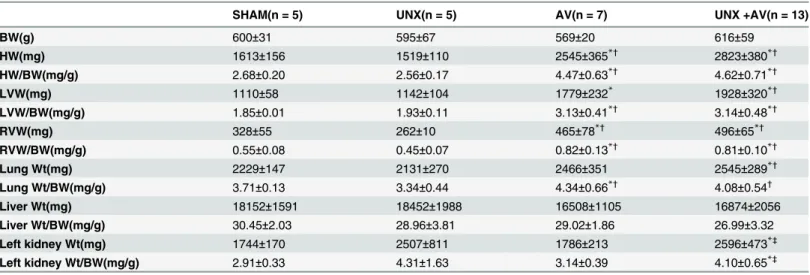
Body weight (BW) and organ weights are shown inTable 1. Eight weeks post various proce-dures, body weight was similar among groups, heart weight and heart/BW ratio, LVW(mg), LVW/BW(mg/g), RVW(mg) and RVW/BW(mg/g) were similar between UNX and Sham groups, while significantly increased in AV and UNX+AV groups compared both Sham and UNX groups. These values (except RVW/BW) also tended to be higher in UNX+AV group compared to AV group. Wet lung weight was significantly higher in UNX+AV groups com-pared to Sham and UNX groups, while wet lung/BW ratio significantly increased in AV and UNX+AV groups compared to Sham and UNX groups. Liver wet weight and liver wet weight/ BW ratio were similar among groups. Left kidney weight was significantly higher in UNX and UNX+AV groups compared to Sham and AV groups.
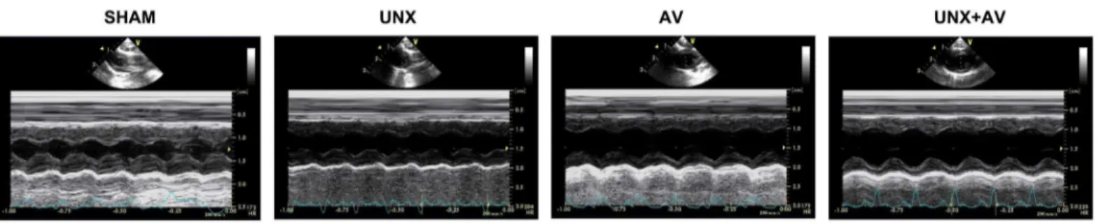
The gross morphological changes of the hearts from various groups are shown inFig 1. As expected, heart enlargement and LV and RV cavity dilation as well as and hypertrophied wall thickness were observed in the vertically cut hearts of the AV group and these changes were more apparent in UNX+AV group while there were no obvious changes in the heart of UNX and Sham groups.
Echocardiography measurements
As shown inFig 2andTable 2, LVEDD and LVESD were significantly higher while LVEF and LVFS values were significantly lower in the AV and UNX+AV groups than in Sham and UNX groups. There is a trend of severer LV remodeling and dysfunction in UNX+AV group com-pared to AV group.
Table 1. Body weight and organ weights.
SHAM(n = 5) UNX(n = 5) AV(n = 7) UNX +AV(n = 13)
BW(g) 600±31 595±67 569±20 616±59
HW(mg) 1613±156 1519±110 2545±365*† 2823±380*†
HW/BW(mg/g) 2.68±0.20 2.56±0.17 4.47±0.63*†
4.62±0.71*†
LVW(mg) 1110±58 1142±104 1779±232* 1928±320*†
LVW/BW(mg/g) 1.85±0.01 1.93±0.11 3.13±0.41*†
3.14±0.48*†
RVW(mg) 328±55 262±10 465±78*† 496±65*†
RVW/BW(mg/g) 0.55±0.08 0.45±0.07 0.82±0.13*†
0.81±0.10*†
Lung Wt(mg) 2229±147 2131±270 2466±351 2545±289*†
Lung Wt/BW(mg/g) 3.71±0.13 3.34±0.44 4.34±0.66*†
4.08±0.54†
Liver Wt(mg) 18152±1591 18452±1988 16508±1105 16874±2056
Liver Wt/BW(mg/g) 30.45±2.03 28.96±3.81 29.02±1.86 26.99±3.32
Left kidney Wt(mg) 1744±170 2507±811 1786±213 2596±473*‡
Left kidney Wt/BW(mg/g) 2.91±0.33 4.31±1.63 3.14±0.39 4.10±0.65*‡
Values are mean±SD. UNX, unilateral nephrectomy; AV, aortocavalfistula; BW, Body weight; HW, Heart weight; LVW, left ventricular weight; RVW, right ventricular weight; Wt, wet weight.
*p<0.05 vs. Sham †p<0.05 vs. UNX ‡p<0.05 vs. AV.
Hemodynamic measurements
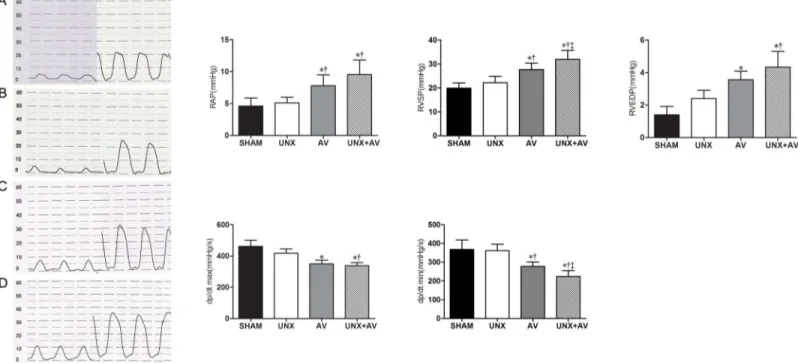
Hemodynamic measurements results are shown in Figs3and4. As shown inFig 3, RAP, RVSP and RVEDP were significantly higher while RV dP/dtmax and RV dP/dtmin were lower in AV and UNX+AV groups compared to Sham and UNX groups, these changes tended to be more apparent in UNX+AV group compared to AV group, but did not reach statistical significance.
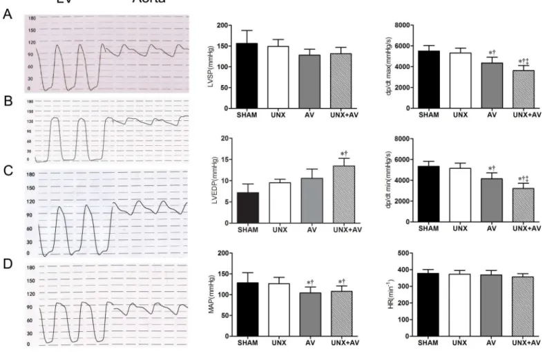
Fig 4shows that LVSP and MAP tended to be lower, LVEDP to be higher in AV and UNX+AV groups compared to Sham and UNX groups. LV dp/dtmax and dP/dtmin were significantly lower in UNX+AV group compared to Sham and UNX groups. Heart rate was similar among groups.
Biochemistry and renal function parameters
Rat brain natriuretic peptide-45 tended to be slightly higher post UNX and AV operations compared to Sham group. Plasma creatinine and cystatin C levels were significantly increased in UNX and UNX+AV groups compared to Sham and AV groups and there was a trend for more apparent changes in UNX+AV group than in UNX group, but these changes did not reach statistical significance (Table 3). GFR tended to be lower in UNX rats and was signifi-cantly reduced in UNX+AV rats compared to Sham rats; ERPF and RBF tended to be lower in UNX and UNX+AV groups and tended to be higher in AV group compared to Sham group; RVR was significantly increased in UNX and UNX+AV groups compared to Sham group; FENa was significantly increased in AV and UNX+AV group compared to Sham group; UV
Fig 1. Gross heart morphology showing ventricular enlargement and cardiac hypertrophy (of rat hearts) in the AV group, especially (of rat hearts) in the UNX+AV group.
doi:10.1371/journal.pone.0134579.g001
Fig 2. Transthoracic echocardiography.Representative B- and M-mode images of the rats from SHAM, UNX, AV and UNX+AV groups.
tended to be lower in UNX group and higher in AV group compared to Sham group; 24 hours albuminuria tended to be higher in UNX+AV group compared to Sham group (Table 3). Renal function was similar one day before AV placement in UNX+AV group and at 9 weeks post operation in UNX group (data not shown).
Renal and cardiac histology
Fig 5Ashowed that the glomerular capillary loops were thin and delicate, endothelial and mesangial cells were normal in Sham group and AV group. In UNX group, there were signs of compensatory glomerular enlargement and the mild mesangial region hyperplasia. In UNX
Table 2. Echocardiographic Parameters.
SHAM (n = 8) UNX (n = 9) AV (n = 12) UNX +AV (n = 16)
LVEDD(mm) 6.378±0.611 5.960±0.659 8.650±0.454†
10.075±0.602*† LVESD(mm) 3.139±0.888 3.162±0.686 4.740±0.724*† 5.748±0.596*†‡
EF(%) 91.50±5.37 86.44±4.22 77.58±3.09* 71.63±2.87*†
FS(%) 59.63±9.49 50.78±5.74 41.83±2.82* 36.50±2.19*†
Values are mean±SD. LVEDD, left ventricular end-diastolic dimension; LVESD, left ventricular end-systolic dimension; EF, ejection fraction; FS, fractional
shortening. *p<0.05 vs. Sham
†p<0.05 vs. UNX
‡p<0.05 vs. AV.
doi:10.1371/journal.pone.0134579.t002
Fig 3. Right heart hemodynamic analysis.[A, Sham (n = 10); B, UNX (n = 10); C, AV (n = 9); D, UNX+AV (n = 15)]. Values are mean±SD. RA, right atrium; RV, right ventricle; RAP, right atrial pressure; RVSP, right ventricular systolic pressure; RVEDP, right ventricular diastolic pressure; dP/dtmax, first derivative of pressure rise; dP/dtmin, first derivative of pressure decrease.*p<0.05 vs. Sham;†p<0.05 vs. UNX;‡p<0.05 vs. AV.
+AV group, there was an area of collagenous sclerosis running across the middle of this glo-merulus implying a focal segmental glomerulosclerosis. Focal segmental glomerulosclerosis quantification showed FGS was significantly increased in both UNX and AV groups and strik-ingly increased in UNX+AV group (Fig 5B).
Fig 6Apresented HE-stained myocardial sections showing cardiomyocyte hypertrophy in the AV group and UNX+AV group,Fig 6Bpresented Masson-stained myocardial section and collagen content was similar among groups.Fig 6Cshowed that cardiomyocyte width from left ventricle andFig 6Dshowed that from right ventricle were significantly increased in AV and UNX+AV groups compared to Sham and UNX groups.
Discussion
The major finding of present study is that chronic volume overload induced cardiac remodel-ing is significantly aggravated, which is joined by a trend of more apparent renal and cardiac dysfunction, in the presence of minor renal dysfunction post unilateral nephrectomy.
Most patients with heart failure have mild or moderate renal dysfunction[13].Both mild and severe renal dysfunctions are closely related to poor outcome in heart failure patients[14]
Fig 4. Left heart hemodynamic analysis.[A, Sham (n = 10); B, UNX (n = 10); C, AV(n = 9); D, UNX+AV (n = 15)]. Values are mean±SD. LV, left ventricle; Aorta, Aorta artery; LVSP, left ventricular systolic pressure; LVEDP, left ventricular diastolic pressure; MAP, mean arterial pressure; dP/dtmax, first derivative of pressure rise; dP/dtmin, first derivative of pressure decrease; HR, heart rate.*p<0.05 vs. Sham;†p<0.05 vs. UNX;‡p<0.05 vs. AV.
and cardiac and renal dysfunction may worsen each other[2]. Previous studies mostly focused the pathogenesis and interactive associations between renal and cardiac dysfunction on disease outcome and therapy goal[15]. Experimental studies already verified the interaction between mild renal dysfunction and ischemic cardiac insult in UNX rats[8,9], while the impact of chronic volume overload on preexisting mild renal dysfunction remains largely unknown.
Results from present study show that UNX alone is only linked with very mild renal dys-function, as shown by light focal glomerulosclerosis, compensatory enlargement of the remain-ing left kidney and slightly but significantly increased plasma creatinine and cystatin C level,
Table 3. Biochemistry and renal function parameters.
SHAM (n = 10) UNX (n = 10) AV (n = 9) UNX +AV (n = 15)
Rat BNP-45 (ng/ml) 0.14±0.02 0.17±0.03 0.23±0.11 0.25±0.06
sCr(mg/L) 4.68±0.87 5.56±0.48* 4.43±0.63† 6.06±0.75*‡
Cys-c(μg/ml) 1.51±0.19 2.66±0.63* 1.80±0.47 2.91±0.66*‡
GFR(ml/min/kg) 0.079±0.041 0.045±0.015 0.093±0.099 0.028±0.010*
ERPF(ml/min/kg) 1.672±0.935 0.871±0.712 4.093±3.629†
0.799±0.318‡ RBF(ml/min/kg) 2.944±1.727 1.468±1.200 6.792±5.795† 1.326±0.512‡ RVR(mmHg/ ml/min/kg) 45.275±24.851 89.681±35.561* 32.853±29.827†
74.259±26.763‡
FENa 0.087±0.055 0.120±0.075 0.535±0.675* 0.263±0.107*
UV(μl/min) 12.670±9.460 6.013±2.151 29.209±33.573† 11.339±3.485 24-h Albumiuria (μg) 155.49±115.25 113.35±127.44 167.52±100.34 291.01±183.13 Values are mean±SD. BNP-45, rat brain natriuretic peptide-45; sCr, plasma creatinine; Cys-c, Cystatin C; GFR, glomerularfiltration rate; ERPF, effective renal plasmaflow; RBF, renal bloodflow; RVR, renal vascular resistance; FENa, fractional excretion of sodium; UV, urine volume
*p<0.05 vs. Sham †p<0.05 vs. UNX ‡p<0.05 vs. AV.
doi:10.1371/journal.pone.0134579.t003
Fig 5. Histological features of renal tissue.[Sham (n = 10); UNX (n = 10); AV (n = 12); UNX+AV (n = 18)]. A. Periodic Acid-Schiff staining (x400) of rat with the highest proteinuria from every experimental group. B. FGS quantification.*p<0.05 vs. Sham;†p<0.05 vs. UNX;‡p<0.05 vs. AV.
and mildly reduced GFR and UV as well as significantly increased RVR. However, chronic vol-ume overload-induced cardiac and renal remodeling tended to be more significant on the basis of mild renal dysfunction post UNX. Thus, mild renal dysfunction is a key determinant for future aggravated cardiac and renal remodeling and function worsening in case of volume overload induced cardiac insult. In a landmark study, Metra and colleagues demonstrated that worsening renal function, characterized by an increase in serum creatinine levels0.3 mg/dL, alone is not an independent determinant of outcomes in patients with heart failure, but it has an additive prognostic value when it occurs in patients with persistent signs of congestion[16]. It is to note that although we evidenced more significant morphological cardiac remodeling in UNX+AV rats than in AV rats, there were mostly just a trend of aggravation on the measured cardiac and renal remodeling and dysfunction parameters in the UNX+AV rats compared to AV rats, the underlying reason might partly explained by the relative short observation period and the limited interval between UNX and AV placement (1 week interval). Future studies with longer observation period and longer interval between UNX and AV placement are
Fig 6. Histological features of cardiac tissue.[Sham (n = 10); UNX (n = 10); AV (n = 13); UNX+AV (n = 19)].A. Hematoxylin and eosin (HE) staining, x200. B. Masson’s trichrome staining, x400. C. Bar graphs of left ventricular cardiomyocyte width. D. Bar graphs of right ventricular cardiomyocyte width.*p<0.05
vs. Sham;†p<0.05 vs. UNX;‡p<0.05 vs. AV.
warranted to verify if there would be significant differences between UNX+AV and AV groups in rats with only mild renal dysfunction. Taken together, our study provoked a potential detri-mental role of slightly increase serum creatinine in case of chronic volume overload in this UNX+AV rat model.
Results from present study are obtained from the rat model of aortocaval fistula, which is a unique model of volume-overload congestive heart failure and cardiac hypertrophy. Creation of aortocaval fistula results in an immediate and sustained decrease in mean arterial pressure together with a substantial increase in venous blood flow to the right heart. These hemody-namic changes could result in compensatory activation of several neuro-hormonal systems as well as adaptive structural alterations in myocardium and vascular system, as shown previously by other investigators[17–19]. Our results showed that UNX resulted in a very mild state of chronic, mild renal function loss. Despite the very mild renal dysfunction observed in this UNX model, additional chronic volume load resulted in greater cardiac and renal remodeling and dysfunction in this model. These results are joined by increased preload of both right and left ventricles as evidenced by higher RVEDP and LVEDP in UNX+AV rats, reduced LV and RV function as shown by reduced EF, FS and LV and RV dP/dtmax and dP/dtmin. Increased RAP also indicated increased right-side volume overload in both AV and UNX+AV rats. Thus, healthy living donors might thus face increased cardiac and renal dysfunction risk in case of future cardiac insult, as in terms of chronic volume load shown in the present study.
Living donor kidney transplantation is regarded as beneficial to allograft recipients and not particularly detrimental to the donors. In 2014, 17105 kidney transplants took place in the U.S. Of these, 11,570 came from deceased donors and 5,535 came from living donors[20].According to our results, healthy living donors might face increased cardiac and renal dysfunction risk in case of future chronic volume overload cardiac insult, as in the case of pregnancy and other high output heart failure situations as in the case of hyperthyroidism[21]and patients with arte-riovenous fistula[22]. Ibrahim and colleagues determined the fetal and maternal outcomes in a large cohort of kidney donors and found that post-donation pregnancies (vs. pre-donation) were associated with a lower likelihood of full-term deliveries and a higher likelihood of fetal loss, with a higher risk of gestational diabetes, gestational hypertension, proteinuria and pre-eclampsia[23].Thus, special medical care and education should be afforded to this population to actively treat and avoid volume overload conditions and related negative renal and cardiac consequences.
Study Limitations
In conclusion, although UNX only induces minor renal dysfunction, additional chronic vol-ume overload placement during the adaptation phase of the remaining kidney is associated with aggravated cardiac remodeling and cardiac and renal dysfunction in UNX rats. Special medical care and education should be afforded to living kidney donors and people with con-genital monokidney, especially in pregnancy and other high-output situations like in patients with hyperthyroidism and arteriovenous fistula to avoid/correct volume overload conditions and to attenuate the negative consequences of volume overload in this special population.
Author Contributions
Conceived and designed the experiments: ZC YG SH. Performed the experiments: JW WZ MZ. Analyzed the data: JW PZ. Contributed reagents/materials/analysis tools: MZ PZ. Wrote the paper: JW SH.
References
1. Dupont M, Shrestha K, Tang WH. Revisiting the cardio-renal hypothesis: the pivotal role of the kidney in congestive heart failure. European journal of heart failure. 2011; 13(8):820–2. Epub 2011/07/28. doi:
10.1093/eurjhf/hfr092PMID:21791537; PubMed Central PMCID: PMCPmc3143833.
2. Metra M, Cotter G, Gheorghiade M, Dei Cas L, Voors AA. The role of the kidney in heart failure. Euro-pean heart journal. 2012; 33(17):2135–42. Epub 2012/08/14. doi:10.1093/eurheartj/ehs205PMID:
22888113.
3. Zunic G, Tomic A, Spasic S. Unilateral nephrectomy causes an early abrupt decrease in plasma argi-nine and simultaneous reduction in glomerular filtration rate in living kidney donors. Clinical biochemis-try. 2013; 46(15):1394–8. Epub 2013/05/15. doi:10.1016/j.clinbiochem.2013.04.031PMID:23669210.
4. Mullins CD, Thomas SK, Pradel FG, Bartlett ST. The economic impact of laparoscopic living-donor nephrectomy on kidney transplantation. Transplantation. 2003; 75(9):1505–12. Epub 2003/06/07. doi:
10.1097/01.tp.0000060280.28204.3cPMID:12792505.
5. Garg AX, Muirhead N, Knoll G, Yang RC, Prasad GV, Thiessen-Philbrook H, et al. Proteinuria and reduced kidney function in living kidney donors: A systematic review, meta-analysis, and meta-regres-sion. Kidney international. 2006; 70(10):1801–10. Epub 2006/09/28. doi:10.1038/sj.ki.5001819PMID:
17003822.
6. Moody WE, Chue CD, Inston NG, Edwards NC, Steeds RP, Ferro CJ, et al. Understanding the effects of chronic kidney disease on cardiovascular risk: are there lessons to be learnt from healthy kidney donors? Journal of human hypertension. 2012; 26(3):141–8. Epub 2011/05/20. doi:10.1038/jhh.2011. 46PMID:21593781.
7. Go AS, Chertow GM, Fan D, McCulloch CE, Hsu CY. Chronic kidney disease and the risks of death, cardiovascular events, and hospitalization. The New England journal of medicine. 2004; 351 (13):1296–305. Epub 2004/09/24. doi:10.1056/NEJMoa041031PMID:15385656.
8. van Dokkum RP, Eijkelkamp WB, Kluppel AC, Henning RH, van Goor H, Citgez M, et al. Myocardial infarction enhances progressive renal damage in an experimental model for cardio-renal interaction. Journal of the American Society of Nephrology: JASN. 2004; 15(12):3103–10. Epub 2004/12/08. doi:
10.1097/01.asn.0000145895.62896.98PMID:15579513.
9. Homma T, Sonoda H, Manabe K, Arai K, Mizuno M, Sada T, et al. Activation of renal angiotensin type 1 receptor contributes to the pathogenesis of progressive renal injury in a rat model of chronic cardiorenal syndrome. American journal of physiology Renal physiology. 2012; 302(6):F750–61. Epub 2011/12/14. doi:10.1152/ajprenal.00494.2011PMID:22160776.
10. Garcia R, Diebold S. Simple, rapid, and effective method of producing aortocaval shunts in the rat. Car-diovascular research. 1990; 24(5):430–2. Epub 1990/05/01. PMID:2142618.
11. Litwin SE, Katz SE, Weinberg EO, Lorell BH, Aurigemma GP, Douglas PS. Serial echocardiographic-Doppler assessment of left ventricular geometry and function in rats with pressure-overload hypertro-phy. Chronic angiotensin-converting enzyme inhibition attenuates the transition to heart failure. Circula-tion. 1995; 91(10):2642–54. Epub 1995/05/15. PMID:7743628.
12. Joles JA, van Goor H, Koomans HA. Estrogen induces glomerulosclerosis in analbuminemic rats. Kid-ney international. 1998; 53(4):862–8. Epub 1998/04/29. doi:10.1111/j.1523-1755.1998.00825.xPMID:
13. Cleland JG, Carubelli V, Castiello T, Yassin A, Pellicori P, Antony R. Renal dysfunction in acute and chronic heart failure: prevalence, incidence and prognosis. Heart failure reviews. 2012; 17(2):133–49. Epub 2012/03/20. doi:10.1007/s10741-012-9306-2PMID:22426973.
14. McAlister FA, Ezekowitz J, Tarantini L, Squire I, Komajda M, Bayes-Genis A, et al. Renal dysfunction in patients with heart failure with preserved versus reduced ejection fraction: impact of the new Chronic Kidney Disease-Epidemiology Collaboration Group formula. Circulation Heart failure. 2012; 5(3):309– 14. Epub 2012/03/24. doi:10.1161/circheartfailure.111.966242PMID:22441773.
15. Shiba N, Shimokawa H. Chronic kidney disease and heart failure—Bidirectional close link and common therapeutic goal. Journal of cardiology. 2011; 57(1):8–17. Epub 2010/10/30. doi:10.1016/j.jjcc.2010. 09.004PMID:21030212.
16. Metra M, Davison B, Bettari L, Sun H, Edwards C, Lazzarini V, et al. Is worsening renal function an omi-nous prognostic sign in patients with acute heart failure? The role of congestion and its interaction with renal function. Circulation Heart failure. 2012; 5(1):54–62. Epub 2011/12/15. doi:10.1161/
circheartfailure.111.963413PMID:22167320.
17. Zucker IH, Earle AM, Gilmore JP. The mechanism of adaptation of left atrial stretch receptors in dogs with chronic congestive heart failure. The Journal of clinical investigation. 1977; 60(2):323–31. Epub 1977/08/01. doi:10.1172/jci108780PMID:874094; PubMed Central PMCID: PMCPmc372372.
18. Flaim SF, Minteer WJ, Zelis R. Acute effects of arterio-venous shunt on cardiovascular hemodynamics in rat. Pflugers Archiv: European journal of physiology. 1980; 385(3):203–9. Epub 1980/06/01. PMID:
7190683.
19. Liu Z, Hilbelink DR, Gerdes AM. Regional changes in hemodynamics and cardiac myocyte size in rats with aortocaval fistulas. 2. Long-term effects. Circulation research. 1991; 69(1):59–65. Epub 1991/07/ 01. PMID:2054942.
20. Organ Procurement and Transplantation Network website. U.S. Department of Helath and Human Ser-vices. Available:http://optn.transplant.hrsa.gov/.
21. Vargas-Uricoechea H, Sierra-Torres CH. Thyroid hormones and the heart. Hormone molecular biology and clinical investigation. 2014; 18(1):15–26. Epub 2014/11/13. doi:10.1515/hmbci-2013-0059PMID:
25389997.
22. MacRae JM, Pandeya S, Humen DP, Krivitski N, Lindsay RM. Arteriovenous fistula-associated high-output cardiac failure: a review of mechanisms. American journal of kidney diseases: the official journal of the National Kidney Foundation. 2004; 43(5):e17–22. Epub 2004/04/28. PMID:15112194.
![Fig 5. Histological features of renal tissue. [Sham (n = 10); UNX (n = 10); AV (n = 12); UNX+AV (n = 18)]](https://thumb-eu.123doks.com/thumbv2/123dok_br/16448405.197328/10.918.83.844.681.1006/fig-histological-features-renal-tissue-sham-unx-unx.webp)
![Fig 6. Histological features of cardiac tissue. [Sham (n = 10); UNX (n = 10); AV (n = 13); UNX+AV (n = 19)].A](https://thumb-eu.123doks.com/thumbv2/123dok_br/16448405.197328/11.918.63.849.123.690/fig-histological-features-cardiac-tissue-sham-unx-unx.webp)
