J of Evolution of Med and Dent Sci/ eISSN- 2278-4802, pISSN- 2278-4748/ Vol. 3/ Issue 39/Aug 28, 2014 Page 10026
A PROSPECTIVE STUDY OF SURGICAL CORRECTION OF CTEV BY
CINCINNATI APPROACH
K. G. Gopalakrishna1, K. S. Manjunath2, Chandrashekar3
HOW TO CITE THIS ARTICLE:
K. G. Gopalakrishna, K. S. Manjunath, Chandrashekar. A Prospective Study of Surgical Correction of CTEV by Cincinnati Approach . Journal of Evolution of Medical and Dental Sciences 2014; Vol. 3, Issue 39, August 28; Page: 10026-10046, DOI: 10.14260/jemds/2014/3301
ABSTRACT: BACKGROUND: Surgical correction of congenital talipes equino varus (ctev) is to address adequately all aspects of this complex foot deformity. Various exposures have been elucidated with varying results and complications. This prospective study discusses the cincinnati approach advocated by Mackay to address the various aspects of clubfoot correction. Objective: To study the adequacy of exposure, wound healing and problems related to Cincinnati approach. To study the effectiveness of primary surgical correction. METHODS: The present prospective study includes treatment of 24 feet in 21 patients with clubfoot treated with posteromedial and lateral soft tissue release by Cincinnati approach and followed up with an average follow up of 6.9 months. 3 (12.5%) were followed up for 1 year and 3 (12.5%) lost for the follow up. 18 (75%) were followed up to 6 months. All were resistant to correction by conservative method. Age at operation averaged 1.6 year, ranged from 9 months to 3 years. RESULTS: Results were evaluated using Laaveg and Ponseti functional rating system of clubfoot. Postoperatively the average arc of movement in ankle joint was 370, which was 50% of normal limb. Inversion, eversion movement of subtalar joint was 230, bimalleolar angle was 770.The functional results were excellent in 6 feet (25%), good in 12 feet (50%), fair in 5 feet (20.83%) and poor in 1 foot (4.17%). In our series complications encountered were post-operative focal necrosis in 3 feet (12.5%) and marginal necrosis in 3 feet (12.5%). CONCLUSION: Cincinnati incision provides an adequate exposure for extensive surgical release under direct vision, there by bony realignment of talus over calcaneus was restored and hence the restoration of normal bimalleolar angle. Skin closure is not found to be a problem in achieving a primary closure. Wound healing leaves only a thin and cosmetically acceptable scar.
KEYWORDS: CTEV, cincinnati incission, subtalar release, bimalleolar angle.
INTRODUCTION: Idiopathic clubfoot is one of the oldest and commonest congenital deformities of mankind. Hippocrate (460 BC -377BC)1 was the first person to describe club foot. The initial treatment of clubfoot is non-operative. It was concluded that most of the club foot can be successfully managed by a series of Plaster casts and wedging without the use anesthetics, or operative procedures with better results.2,3
In neglected, resistant, recurrent, relapsed, failed conservatively treated CTEV pathological contractures of the soft tissues prevented the reduction of the navicular on the head of talus and calcaneum and surgical correction becomes necessary.4
The comprehensive soft tissue release in current favor is posteromedial release of Turco. Mukhopadhyay procedure with its variants and circumferential release as described by McKay, Carrol and Simons, etc. are some of more than hundred surgeries described.
J of Evolution of Med and Dent Sci/ eISSN- 2278-4802, pISSN- 2278-4748/ Vol. 3/ Issue 39/Aug 28, 2014 Page 10027 incomplete soft tissue release, incomplete peritalar release, incomplete subtalar release and incomplete correction of rotation deformity of calcaneum. This could lead on to either persistence or recurrence of deformity.
To predicate the above mentioned problem circumferential Cincinnati approach has been practiced worldwide. A significantly lower incidence of wound complications was seen in the Cincinnati treatment group when compared with the modified Turco.5 so we intend to study the surgical management of CTEV by Cincinnati approach in light of the published literature.
OBJECTIVES OF STUDY:
RELATED TO APPROACH: To study the adequacy of exposure, wound healing and problems related to Cincinnati approach.
RELATED TO PROCEDURE: To study the effectiveness of primary surgical correction.
Surgery in the treatment of clubfoot must be tailored to the age of the child and to the deformity to be corrected.
A modified McKay procedure through a transverse circumferential (Cincinnati) incision is our preferred technique
Any approach should be able to address the release in all quadrants, which are as follows: Plantar: Plantar fascia, abductor hallucis, flexor digitorum brevis, long and short plantar ligaments
Medial: Medial structures, tendon sheaths, talonavicular and subtalar release, tibialis posterior, FHL, and FDL lengthening
Posterior: Ankle and subtalar capsulotomy, especially releasing post talofibular and tibiofibular ligaments and the calcaneofibular ligaments
Lateral: Lateral structures, peroneal sheath, calcaneocuboid joint, and completion of talonavicular and subtalar release
Any approach should afford adequate exposure. Structures to be released or lengthened are the following:
Achilles tendon.
Tendon sheaths of the muscles crossing the subtalar joint. Posterior ankle capsule and deltoid ligament.
Inferior tibiofibular ligament. Fibulocalcaneal ligament.
Capsules of the talonavicular and subtalar joints.
Division of associated ligaments around the subtalar joint. Plantar fascia and intrinsic muscles.
J of Evolution of Med and Dent Sci/ eISSN- 2278-4802, pISSN- 2278-4748/ Vol. 3/ Issue 39/Aug 28, 2014 Page 10028 Hospitals, Bangalore between November 2007 to October 2009 A prior consent was obtained from all the patients and the study was approved by the Ethical Committee of the Hospital.
METHODOLOGY: Required data was collected from patients admitted in Victoria Hospital, Bowring and Lady Curzon hospitals. All patients included in study were assessed pre-operatively and post operatively (clinical and functional) as per Laaveg and Ponseti functional rating system score.
Children with idiopathic CTEV, more than 6 months of age (Neglected CTEV)-<3years, Rigid CTEV, partially corrected CTEV (failed conservative)/recurrent CTEV were included for the study. X-rays of foot, routine Blood investigations, weight recording done. Cases were followed up at 2 weeks, 6 weeks, 6months and 1year.
EXCLUSION CRITERIA: less than 6 months
Flexible CTEV.
Aquired talipes equino varus (trauma, burns, neurogenic, muscular dystrophies-, AMC, Teratological-etc).
>3 years where bony procedures were needed.
OBSERVATION AND RESULTS: The present study includes treatment of 24 feet in 21 patients with clubfoot treated with posteromedial and lateral soft tissue release by Cincinnati approach from the period November 2007 to October 2009 and followed up with an average follow up of 6.9 months. 3 (12.5%) were followed up for 1 year and 3 (12.5%) lost for the follow up. 18 (75%) were followed up to 6 months. Age at operation averaged 1.6 year ranged from 9 months to 3 years.
Age distribution:
Age in years Total unilateral Bilateral
9 months _1 year 7 5 2
1.1 _2 year 9 8 1
2.1_3 year 5 5 0
Total 21 18 3
Table 1: Showing age distribution
Sex distribution:
Sex No. of cases Percentage
Male 15 71.4%
Female 6 28.6%
Total 21 100
Table 2: showing sex distribution
Side affected No. of cases Percentage
Unilateral 18 86%
Bilateral 3 14%
Total 21 100
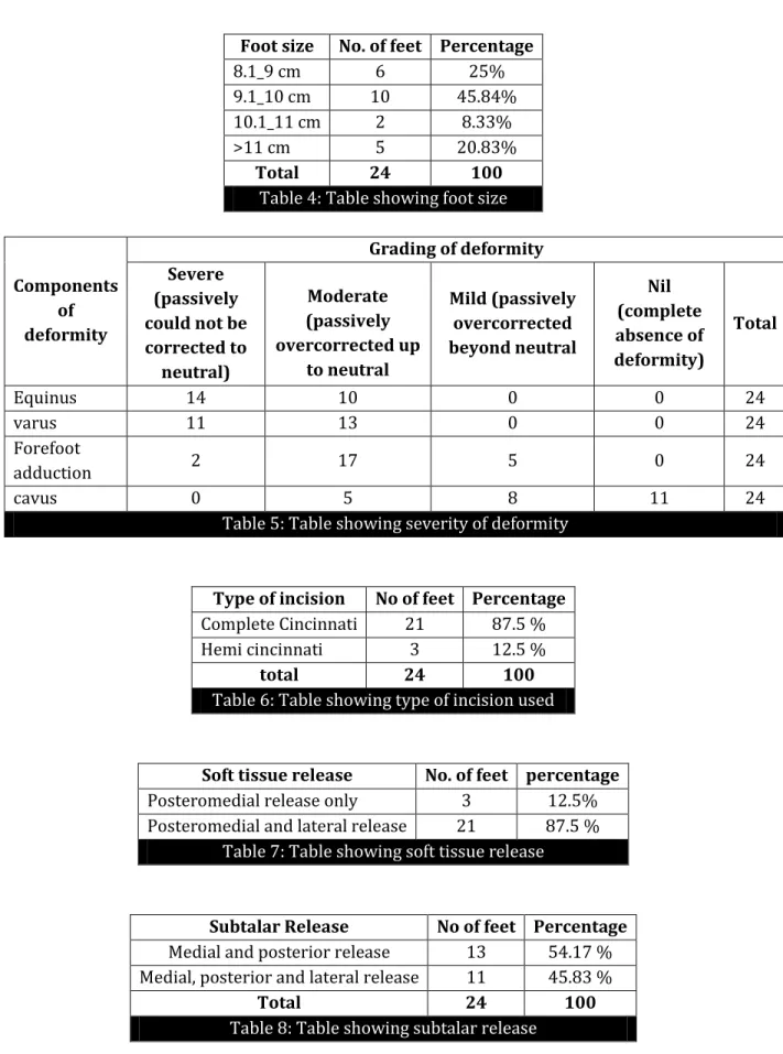
J of Evolution of Med and Dent Sci/ eISSN- 2278-4802, pISSN- 2278-4748/ Vol. 3/ Issue 39/Aug 28, 2014 Page 10029 Foot size No. of feet Percentage
8.1_9 cm 6 25%
9.1_10 cm 10 45.84%
10.1_11 cm 2 8.33%
>11 cm 5 20.83%
Total 24 100
Table 4: Table showing foot size
Components of deformity
Grading of deformity Severe
(passively could not be corrected to neutral) Moderate (passively overcorrected up to neutral Mild (passively overcorrected beyond neutral Nil (complete absence of deformity) Total
Equinus 14 10 0 0 24
varus 11 13 0 0 24
Forefoot
adduction 2 17 5 0 24
cavus 0 5 8 11 24
Table 5: Table showing severity of deformity
Type of incision No of feet Percentage
Complete Cincinnati 21 87.5 %
Hemi cincinnati 3 12.5 %
total 24 100
Table 6: Table showing type of incision used
Soft tissue release No. of feet percentage
Posteromedial release only 3 12.5%
Posteromedial and lateral release 21 87.5 % Table 7: Table showing soft tissue release
Subtalar Release No of feet Percentage Medial and posterior release 13 54.17 % Medial, posterior and lateral release 11 45.83 %
Total 24 100
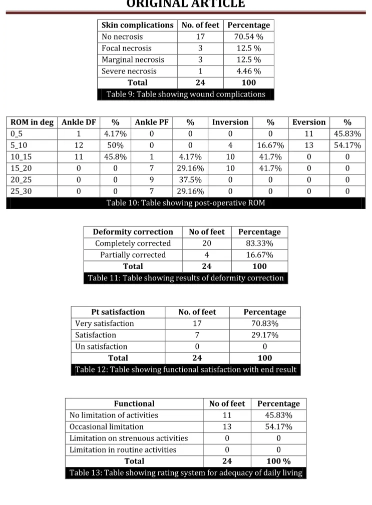
J of Evolution of Med and Dent Sci/ eISSN- 2278-4802, pISSN- 2278-4748/ Vol. 3/ Issue 39/Aug 28, 2014 Page 10030 Skin complications No. of feet Percentage
No necrosis 17 70.54 %
Focal necrosis 3 12.5 %
Marginal necrosis 3 12.5 %
Severe necrosis 1 4.46 %
Total 24 100
Table 9: Table showing wound complications
ROM in deg Ankle DF % Ankle PF % Inversion % Eversion %
0_5 1 4.17% 0 0 0 0 11 45.83%
5_10 12 50% 0 0 4 16.67% 13 54.17%
10_15 11 45.8% 1 4.17% 10 41.7% 0 0
15_20 0 0 7 29.16% 10 41.7% 0 0
20_25 0 0 9 37.5% 0 0 0 0
25_30 0 0 7 29.16% 0 0 0 0
Table 10: Table showing post-operative ROM
Deformity correction No of feet Percentage
Completely corrected 20 83.33%
Partially corrected 4 16.67%
Total 24 100
Table 11: Table showing results of deformity correction
Pt satisfaction No. of feet Percentage
Very satisfaction 17 70.83%
Satisfaction 7 29.17%
Un satisfaction 0 0
Total 24 100
Table 12: Table showing functional satisfaction with end result
Functional No of feet Percentage
No limitation of activities 11 45.83%
Occasional limitation 13 54.17%
Limitation on strenuous activities 0 0
Limitation in routine activities 0 0
Total 24 100 %
J of Evolution of Med and Dent Sci/ eISSN- 2278-4802, pISSN- 2278-4748/ Vol. 3/ Issue 39/Aug 28, 2014 Page 10031
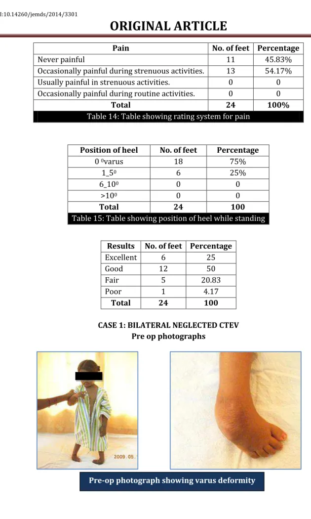
Pain No. of feet Percentage
Never painful 11 45.83%
Occasionally painful during strenuous activities. 13 54.17%
Usually painful in strenuous activities. 0 0
Occasionally painful during routine activities. 0 0
Total 24 100%
Table 14: Table showing rating system for pain
Position of heel No. of feet Percentage
0 0varus 18 75%
1_50 6 25%
6_100 0 0
>100 0 0
Total 24 100
Table 15: Table showing position of heel while standing
Results No. of feet Percentage
Excellent 6 25
Good 12 50
Fair 5 20.83
Poor 1 4.17
Total 24 100
CASE 1: BILATERAL NEGLECTED CTEV Pre op photographs
J of Evolution of Med and Dent Sci/ eISSN- 2278-4802, pISSN- 2278-4748/ Vol. 3/ Issue 39/Aug 28, 2014 Page 10032
Pre op showing equinus deformity intra op showing lateral subtalar release
Wound at second week Wound at second week
A/K POP CAST application with corrective shoes
J of Evolution of Med and Dent Sci/ eISSN- 2278-4802, pISSN- 2278-4748/ Vol. 3/ Issue 39/Aug 28, 2014 Page 10033 CASE 2: BILATERAL CTEV
Pre op photographs
Hemi Cincinnati incision was used Skin condition at 2 weeks
J of Evolution of Med and Dent Sci/ eISSN- 2278-4802, pISSN- 2278-4748/ Vol. 3/ Issue 39/Aug 28, 2014 Page 10034 CASE 3: NEGLECTED CTEV
With corrective shoes at 18 months
At 18 months showing corrected equinus Heel position at 18 months
J of Evolution of Med and Dent Sci/ eISSN- 2278-4802, pISSN- 2278-4748/ Vol. 3/ Issue 39/Aug 28, 2014 Page 10035
Marginal wound necrosis at 2 weeks
At 6 months
J of Evolution of Med and Dent Sci/ eISSN- 2278-4802, pISSN- 2278-4748/ Vol. 3/ Issue 39/Aug 28, 2014 Page 10036
CASE 4: BILATERAL CTEV
With corrective shoes
Pre-operative photographs
J of Evolution of Med and Dent Sci/ eISSN- 2278-4802, pISSN- 2278-4748/ Vol. 3/ Issue 39/Aug 28, 2014 Page 10037
CASE 5: BILATERAL NEGLECTED CTEV
Corrected heel varus
J of Evolution of Med and Dent Sci/ eISSN- 2278-4802, pISSN- 2278-4748/ Vol. 3/ Issue 39/Aug 28, 2014 Page 10038
Pre-operative photographs
Severe skin necrosis at 2 weeks
J of Evolution of Med and Dent Sci/ eISSN- 2278-4802, pISSN- 2278-4748/ Vol. 3/ Issue 39/Aug 28, 2014 Page 10039
DISCUSSION:
Approach and age: CTEV is an abnormality involving the ankle and foot, which causes adduction, supination, varus and equinus deformity. The long term aim of the management is to achieve a functional, pain free, cosmetically acceptable, and mobile as close to normal feet. Surgical treatment is usually required in failed conservatively managed clubfeet, neglected CTEV, rigid CTEV etc.
The general principle of surgical procedure is to prevent multiple procedures to achieve full correction because complications exponentially increase with the number of interventions. Hence a combination of right approach and an extensive soft tissue release including posteromedial release, lateral release, and subtalar release will achieve the goal of the surgical treatment.
In our study we have adopted Cincinnati approach with posteromedial and lateral soft tissue release and subtalar release. Though better results were obtained by posterior and posteromedial soft tissue release in younger children it has been suggested that early surgical treatment lead to severe fibrosis and development of rigid foot. Thus surgery should better be executed between 1 and 2 years of child age6, 7, 8.
In our study we have 14 children were above the age of 1 year and remaining 7 children were almost nearer to 1 year. 5 children were operated between 2-3 years of age. They belonged to severely neglected CTEV. DePuy and Drennan divided 44 feet treated by PMR in 3 groups which were operated on 4, 9 and 16 months. They did not find significant functional or radiographic difference between the groups. However in younger group less tarsal bone deformity was observed than in older groups.9
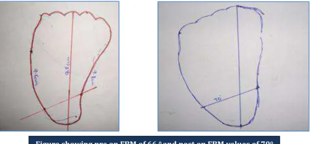
Foot Size: Simons recommended that the size of the foot rather than the age of the patient be used to determine the optimum time to perform the surgery. He stated that the foot should be >=8 cm long at the time of surgery. 10The longitudinal length of the foot was measured by taking foot tracing. The bimalleolar plane was marked by connecting lateral and medial malleolar marking. The longitudinal plane of the foot drawn from second toe to the tip of the heel intersecting bimalleolar line is measured for the foot size. Normal bimalleolar angle is 760.11
J of Evolution of Med and Dent Sci/ eISSN- 2278-4802, pISSN- 2278-4748/ Vol. 3/ Issue 39/Aug 28, 2014 Page 10040 Deformity: The deformity was very severe in children above the age of one year. 7 feet in 6 children were less than 1 year and deformity is of less severe. The poor result was noted in less deformed foot. However the fair results were observed in both mild and severely deformed feet. Our study is not very conclusive about the correlation of severity of deformity and the end result.
Severe deformities pose the problem of severe skin contracture on the medial side of foot and ankle, which is notorious for poor nutrition and delayed healing, there by high incidence of wound necrosis. The severe deformities also encounter cavus deformity of foot due to severely contracted plantar fascia and instrinsic muscles of the foot increasing the chances of poor outcome.12
Incision: Incision to CTEV falls in to three categories. Turcos posteromedial incision which is oblique and hockey stick like. Since the Turcos incision cuts across the skin crease it causes non-acceptable skin necrosis and ugly scar. This incision being eccentrically placed exposure and visualization of other side of ankle and subtalar joint is difficult there by an incomplete soft tissue release.
Carolls incision includes two separate incisions, a curvilinear medial incision and posterolateral incision to allow adequate exposure for plantar, lateral, medial and posterior structures. He emphasized the release of plantar fascia and capsulotomy of calcaneo cuboid joint were critical in achieving correction.10
The Cincinnati incision is based on thorough study of anatomy of the foot and ankle. The obvious goal of a good surgical approach is to provide adequate exposure of the pathologically involved structures while at the same time minimizing morbidity resulting from damage to the blood vessels, nerves, tendons, and articular surfaces in the area and allows the surgeon to correct a deformity in all planes simultaneously.
Satisfactory reduction and stabilization of the tarsal bones is easily accomplished under direct vision. The Cincinnati incision also proved to be more acceptable cosmetically than other incisions as it often heals with thin scar. The problems with Cincinnati incision have been minimum like marginal skin necrosis and limited exposure of tendo Achilles.13
We have used complete Cincinnati incision in 21 feet (87.5%) and hemi Cincinnati incision in 3 feet (12.5%). We have observed the exposure is very adequate to visualize the medial, posterior and lateral subtalar release under direct vision. The only problem we encountered during the procedure is the limited exposure for tendo Achilles, there by requiring the need of excessive retraction posteriorly for TA lengthening. We have achieved primary wound closure in all cases without any problem.
Skin Necrosis: Marginal skin edge necrosis is seen in 3 feet (12.5%). Small area of focal necrosis in 3 feet (12.5%). 1 foot (4.46%) had severe necrosis. The child with severe necrosis had severe rigid CTEV. The severe and rigid deformity, subcutaneous thicker suture material could have been the reason for severe necrosis. However the wound was completely healed in 10 days of time without eventually affecting the outcome of the functional result. The children with marginal necrosis and focal necrosis host no problem and went on healing without any delay. 71% of feet healed without any delay.
J of Evolution of Med and Dent Sci/ eISSN- 2278-4802, pISSN- 2278-4748/ Vol. 3/ Issue 39/Aug 28, 2014 Page 10041 skin necrosis in 15% and deep necrosis including Achilles tendon in 6%. Dimeglo reported no necrosis in 91 cases.6,15
Zhon-Liau Lee has reported skin problem in 11 feet in total of 60 feet (18.3%). Among them 7 feet had mild patchy necrosis, 3 feet had moderate necrosis and one foot had extensive skin necrosis, wound dehiscence and deep infection.16 In a retrospective study by Hsu, Wellington K et al., of 217 patients who underwent primary PMR using modified Turco or Cincinnati incision, a significantly low incidence of wound complication is seen in Cincinnati incision than Turcos incision. (6.9% vs 19.6%).5
There are various recommendation to prevent wound necrosis17,18,13,19,20.
Benjamin Joseph et al., using hemi Cincinnati incision for posteromedial soft tissue release reported that 42 feet could be put in to neutral plantigrade position at end of operation and found satisfactory healing without any wound necrosis. Wound closure by tension had led on to superficial dehiscence in 14 % of case. Minor degree of wound dehiscence may be avoided if the foot is held in inversion postoperatively.21
In our study we have noticed 2 children had very mild wound dehiscence less than 2 mm. One child had wound dehiscence more than 2 mm. However all the wounds healed without posing any problem. We have noticed retaining subcutaneous fat tissue; sharp dissection without undermining the tissue plane, good haemostasis, tension free closure facilitates better healing without any necrosis.
Evaluation of ROM: In Mc. Kay evolution rating system ankle arc of motion of 400-50 0 scores better than arc of movement 350 - 400. Arc of movement less than 350 face poorly11. Indeed the radiographic studies shows, most motion of plantar flexion and dorsiflexion occur not in true ankle joint but at the mid tarsal joint.22
Boone and Ayen reported an estimate of 710 of total ankle motion for normal male children23. Gianneestros considered 600 to be normal passive ankle motion in children24. Jerry B Magone et. al., report 510 as the average ankle arc of movement in normal feet in their study.22 Stauffer reported the average ROM during walking gait is 24.40.25
Simons was the only investigator who compared radiographic true ankle motion in clubfeet pre operatively and post operatively. The average pre-operative ROM was 310 and post-operative ROM was 290. But the arc of motion directed more towards dorsiflexion by 100. In our study a significantly improved range of ankle motion from preoperative range of 260 to post-operative range of 370 of arc of motion as been giving good functional result.
Subtalar Release: Turcos posteromedial release though widely used deformity may persist or recur as the calcaneus has not been fully freed to allow it to rotate beneath the talus. Mc Kay described the concept of calcaneal rotation and subsequently reported by Ghali et al., confirmed the correctness of Mc Kay concept. The posteromedial release does not permit full correction. Only the complete subtalar release accomplishes the full de rotation of calcaneus in single procedure. The complete subtalar release was performed in four basic stages:
J of Evolution of Med and Dent Sci/ eISSN- 2278-4802, pISSN- 2278-4748/ Vol. 3/ Issue 39/Aug 28, 2014 Page 10042 The optional additional procedures are calcaneo cuboid capsulotomy, calcaneo cuboid osteotomy and plantar release.26
Excellent and good short-term results have been reported in congenital clubfoot in 71% of the cases by McKay, in 72% of the cases by Simons, in 69% of the cases by Rumyantsev and Ezrohi, in 83% of the cases by Centel et al., in 63% of the cases by Magone et al., and in 84% of the cases by Turco, which were treated by extensive surgical dissection. Denis reports 76.6% of good and excellent results in extensive soft tissue release.
Dobbs et al, has evaluated the long term result of 60 feet with extensive soft tissue release and 14 with limited soft tissue release. All the 14 feet with limited release showed poor result and some of the extended released feet also had poor result. They observed and found that the poor outcome could be significantly correlated with inadequate surgical release. 67% of their poor outcome was noticed and found to be significantly correlated with inadequate surgical release.6
Bimalleolar angle/axis: Measurement of foot bimalleolar angle is an objective, simple and effective method for foot classification prior to treatment and evaluation of its results in congenital clubfoot. The feet in CTEV were classified on basis of bimalleolar angle into four types:
Type I 750-850
Type II 700-740 and 860-900
Type III 650-690 and >900
Type IV < 650.
Deniz et al, in his long term evaluation study found that all type IV feet (less than 650) had poor functional score. A.K.Jain et al, in their study on evaluation of FBM in management of CTEV have observed the mean FBM angle of 73.20 in grade I deformity (mild CTEV), mean FBM of 66.60 in grade II deformity (moderate CTEV), mean FBM of 54.70in grade III deformity (severe deformity).27
In our study we had 1 poor result in type IV feet with bimalleolar angle less than 650. We had average pre-operative bimalleolar angle of 730 and average post-operative bimalleolar angle of 770.
J of Evolution of Med and Dent Sci/ eISSN- 2278-4802, pISSN- 2278-4748/ Vol. 3/ Issue 39/Aug 28, 2014 Page 10043 The fundamental principles of soft tissue release are:
1. To achieve bony realignment in talo navicular articulation and in subtalar articulation, 2. Proper alignment of bi malleolar axis.
3. Improve the range of motion.
In our study 5 feet with fair functional result had only medial and posterior soft tissue release. In all the feet with extensive soft tissue release, result was good to excellent.
Position of heel after surgery reflect optimised bimalleolar axis. Heel position of 0-50 varus gives an optimal correction. Valgus position of heel tends to reduce the bimalleolar axis reflecting the poor result. However certain degrees of varus position of heel also suggest non-optimal bimalleolar axis and resultant poor function. Heel position can be graded as normal neutral heel, heel in mild varus (1-50), heel in moderate varus (6-100) and heel in valgus (>50 valgus). Heel in moderate varus and heel in valgus tends to give poor results.
In our study we have 75% of feet were of 00 varus and 25% of feet having 0-50 varus reflecting good restoration of bimalleolar axis. This again reflects adequate soft tissue release medially, posteriorly and laterally.
Almost all patients in our study never had intoeing gait. Intoeing gait without metatarsus adductor deformity could be due to posterior displacement of lateral malleolus reducing bimalleolar axis. This intoeing gait could be prevented by
1. The foot bimalleolar axis should be intraoperatively adjusted to 900 as suggested by Mc Kay. 2. Foot should be casted in external rotation as recommended by Caroll.
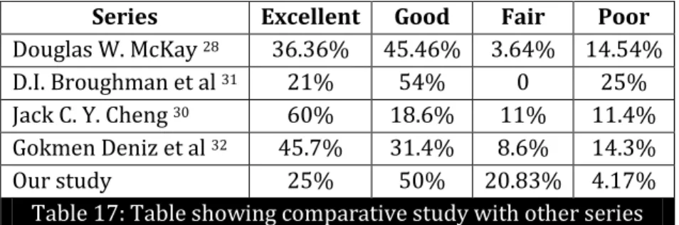
RESULTS: In a study by Douglas W.Mc Kay28, of 55 feet he found excellent in 36.36%, good in 45.46%, fair in 3.64% and poor result in 14.54%. D.I. Broughman et al 29 reported that in his series with 32 feet he found excellent in 21%, good in 54% and poor in 25% of feet. In a study by Jack C. Y. Cheng, 30 of 70 feet he reported excellent in 60%, good in 18.6%, fair in 11% and poor in 11.4%. Gokmen Deniz et al, 6 reported that in his series with 35 feet he found excellent in 16 feet (45.7%), good in 11 feet(31.4%), moderate in 3 feet(8.6%) and poor in 5 feet (14.3%). The functional results were evaluated at the end of the study showed excellent in 6 feet (25%), good in 12 feet (50%), fair in 5 feet (20.83%) and poor in 1 foot (4.17%).
Series Excellent Good Fair Poor
Douglas W. McKay 28 36.36% 45.46% 3.64% 14.54%
D.I. Broughman et al 31 21% 54% 0 25%
Jack C. Y. Cheng 30 60% 18.6% 11% 11.4%
Gokmen Deniz et al 32 45.7% 31.4% 8.6% 14.3%
Our study 25% 50% 20.83% 4.17%
Table 17: Table showing comparative study with other series
J of Evolution of Med and Dent Sci/ eISSN- 2278-4802, pISSN- 2278-4748/ Vol. 3/ Issue 39/Aug 28, 2014 Page 10044 angle. Skin closure is not found to be a problem in achieving a primary closure. Wound healing leaves only a thin and cosmetically acceptable scar.
However the only problem noticed is limited exposure of ten do Achilles tendon for z-plasty. Complete subtalar release is essential to effectively correct the deformity and proper realignment of tarsal bones. Post-operative marginal wound necrosis and wound dehiscence is of no consequences.
BIBLIOGRAPHY:
1. Steven J. De Valentine, Timothy J.Blakeslee. Congenital Talipes Equinovarus . -146.
2. Cummings. J. Lovel WW, Current concept operative treatment of congenital idiopathic
clubfoot J Bone Joint Surg., 1988, 70-A, 1108.
3. Ferlic, Randolph J MD Partial wound closure after surgical correction of equinovarus deformity. Journal of Pediatric Orthopaedics vol 17 (4) 1997. 486-489.
4. Turco Vincent. J, Surgical correction of Residual congenital clubfeet with one stage postero
medial release with internal fixation J. Bone Joint Surg., 1971, 53-A, 4770.
5. Nimityongskul P, Anderson LD, Herbert DE. Surgical treatment of clubfoot: a comparison of two techniques. Foot Ankle. 1992 Mar-Apr; 13 (3): 116-24.
6. Franke J, Hein G. Our experiences with the early operative treatment of congenital clubfoot. J Pediatr Orthop 1988; 8: 26-30.
7. Main BJ, Crider RJ, Polk M, Lloyd-Roberts GC, Swann M, Kamdar BA. The results of early operation in talipes equino-varus. A preliminary report. J Bone Joint Surg [Br] 1977; 59:337-41. 8. DePuy J, Drennan JC. Correction of idiopathic clubfoot: a comparison of results of early versus
delayed posteromedial release. J Pediatr Orthop 1989; 9: 44-8.
9. Kalenderer O, Aguş H, Ak M, Ozluk S. Correlation of clinical and radiologic results of complete subtalar release in congenital clubfoot. [Article in Turkish] Acta Orthop Traumatol Turc 2003; 37:368-73.
10.Peter L. Williams. Grays Anatomy. In: Roger W. Saamers (ed), Skeletal System, chapter 6, . In: Stanley salmons (ed), Muscle, chapter 7, 38th edition, Churchill Livingstone (ELBS), 1995: 712-736, 886-894.
11.Simons, G. W., Complete subtalar release in clubfeet J. Bone Joint Surg., 1985, 67-A, 1044-1055.
12.Simons. G. W., Analytical radiography of clubfeet J. Bone Joint Surg., 1977, 485.
13.Rai P. K. and Sharma O. P., Correction of clubfoot by combined posteromedial and subtalar
release Ind. J. Ortho, 1986, 14, 94-96.
14.Karakurt L, Yilmaz E, Inci M, Serin E, Ozturk M. Early results of complete subtalar release in congenital clubfoot deformity. [Article in Turkish] Acta Orthop Traumatol Turc 2003; 37: 53-62. 15.Zhon-Liau Lee, Chung-Hsiung Shih, Skin complications of surgical treatment for talipes
equinovarus using Cincinnati incision. J. Orthop Surg ROC 14: 272-275, 1997.
16.Rosselli P, Reyes R, Medina A, Cespedes LJ. Use of a soft tissue expander before surgical treatment of clubfoot in children and adolescents.J PaedOrthop 2005; 25: 353-6.
17.John P. Lubicky, M.D., and Haluk Altiok, M. D. Regional Fasciocutaneous Flap Closure for Clubfoot Surgery.Journal of Pediatric Orthopaedics 2001; 21;50-54
J of Evolution of Med and Dent Sci/ eISSN- 2278-4802, pISSN- 2278-4748/ Vol. 3/ Issue 39/Aug 28, 2014 Page 10045 19.Uglow MG, Clarke NM. Relapse in staged surgery for congenital talipes equinovarus. J Bone Joint
Surg [Br] 2000; 82: 739-43.
20.Magone JB, Torch MA, Clark RN, Kean JR. Comparative review of surgical treatment of the idiopathic clubfoot by three different procedures at Columbus Children’s Hospital. J Pediatr Orthop 1989; 9: 49-58.
21.Joshi B.B. Correction of congenital talipes equinovarus by controlled differential fractional
distraction using Joshi’s external stabilisation system, Published by JESS Research and
Development Centre. 2001, first edition, 1-53.
22.Boone DC, Ayen SP. Normal range of motion of joints in male subjects. J Bone Joint Surg(Am) 1979; 61: 756-9.
23.Giannestras NJ. Foot disorders. Medical and surgical management. Philadelphia: Lea & Febiger, 1967.
24.Stauffer RN, Chao EYS, Brewster RC. Force and motion analysis of the normal diseased and prosthetic ankle joint. Clin Orthop 1977; 127:189-96.
25.Jain AK, Zulfiqar A M, Kumar S, Dhammi IK. Evaluation of foot bimalleolar angle in the management of congenital talipes equinovarus. J Pediatr Orthop 2001; 21: 55-9.
26.Wellington K. Hsu, MD, Nitin N. Bhatia, MD, Alexander Raskin, MD, and Norman Y. Otsuka, MD Wound Complications From Idiopathic Clubfoot Surgery A Comparison of the Modified Turco and the Cincinnati Treatment Methods.Journal of Pediatric Orthopaedics 2007; 27 (3) 329-332. 27.Jack C. Y. Cheng. Subtalar Realignment in Congenital Clubfoot Using the Cincinnati Approach.
Operative Orthopaedic and Traumatology 1997; 9: 120-131
28.McKay, D. W.: New concept of and approach to clubfoot treatment: section III- evaluation and results. J. pediat. Orthop. 3 (1983), 141-148.
29.Joseph B, Ajith K, Varghese RA. Evaluation of the hemi-Cincinnati incision for posteromedial soft-tissue release in clubfoot. J Pediatr Orthop. 2000; 20 (4): 524-528.
30.Terry Canals S, Campbells operative orthopaedics vol 2 11thedition 2008, Mosby publishers, 1079-1100.
31.Brougham DI, Nicol RO. Use of the Cincinnati incision in congenital talipes equinovarus. J Pediatr Orthop. 1988; 8 (6): 696-698.
J of Evolution of Med and Dent Sci/ eISSN- 2278-4802, pISSN- 2278-4748/ Vol. 3/ Issue 39/Aug 28, 2014 Page 10046
AUTHORS:
1. K. G. Gopalakrishna 2. K. S. Manjunath 3. Chandrashekar
PARTICULARS OF CONTRIBUTORS:
1. Assistant Professor, Department of
Orthopaedics, Bangalore Medical College and Research Institute.
2. Professor and HOD, Department of
Orthopaedics, Bangalore Medical College and Research Institute.
3. Resident, Department of Orthopaedics, Bangalore Medical College and Research Institute.
NAME ADDRESS EMAIL ID OF THE CORRESPONDING AUTHOR:
Dr. K. G. Gopalakrishna, #106, Ideal Apts, 16th Cross,
Rajarajeshwari Nagar, Bangalore – 98. Email: [email protected]