Analgesic and Anti-Inflammatory Properties
of Gelsolin in Acetic Acid Induced Writhing,
Tail Immersion and Carrageenan Induced
Paw Edema in Mice
Ashok Kumar Gupta1, Devraj Parasar1, Amin Sagar2, Vikas Choudhary2, Bhupinder Singh Chopra1, Renu Garg2, Ashish2, Neeraj Khatri1*
1Division of Animal Facility, Council of Scientific and Industrial Research—Institute of Microbial Technology,
Chandigarh, India,2Division of Protein Science & Engineering, Council of Scientific and Industrial Research-Institute of Microbial Technology, Chandigarh, India
*neeraj@imtech.res.in
Abstract
Plasma gelsolin levels significantly decline in several disease conditions, since gelsolin gets scavenged when it depolymerizes and caps filamentous actin released in the circula-tion following tissue injury. It is well established that our body require/implement inflamma-tory and analgesic responses to protect against cell damage and injury to the tissue. This study was envisaged to examine analgesic and anti-inflammatory activity of exogenous gelsolin (8 mg/mouse) in mice models of pain and acute inflammation. Administration of gelsolin in acetic acid-induced writhing and tail immersion tests not only demonstrated a significant reduction in the number of acetic acid-induced writhing effects, but also exhibited an analgesic activity in tail immersion test in mice as compared to placebo treated mice. Additionally, anti-inflammatory function of gelsolin (8 mg/mouse) compared with anti-inflam-matory drug diclofenac sodium (10 mg/kg)] was confirmed in the carrageenan injection induced paw edema where latter was measured by vernier caliper and fluorescent tomogra-phy imaging. Interestingly, results showed that plasma gelsolin was capable of reducing severity of inflammation in mice comparable to diclofenac sodium. Analysis of cytokines and histo-pathological examinations of tissue revealed administration of gelsolin and diclo-fenac sodium significantly reduced production of pro-inflammatory cytokines, TNF-αand
IL-6. Additionally, carrageenan groups pretreated with diclofenac sodium or gelsolin showed a marked decrease in edema and infiltration of inflammatory cells in paw tissue. Our study provides evidence that administration of gelsolin can effectively reduce the pain and inflam-mation in mice model.
OPEN ACCESS
Citation:Gupta AK, Parasar D, Sagar A, Choudhary V, Chopra BS, Garg R, et al. (2015) Analgesic and Anti-Inflammatory Properties of Gelsolin in Acetic Acid Induced Writhing, Tail Immersion and Carrageenan Induced Paw Edema in Mice. PLoS ONE 10(8): e0135558. doi:10.1371/journal. pone.0135558
Editor:Prasun K Datta, Temple University, UNITED STATES
Received:April 9, 2015
Accepted:July 24, 2015
Published:August 14, 2015
Copyright:© 2015 Gupta et al. This is an open access article distributed under the terms of the
Creative Commons Attribution License, which permits unrestricted use, distribution, and reproduction in any medium, provided the original author and source are credited.
Data Availability Statement:All relevant data is within the paper.
Introduction
In today’s competitive world, people are always in race to surge ahead of each other. In doing
so, we compromise with our health and suffer with pain viz. headache, back pain etc. Many of these pains are commonly due to our lifestyle and occupational hazards. In order to relieve our-selves from the pain, we rely on pain killer. Worldwide scientific research has been focused on inflammation because of its repercussion in practically all human and animal diseases. Dis-covery of alternative therapies for treatment of inflammation is continuing throughout the world, since many of the presently existing analgesic and anti-inflammatory drugs have numerous detrimental effects such as gastro-intestinal ulcers, bleeding, and renal disorders etc
[1]. Inflammation is a defensive biological response of body to cell damage and injury to the
tissue and is characterized by redness, pain, swelling, heat and loss of function at the site of injury [2,3,4,5,6,7,8,9]. Inflammation usually involves pain and edema at the site of injury due
to release of many pro-inflammatory mediators [10] along with leakage of fluid from the
vascu-lar tissues due to increase in the permeability of the vessel walls, migration of inflammatory cells, tissue damage and healing.
All types of pain, whether it is acute or chronic, peripheral or central, initiate from
inflam-mation [11]. During inflammation, many pro-inflammatory mediators such as interleukin 6
(IL-6), IL-12, interferon (INF-γ), tumor necrosis factor (TNF), cyclooxygenase-2 (COX-2) and
inducible nitric oxide synthase (iNOS) are secreted [2,12].
Gelsolin (GSN), an actin-scavenging protein, is responsible for depolymerizing and capping of actin filaments, which are normally released in circulation upon cell death. For its actin activity, gelsolin is dependent on free calcium, pH and PIP2. Gelsolin can be present both as an intracellular (cytoplasmic gelsolin, cGSN) and a secreted protein (plasma gelsolin, pGSN), which are encoded by the same gene on chr 9. pGSN is 85.7 kDa protein and is identical to cGSN albeit an additional 23 amino acid sequence present in pGSN at the N- terminus, which
is processed to enable its secretion [13]. Many studies have shown that pGSN levels falls by 20–
50% in a wide variety of diseases such as sepsis, major trauma, acute liver injury, myocardial infarction, multiple organ dysfunction syndromes (MODS), lung injury, rheumatoid arthritis,
hemodialysis, multiple sclerosis, Alzheimer’s disease, Tick-Borne Encephalitis and Lyme
neu-roborreliosis[14,15]. In addition, decreased levels of pGSNhave been observed in several other
disease conditions that include Malaria [16], allogenic stem cell transplantation [17], hyperoxia in mice [18], oleic acid induced lung injury [19], cecal ligation/puncture model of sepsis, lipopolysacacharide (LPS, endotoxin) challenge [20,21] murine stroke [22], diabetes [23]etc. Currently, initial clinical trials are underway to explore the therapeutic potential of gelsolin in inflammation in humans. These are based on promising results from animal experiments,
where markedly improved outcomes have been noted (for example, in sepsis, burn [24] brain
inflammation [25] and diabetes in mice [23] and rats) after exogenous administration of
gelso-lin, thereby advocating a need for the“gelsolin replacement therapy”.
Questioning whether gelsolin repletion improves condition via analgesic mode also, we studied the analgesic and anti-inflammatory role of gelsolin in mice model of pain and carra-geenan-induced paw edema. Gelsolin is generally regarded as an injury recovery protein due to its interference with fibrosis process in transgenic GSN-null mice [26]. In addition, gelsolin is reported to bind the inflammatory mediators such as platelet activating factor (PAF), lysopho-sphatidic acid (LPA). Thus, gelsolin acts as a buffering agent in inflammation, which by
seques-tering the bioactive mediators of inflammation [27,28]localizes the inflammatory and immune
reactions. Although the above studies have established the role of gelsolin in inflammation; using different animal models we first time demonstrate the beneficial effects of exogenous gel-solin and propose its therapeutic role as an analgesic as well as anti-inflammatory agent.
Materials and Methods
Drugs, Chemicals and Reagents
Expression and purification of the recombinant human gelsolin (rhuGSN) has been described
earlier [29]. For animal experiments,λcarrageenan and diclofenac sodium were purchased
from Himedia, Evans Blue dye was procured from Sigma and MMPSense for Fluorescence Imaging was procured from PerkinElmer life Sciences, Waltham, MA. CBA-Flex kit for analy-sis of various cytokines was procured from BD Biosciences.
Experimental Mice
Female BALB/c mice of 6–8 weeks of age were procured from the Animal Facility of the
Insti-tute of Microbial Technology (IMTECH). The protocol was approved by the Animal Ethics Committee of IMTECH, Chandigarh wide approval number IAEC/13/23. All experiments on animals were performed as per the guidelines of the Committee for the Purpose of Supervision of Experiments on Animals (CPCSEA), national regulatory body for experiments on animals, Ministry of Environment & Forests, India. In all animal experiments, mice were divided into 3 groups, each group consisting of 6 mice unless mentioned otherwise.
Analgesic activity of gelsolin
Analgesic activity of gelsolin was evaluated using acetic acid-induced writhing test and tail immersion test in mice.
Acetic acid writhing test. Acetic acid-induced writhing test was performed as reported
previously [30]. Briefly, mice were treated with diclofenac sodium (20 mg/kg, i.p.), a standard
analgesic drug, rhuGSN and the vehicle 30 min before intra-peritoneal injection of 0.6%, 10 ml/kg body weight acetic acid. Number of abdominal constrictions i.e. writhes were counted for each group of mice starting from 5 minutes after the injection of acetic acid up to 20 min-utes and expressed as percent protection. The percentage protection against acetic acid was cal-culated using the following formula:
%Protection¼Nc Nt
Nc
100
Where Ncis number of writhings in control, and Ntis the number of writhings in test animals
Tail immersion test. To evaluate the analgesic activity of gelsolin, tail immersion test in
mice was performed as per the method described by Aydin [31]. The lower 5 cm section
of tail of mice was immersed in a beaker in which temperature of water was maintained at 55 ± 0.5°C (15). The time, in seconds, for tail withdrawal from the water was recorded as the reaction time; with a cut-off time for immersion set at 10 seconds. The reaction time was measured 30 minutes before and after intra-peritoneal administration of diclofenac sodium (20 mg/kg), the standard analgesic drug, PBS (10 ml/kg) and subcutaneous injection of rhuGSN (8 mg/mouse) and thereafter every 30 minutes up to 120 minutes.
Anti-inflammatory activity of gelsolin in vitro and in vivo
Evaluation ofin vitroanti-inflammatory activity. In vitro anti-inflammatory activityof gelsolin was examined by Egg Albumin Denaturation test. The reaction mixture (5 ml com-prised of 0.2 ml of egg albumin, 2.8 ml of phosphate buffered saline (PBS, pH 6.4) and 2 mL
of different concentrations of rhuGSN (20, 50 and 100μg/ml). Comparable volume of
incuba-measured at 660 nm (CECIL Aquarius CE 7500) using vehicle as blank. Diclofenac sodium at
the final concentration of (20, 50 and 100μg/ml) was used as standard drug and treated
simi-larly for measurement of absorbance.
The percentage inhibition of protein denaturation was calculated using the following for-mula:
%Inhibition¼Ac At
Ac
100
Where Acand Atare absorbance values in control and test samples, respectively.
Carrageenan-induced paw edema in mice. Carrageenan-induced paw edema in mice is a well established model of acute inflammation for screening of anti-inflammatory agents. Anti-inflammatory activity of rhuGSN was studied using carrageenan induced paw edema in mice
as per previously described method [32]. Briefly, inflammation was induced in right hind paw
of mice by subcutaneous injection of 0.1 ml of 1%λcarrageenan. The mice were pretreated
with intra-peritoneal injection of standard drug, diclofenac sodium (10 mg/kg) and placebo (10 ml/kg) and the drug, rhuGSNsubcutaneously (8 mg/mouse) one hour before administra-tion of carrageenan. Edema in the paw was measured at hourly interval starting from zero hour up to 5 hours using vernier caliper and compared with the controls. The inhibitory effect was determined by using following formula:
%Inhibition¼ðCt C0Þcontrol ðCt C0Þtreated ðCt C0Þcontrol
100
Where (Ct−C0)controlis the difference in the size of paw at 5 hours in control mice, and (Ct− C0)treatedis the difference in the size of paw at 5 hours in mice treated either with the standard
drug or gelsolin.
Measurement of cytokines. Blood samples were collected from retro orbital plexus of
mice for harvesting serum. Levels of serum cytokines such as TNF-αand IL 6 were measured
by CBA-Flex kitusingBD FACSCalibur according to manufacturer’s instructions (BD
Biosciences).
Histopathology of paws. To further confirm the inflammatory changes in paws of mice after injection of carrageenan, mice were sacrificed and paws of all groups of mice were taken, fixed in 10% buffered formalin solution and stained with hematoxylin and eosin (H & E) stain to ascertain the degree of inflammation.
In vivo FMT tomographic imaging of carrageenan induced paws. The fluorescence
imaging of inflamed paws of different groups of mice was performed with the help of thein
vivoimager FMT 2500 Lx (Perkin Elmer Life Sciences, Waltham, MA). MMPSense 750
(cathepsin-specific activable probes) was used for visualizing inflammatory responses. The probe was injected intravenously 6 hours prior to imaging. Later on, hairs were removed by hair clipper and depilatory cream. The animals were imaged under a given laser wave length for excitation and emission fluorescence. All procedures were performed under gas anesthesia (isoflurane). The intensity of fluorescence was directly proportional to the severity of the inflammation. Image processing and analysis was performed using TrueQuant software.
Evans blue dye extravasations in carrageenan induced paw edema in mice. The experi-ments to measure increase in vascular permeability following inflammation of paw of mice were performed by Evans blue dye extravasations according to procedures described in
previ-ous studies [33,34]. Paw edema in mice was induced as described above. Mice of different
injection for measuring extravasations of dye in hind paw tissues of mice as marker of inflam-mation. Photographs of paws of mice were taken 30 minutes after injection of Evans blue dye. After 4 hr of carrageenan injection, mice were sacrificed by cervical dislocation and paw tissues were dissected and an equal weight of paw from each mice was sliced, homogenized in 1 ml solution of acetone and 1% sodium sulfate in a ratio of 4:1 and incubated for 24 hr at 37°C to allow extraction of Evans blue from the tissues. Supernatants were collected after centrifuging the solution at 2000 rpm for 10 min and the absorbance was checked at 620 nm using a micro-plate spectrophotometer (PowerWave HT, Biotek). The per cent inhibitory effect of standard drug and rhuGSN in comparison to the control was calculated according to the following formula:
%Inhibition¼Abscontrol Abstreated
Abscontrol
100
Where Abscontrolis the absorbance of supernatants of control mice, and Abstreatedis the
absor-bance of supernatants of paw of mice treated either with the standard drug or rhuGSN.
Statistical analysis. The results are expressed as Mean ± S.D. One way ANOVA followed by Dunnett and Bonferroni tests were used for analyzing the data using GraphPadInStat 3. A value ofp<0.05 was considered statistically significant (p<0.05,p<0.01,p<0.001).
Results
Analgesic activity of rhuGSN
Effect of rhuGSN on acetic acid-induced writhing response in mice. We found that rhuGSN and diclofenac sodium significantly reduced the writhing number as compared to
control mice treated with placebo only (p0.01) (Table 1). The percentage of inhibition of
writhing was 54.24 and 58.47 in rhuGSN- and diclofenac sodium-treated mice, respectively (Fig 1).
Effect of rhuGSN on tail immersion test in mice. Tail immersion test was used to investi-gate the effect of central acting analgesic drugs in increasing the reaction time of mice in
response to hot water. As shown inFig 2, rhuGSN and diclofenac sodium produced a
signifi-cant analgesic activity from 60 minutes onwards and maximum effect was observed at 120
minutes after administration of these drugs (Table 2). Maximum inhibition of thermal stimuli
shown by rhuGSN and diclofenac sodium was 5.53 ± 0.30 and 7.64± 0.15 seconds respectively. The effect observed was statistically significant when compared with the mice of control group (p<0.01).
Anti-inflammatory activity of gelsolin
In vitrosystem. Denaturation of the tissue proteins is one of the well-documented causes
of inflammation andin vivodenaturation of proteins has been shown in rheumatoid arthritis
due to release of auto-antigens [35]. In this experiment, we explored the anti-inflammatory
activity of gelsolinin vitroagainst denaturation of egg albumin induced by heat treatment. As
Table 1. Protective effect of rhuGSN on writhing induced by acetic acid in mice.
S. No. Groups Dose No. of writhes % Protection
1 Control (Saline) 0.2 ml/mouse, ip 23.67±1.21
-2 Standard (Diclofenac Sod.) 20 mg/kg, ip 9.83±0.98*** 58.47%
3 Test (rhuGSN) 8 mg/Mouse, sc 10.83±0.98*** 54.24%
shown inFig 2, rhuGSN exhibited a mean inhibition of protein denaturation of 64.5, 51.4, and
25.9% for doses of 100, 50 and 20μg/mL correspondingly, however, for diclofenac sodium
these values were 77.4, 65.6 and 34.4%, respectively for similar doses (Table 3andFig 3).
Carrageenan-induced paw edema in mice. In our studies, subcutaneous injection of car-rageenan caused an increase in paw size in mice due to edema, thus indicating acute
inflamma-tion of paw.Fig 4shows the thickness of paws in different groups of mice following treatment
with rhuGSN and diclofenac sodium. Diclofenac sodium and rhuGSN exhibited 56.0% and 45.3% inhibition, respectively in comparison to the mice treated with the placebo at 5 hours (P0.01) (Table 4).
Photographs of the paw of various groups of mice i.e. normal control or carrageenan induced paw edema mice treated with the placebo or rhuGSN or diclofenac sodium have been
shown inFig 5A. These images clearly showed significant reduction in paw thickness in
diclo-fenac sodium and rhuGSN treated groups as compared to control group.
Histo-pathological examination of paw. To assess the anti-inflammatory effect of rhuGSN histologically, paw tissues from all the groups of mice were examined by H&E stain-ing. The control groups not induced with inflammation showed normal tissue. At the same time, the mice injected with carrageenan exhibited an intense edema, characterized by epithe-lial and conjunctive tissue blisters with substantial number of infiltrated inflammatory cells, mainly neutrophils. In contrast, carrageenan groups pretreated with diclofenac sodium (10 mg/kg) or rhuGSN (8 mg/mouse) showed a significant decrease in edema as well as decrease in the infiltration of inflammatory cells (Fig 5B).
In vivoFMT tomographic imaging of carrageenan induced paw. Imaging of paws of
mice of carrageenan group showed maximum intensity of fluorescence, which is directly pro-portional to the severity of inflammation compared to control group. On the other hand, inten-sity of fluorescence was decreased in mice pretreated with either diclofenac sodium orrhuGSN,
indicating recovery of mice from inflammation following treatment with the drugs (Figs5C
and6).
Fig 1. Analgesic effect of rhuGSN on acetic acid writhing in mice (N = 6).
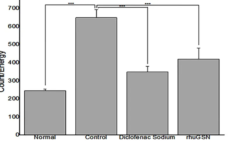
Evans blue dye extravasation in carrageenan induced paw edema in mice. Our results demonstrated that acute inflammation caused by carrageenan injection increases the vascular permeability, which can be adjudged by checking the evans blue extravasation in the tissues (Fig 5D). Absorbance data of evans blue at 620 nm clearly showed that treatment with Diclofe-nac sodium (10mg/kg) and rhuGSN (8mg) 1 hr prior carrageenan strikingly inhibited extrava-sation of Evans blue dye and exhibited 32% and 29% inhibition, respectively in comparison to the mice treated with the placebo (Fig 7).
Table 2. Analgesic effect of rhuGSN on tail withdrawal reflex induced by immersion of tail of mice in hot water.
Groups Parameter 0 min 30 min 60 min 90 min 120 min
Control (Saline) 0.2 ml / mouse, ip Mean Reaction Time (sec.±SD) 2.36±0.11 2.36±0.28 2.24±0.25 2.36±0.25 2.23±0.22
Standard (Diclofenac Sod.) 20 mg/kg, ip Mean Reaction Time (sec.±SD) 2.26±0.14 5.14±0.29** 6.25±0.48** 7.03±0.40** 7.64±0.15***
% inhibition - 54.08 64.14 66.42 70.08
Test (rhuGSN) 8 mg / mouse, sc Mean Reaction Time (sec.±SD) 2.21±0.09 2.86±0.35* 4.29±0.15** 4.99±0.23** 5.53±0.30**
% inhibition - 17.48 44.98 52.7 59.67
Fig 2. Analgesic effect of rhuGSN on tail immersion in mice (N = 6).
Effect of gelsolin on expression of IL 6 and TNF-α. As compared with normal mice, the
levels of IL 6 and TNF-αwere significantly increased (p<0.05) in the serum of mice in which
acute inflammation was induced in the paw by injection of carrageenan. However, when we compared cytokine levels of control mice treated with placebo with the mice treated either with rhuGSN or diclofenac sodium, the results showed that rhuGSN and diclofenac sodium
signifi-cantly (p<0.05) reduced the expression of these pro-inflammatory cytokines (Fig 8A and 8B).
Discussion
Non-steroidal anti-inflammatory drugs (NSAIDs) are commonly used for treatment of pain and inflammation. However, prolonged use of these drugs leads to gastro-intestinal ulcers,
Table 3. Effect of rhuGSN on protein denaturationin vitro.
S. No. % inhibition at concentration Control Diclofenac sodium rhuGSN
1 20μg/ml - 34.38±0.81** 25.89±0.76**
2 50μg/ml - 65.60±0.83** 51.43±1.29**
3 100μg/ml - 77.37±0.60** 64.50±0.75**
doi:10.1371/journal.pone.0135558.t003
Fig 3. Effect of rhuGSN and diclofenac sodium on protein denaturation.Comparison curve of diclofenac sodium and rhuGSN have been plotted.
bleeding, and renal disorders [1]. Thus, there is a need for discovery of new anti-inflammatory and analgesic drugs without these side effects. Gelsolin levels have been reported to decline in
many diseases of humans as well as in animals [14–23] and there are several reports in the
liter-ature regarding use of gelsolin for treatment of sepsis, burn, brain inflammation, diabetes [23–
25]. In this study, we have explored analgesic and anti-inflammatory activities of rhuGSN in
in-vitrosystem as well as by using standard animal models. It is pertinent to mention outright that we used recombinant human gelsolin (rhuGSN) for our experiments because of the fact
Fig 4. rhuGSN ameliorated the symptoms of acute inflammation induced by carrageenan.Measurement of edema of paw in mice (N = 6) of different treatment groups.
doi:10.1371/journal.pone.0135558.g004
Table 4. Anti-inflammatory activity of rhuGSN using carrageenan induced rat paw edema in mice.
Groups Dose 0 min 1 hr 2 hrs 3 hrs 4 hrs 5 hrs %
Inhibition
Control (Saline) 0.2 ml/ mouse, ip
2.36±0.1 3.84±0.10 4.01±0.04 4.20±0.02 4.39±0.14 4.59±0.18
-Standard (Diclofenac sod.)
10 mg/kg, ip 2.33±0.09 3.24±0.17*** 3.29±0.28*** 3.39±0.26*** 3.42±0.24*** 3.31±0.15*** 56.05
Test (rhuGSN) 8 mg/mouse, sc
that mice and human GSN are 96% identical. For studying analgesic activity, we used two mice models in order to evaluate both the peripheral and central analgesic effect of rhuGSN.
Acetic acid induced writhing experiment was done to evaluate the peripheral analgesic property of test drugs [30]. It is well known that acetic acid in some way is responsible for secretion of endogenous mediators of pain thereby stimulating the neurons responsible for
pain sensation, which are responsive to anti-inflammatory drugs [36]. In this study, gelsolin
showed significant analgesic activity in the acetic acid-induced writhing test in mice.
In the tail immersion test, we evaluated the central analgesic property of rhuGSN. This test causes centrally mediated pain at the supra-spinal level. Increment in the response time in tail withdrawal was observed for evaluating central analgesic activity. Results of tail immersion test in our study demonstrated that administration of rhuGSN to the mice resulted in a signifi-cantly prolonged tail withdrawal reflex time in response to heat stimuli.
Taken together these two experimental results, we conclude that rhuGSN can exert both peripheral as well as central analgesic effect possibly by blocking release of endogenous
inflam-matory mediators like histamine, serotonin, prostaglandinetc. Of course, delineating the
precise mechanism would be part of our future interest.
the hind paws of normal, carrageenan treated (Control), diclofenac sod. (carrageenan + diclofenac sodium) and rhuGSN (carrageenan + rhuGSN mice) showing leakage of Evans Blue dye (D)
doi:10.1371/journal.pone.0135558.g005
We also studied the anti-inflammatory activity of rhuGSN on protein denaturation
experi-mentsin vitroas well as in carrageenan-induced paw edema in mice. Denaturation of proteins
has been clearly established reason of inflammation and rheumatoid arthritis and this denatur-ation perhaps is because of the change in electrostatic, hydrogen, hydrophobic and disulphide
bonding[37]. In our experiment, rhuGSN showed significant inhibition of heat induced
dena-turation of protein and may possibly be the explanation of this anti-inflammatory activity. Carrageenan-induced paw edema is generally used model for evaluating the anti-inflamma-tory activities of new compounds. This type of inflammation is biphasic, the initial phase is due to release of histamine, 5-hydroxytryptamine, leukotriens, kinins and cyclooxygenases in the first hour of the administration of carrageenan, and the delayed phase has been linked to pro-duction of prostaglandins, bradykinin, neutrophil infiltration etc. [38]. In present investigation, gelsolin significantly ameliorated paw edema induced by carrageenan after 5 hours. This result suggests that anti-inflammatory effect of gelsolin may be due to the inhibition of cyclooxygen-ase synthesis and this effect is similar to that produced by non steroidal anti-inflammatory drugs such as diclofenac sodium. However, exact mechanism how gelsolin is inhibiting cyclo-oxygenase synthesis would be part of our future studies.
Upon tissue injury the blood vessels in the damaged area constrict momentarily (vasocon-striction), followed by, the dilation of blood vessels (vasodilation), increasing blood flow into the area, which may last from 15 minutes to several hours. The walls of these blood vessels, which normally allow only water and salts to pass through, become permeable resulting in pro-tein-rich fluid, called exudate, to exit into the tissues. This is followed by emigration of white blood cells into the extravascular space of the tissue. It is well established that the extravasation of fluid and proteins in interstitial spaces lead to formation of edema. Evans blue dye extravasa-tion assay is done routinely to evaluate degree of vascular permeability following inflammaextravasa-tion
caused by injection of carrageenan [33,34,39]. In our experiment we observed that rhuGSN
could significantly reduce exudation of plasma proteins in paw of mice resulting in decrease in
Fig 7. Quantification of dye extracted from carrageenan induced paw edema in mice (N = 3) following treatmentwith diclofenac sodium, rhuGSN and control.
Fig 8. TNF-α(A) and IL-6 levels (B) in serum of mice (N = 3) are expressed as picogram/milliliter and depict the change in their levels following
degree of edema in comparison to the mice treated with the placebo. This experiment further confirmed the anti-inflammatory effect of gelsolin in carrageenan-induced paw edema in mice.
Several reports in the literature suggested that many cytokines for instance TNF-α, IL-1β,
IL-2, IL-6, and PGE2play role in during inflammation [40]. Out of these cytokines, TNF-αis
most important player in inflammatory reactions, generating native protective responses by stimulating T cells and macrophages and release of kinins and leukotrienes, and further
activat-ing production of additional inflammatory cytokines [41,42]. Interleukin-6 (IL-6) is another
important cytokine which is released by variety of cells at the site of injury [43]. Our data has shown that pretreatment of carrageenan mice either with rhuGSN or with diclofenac sodium
reduced the TNF-αand IL-6 levels in plasma as compared to the mice treated with the placebo
thus, indicating the anti-inflammatory activity of gelsolin, These results are in confirmation with earlier findings where gelsolin treatment down regulated these pro-inflammatory
cyto-kines in sepsis, burn and inflammation of brain [24–25]. Besides these diseases, exogenous
administration of gelsolin has also shown protective effect in treatment of diabetes and stroke in mice and rats [23,44].
Though these experiments evidently establishes analgesic and anti-inflammatory function of rhuGSN in these mouse models, however, the mechanism of action of pGSN of this protec-tion remains to be explored in depth. The probable mechanism proposed in the literature involves actin depolymerization. Following any injury, actin is released from injured tissues in the circulation and gelsolin binds to this actin and after severing, remove it from the circulation
and thus, limits the inflammation [20]. In addition, pGSN may also play an important role in
regulating inflammation as gelsolin has been reported to act as a buffering agent in inflamma-tion by binding to the inflammatory mediators such as platelet activating factor (PAF), lyso-phosphatidic acid (LPA) [27,28].
In summary, we conclude that rhuGSN can exert analgesic anti-inflammatory activity by actin scavenging from the site of injury and by down regulating the expression of
pro-inflam-matory cytokines such as TNF-αand IL-6.
Acknowledgments
Authors acknowledge consistent support from faculty and staff of CSIR-IMTECH. We thank Animal Facility of the institute for providing mice required for experiments. The authors are thankful to Dr. Pradip Sen for critical reading of the manuscript. This is IMTECH communica-tion number 022/2015.
Author Contributions
Conceived and designed the experiments: NK A. Performed the experiments: AKG DP BSC. Analyzed the data: NK A AKG DP BSC. Contributed reagents/materials/analysis tools: AS VC AKG RG. Wrote the paper: NK A.
References
1. Sostres C, Gargallo CJ, Arroyo MT, Lanas A (2010) Adverse effects of non-steroidal anti-inflammatory drugs (NSAIDs, aspirin and coxibs) on upper gastrointestinal tract. Best practice & research Clinical gastroenterology 24: 121–132.
2. Chiu YJ, Huang TH, Chiu CS, Lu TC, Chen YW, et al. (2012) Analgesic and Antiinflammatory Activities of the Aqueous Extract from Plectranthus amboinicus (Lour.) Spreng. Both In Vitro and In Vivo. Evi-dence-based complementary and alternative medicine: eCAM 2012: 508137. doi:10.1155/2012/ 508137PMID:21915187
4. Mothana RA (2011) Anti-inflammatory, antinociceptive and antioxidant activities of the endemic Soqo-traen Boswellia elongata Balf. f. and Jatropha unicostata Balf. f. in different experimental models. Food and chemical toxicology: an international journal published for the British Industrial Biological Research Association 49: 2594–2599.
5. Choi JH, Cha DS, Jeon H (2012) Anti-inflammatory and anti-nociceptive properties of Prunus padus. Journal of ethnopharmacology 144: 379–386. doi:10.1016/j.jep.2012.09.023PMID:23010365
6. Nathan C (2002) Points of control in inflammation. Nature 420: 846–852. PMID:12490957
7. Ferrero-Miliani L, Nielsen OH, Andersen PS, Girardin SE (2007) Chronic inflammation: importance of NOD2 and NALP3 in interleukin-1beta generation. Clinical and experimental immunology 147: 227–
235. PMID:17223962
8. Lawrence T, Fong C (2010) The resolution of inflammation: anti-inflammatory roles for NF-kappaB. The international journal of biochemistry & cell biology 42: 519–523.
9. Libby P (2007) Inflammatory mechanisms: the molecular basis of inflammation and disease. Nutrition reviews 65: S140–146. PMID:18240538
10. Osadebe PO, Okoye FB (2003) Anti-inflammatory effects of crude methanolic extract and fractions of Alchornea cordifolia leaves. Journal of ethnopharmacology 89: 19–24. PMID:14522428
11. Omoigui S (2007) The biochemical origin of pain: the origin of all pain is inflammation and the inflamma-tory response. Part 2 of 3—inflammatory profile of pain syndromes. Medical hypotheses 69: 1169–
1178. PMID:17728071
12. Moncada S, Palmer RM, Higgs EA (1991) Nitric oxide: physiology, pathophysiology, and pharmacol-ogy. Pharmacological reviews 43: 109–142. PMID:1852778
13. Kwiatkowski DJ, Stossel TP, Orkin SH, Mole JE, Colten HR, et al. (1986) Plasma and cytoplasmic gel-solins are encoded by a single gene and contain a duplicated actin-binding domain. Nature 323: 455–
458. PMID:3020431
14. Peddada N, Sagar A, Ashish, Garg R (2012) Plasma gelsolin: a general prognostic marker of health. Med Hypotheses 78: 203–210. doi:10.1016/j.mehy.2011.10.024PMID:22082609
15. Khatri N, Ashish and Garg V (2014) Reviewing biomedical role of plasma gelsolin. The Pharma Innova-tion Journal 3: 16–20.
16. Smith DB JP, Sherwood JA, Howard RJ, Lind SE (1988) Decreased plasma gelsolinlevels in patients with Plasmodium falciparum malaria: a consequence of hemolysis? Blood 72: 214–218. PMID:
2839253
17. DiNubile MJ, Stossel TP, Ljunghusen OC, Ferrara JL, Antin JH (2002) Prognostic implications of declin-ing plasma gelsolin levels after allogeneic stem cell transplantation. Blood 100: 4367–4371. PMID:
12393536
18. Christofidou-Solomidou M, Scherpereel A, Solomides CC, Christie JD, Stossel TP, et al. (2002) Recombinant plasma gelsolin diminishes the acute inflammatory response to hyperoxia in mice. Jour-nal of investigative medicine: the official publication of the American Federation for Clinical Research 50: 54–60.
19. Smith DB, Janmey PA, Lind SE (1988) Circulating actin-gelsolin complexes following oleic acid-induced lung injury. The American journal of pathology 130: 261–267. PMID:2829631
20. Lee PS, Waxman AB, Cotich KL, Chung SW, Perrella MA, et al. (2007) Plasma gelsolin is a marker and therapeutic agent in animal sepsis. Critical care medicine 35: 849–855. PMID:17205019
21. Cohen TS, Bucki R, Byfield FJ, Ciccarelli NJ, Rosenberg B, et al. (2011) Therapeutic potential of plasma gelsolin administration in a rat model of sepsis. Cytokine 54: 235–238. doi:10.1016/j.cyto.
2011.02.006PMID:21420877
22. Endres M, Fink K, Zhu J, Stagliano NE, Bondada V, et al. (1999) Neuroprotective effects of gelsolin dur-ing murine stroke. The Journal of clinical investigation 103: 347–354. PMID:9927495
23. Khatri N, Sagar A, Peddada N, Choudhary V, Chopra BS, et al. (2014) Plasma gelsolin levels decrease in diabetic state and increase upon treatment with F-actin depolymerizing versions of gelsolin. Journal of diabetes research 2014: 152075. doi:10.1155/2014/152075PMID:25478578
24. Rothenbach PA, Dahl B, Schwartz JJ, O'Keefe GE, Yamamoto M, et al. (2004) Recombinant plasma gelsolin infusion attenuates burn-induced pulmonary microvascular dysfunction. Journal of applied physiology 96: 25–31. PMID:12730154
26. Witke W SA, Hartwig JH, Azuma T, Stossel TP, et al. (1995) Hemostatic, inflammatory, and fibroblast responses are blunted in mice lacking recombinant human gelsolin (rhuGSN). cell 81 41–51. PMID:
7720072
27. Osborn TM, Dahlgren C, Hartwig JH, Stossel TP (2007) Modifications of cellular responses to lysopho-sphatidic acid and platelet-activating factor by plasma gelsolin. American journal of physiology Cell physiology 292: C1323–1330. PMID:17135294
28. Goetzl EJ, Lee H, Azuma T, Stossel TP, Turck CW, et al. (2000) Gelsolin binding and cellular presenta-tion of lysophosphatidic acid. The Journal of biological chemistry 275: 14573–14578. PMID:10799543
29. Peddada N, Sagar A, Rathore YS, Choudhary V, Pattnaik UB, et al. (2013) Global shapes of F-actin depolymerization-competent minimal gelsolins: insight into the role of g2-g3 linker in pH/Ca2+ insensi-tivity of the first half. The Journal of biological chemistry 288: 28266–28282. doi:10.1074/jbc.M113.
463224PMID:23940055
30. Koster R, Anderson M and De Beer J (1959) Acetic acid for analgesic screening. Federation Proceed-ings 18: 412–417.
31. Aydin S, Demir T, Ozturk Y, Baser KH (1999) Analgesic activity of Nepeta italica L. Phytotherapy research: PTR 13: 20–23. PMID:10189945
32. Winter CA, Risley EA, Nuss GW (1962) Carrageenin-induced edema in hind paw of the rat as an assay for antiiflammatory drugs. Proceedings of the Society for Experimental Biology and Medicine Society for Experimental Biology and Medicine 111: 544–547.
33. Mahmoodi M, Hadad MK, Shamsizadeh A, Azarang A, Rayeni RA (2009) Effect of trifluoperazine on carrageenan-induced acute inflammation in intact and adrenalectomized rats. International journal of physiology, pathophysiology and pharmacology 1: 150–153. PMID:21383884
34. Han ED, MacFarlane RC, Mulligan AN, Scafidi J, Davis AE 3rd (2002) Increased vascular permeability in C1 inhibitor-deficient mice mediated by the bradykinin type 2 receptor. The Journal of clinical investi-gation 109: 1057–1063. PMID:11956243
35. Vane JR, Botting RM (1995) New insights into the mode of action of anti-inflammatory drugs. Inflamma-tion research: official journal of the European Histamine Research Society [et al] 44: 1–10.
36. Arslan R, Bektas N, Ozturk Y (2010) Antinociceptive activity of methanol extract of fruits of Capparis ovata in mice. Journal of ethnopharmacology 131: 28–32. doi:10.1016/j.jep.2010.05.060PMID:
20595018
37. Mizushima Y (1966) Screening test for anti-rheumatic drugs. Lancet 2: 343.
38. Brooks PM, Day RO (1991) Nonsteroidal antiinflammatory drugs—differences and similarities. The
New England journal of medicine 324: 1716–1725. PMID:2034249
39. Emanueli C, Grady EF, Madeddu P, Figini M, Bunnett NW, et al. (1998) Acute ACE inhibition causes plasma extravasation in mice that is mediated by bradykinin and substance P. Hypertension 31: 1299–
1304. PMID:9622145
40. Delgado AV, McManus AT, Chambers JP (2003) Production of tumor necrosis factor-alpha, interleukin 1-beta, interleukin 2, and interleukin 6 by rat leukocyte subpopulations after exposure to substance P. Neuropeptides 37: 355–361. PMID:14698678
41. Yun KJ, Kim JY, Kim JB, Lee KW, Jeong SY, et al. (2008) Inhibition of LPS-induced NO and PGE2 pro-duction by asiatic acid via NF-kappa B inactivation in RAW 264.7 macrophages: possible involvement of the IKK and MAPK pathways. International immunopharmacology 8: 431–441. doi:10.1016/j.intimp.
2007.11.003PMID:18279797
42. Huang MH, Huang SS, Wang BS, Wu CH, Sheu MJ, et al. (2011) Antioxidant and anti-inflammatory properties of Cardiospermum halicacabum and its reference compounds ex vivo and in vivo. Journal of ethnopharmacology 133: 743–750. doi:10.1016/j.jep.2010.11.005PMID:21073940
43. Yu XZ M., Witschi H., and Pinkerton K. E. (2002) The role of interleukin-6 in pulmonary inflammation and injury induced by exposure to environment air pollutants. Toxicological Sciences 68: 488–497.
PMID:12151646