Iranian Journal of Basic Medical Sciences
ijbms.mums.ac.ir
Phenolic contents and bioactivities of pericarp and seeds of
Pleiogynium solandri
(Benth.) Engl. (Anacardiaceae)
Ataa Said
1, Elsayed A Abuotabl
2, Gehan Fawzy Abdel Raoof
1*, Antje Huefner
3, Somaia A Nada
4 1 Pharmacognosy Department, National Research Center, Dokki, Cairo, Egypt2 Pharmacognosy Department, Faculty of Pharmacy, Cairo Univ. Egypt
3 Institute for Pharmaceutical Chemistry and Pharmaceutical Technology, University of Graz, Schubertstr.1, A8010 Graz, Austria
4 Pharmacology Department, National Research Center Dokki, Cairo, Egypt
A R T I C L E I N F O A B S T R A C T
Article type:
Original article Objective(s):of many of synthetic drugs. Methanol extracts of both pericarp and seeds of This study aimed to develop drugs from natural sources to overcome the side effects Pleiogynium solandri
were used to investigate antioxidant, hepatoprotective, and renal function protective, analgesic, and anti-inflammatory effects and to determine the chemical composition of the extract responsible for bioactivity.
Materials and Methods: Methanol (70%) extracts of the seeds and pericarps of P. solandri were prepared. Hot plate method was used to test analgesic activity, carrageenan-induced paw inflammation method was used to test anti-inflammatory activity, and colorimetric methods were used to test antioxidant, hepatoprotective (by determination of serum alanine and aspartate aminotransferase activities), and renal function protective effects (by measuring uric acid and creatinine levels). Chromatographic methods and means of 1H-NMR, 13C –NMR, and UV spectra were used for isolation and identification of the responsible compounds.
Results:In this study for the first time,four phenolic compounds were isolated from the pericarp of P. solandri which were identified as catechin, quercetin, quercetrin and rutin. Methanolic extract of both seeds and pericarp of P. solandri showed strong antioxidant effect, hepatoprotective, renal function protective, analgesic, and anti-inflammatory effects. However, seed extract had lower effect than pericarp in a dose dependent manner.
Conclusion: This study showed that methanol extract of pericarp of P. solandri is more powerful than that of the seed regarding its antioxidant, hepato-protective; renal function protective, analgesic, and anti-inflammatory effects. The phenolic compounds isolated from the methanol extract of pericarp were responsible for bioactivity.
Article history: Received: Jan 14, 2014 Accepted: Jul 11, 2014
Keywords:
Analgesic Anti-inflammatory Antioxidant Hepatorenal Pleiogynium Phenolic
►
Please cite this paper as:Said A, Abuotabl EA, Fawzy Abdel RaoofG, Huefner A, Nada SA.Phenolic contents and bioactivities of pericarp and seeds of Pleiogynium solandri (Benth.) Engl. (Anacardiaceae). Iran J Basic Med Sci 2015; 18:164-171.
Introduction
Nowadays, the development of drugs from natural sources is recommended to overcome the side effects of many of the synthetic drugs. Recent research on medicinal plants has generated a great deal of information about the biologically active chemical components that are responsible for the claimed medicinal effects.
Pleiogynium solandri (Benth.) Engl. (Anacardiaceae), is an evergreen tree indigenous to tropical and subtropical regions; it is known in Arabic as Gambozia and is cultivated in Egypt as an ornamental plant, which
has many synonyms as Pleiogynium timorense and
Pleiogynium cerasiferum (1, 2). Plant family of
Anacardiaceae contains plants with edible fruits and seeds (3) e.g. mango, red mombin, and other species of Spondias genus. P. solandri is among plants which have edible fruits used in preparation of jellies, jams, and
preserves (4). Previous phytochemical studies that resulted in the isolation of quercetin, myricetin,
rutin, quercitrin, hyperin, lupeol, -sitosterol from
the leaves, and aqueous and alcoholic extracts of the leaves showed good antimicrobial activity against
Staphylococcus aureus and Bacillus subtilis (5). The ethanolic extract of the leaves showed signifycant hypoglycemic, antioxidant and
anti-inflammatory properties, additionally, twelve phenolic compounds were isolated from the plants leaves including kaempferol, gallic acid, kaempferol-
3-O- -D-galactopyranoside, kaempferol-3-O-
-D-glucopyranoside, quercetin 3-O- -d-galactopyranoside,
quercetin 3-O- -D-glucopyranoside, kaempferol
3-O- -D-6"-methylglucuronopyranoside, kaempferol
3-O- -d-glucuronopyranoside, myricetin-3-O-
-L-rhamnopyranoside, 3,5-di-O-galloylquinic acid, 1,4,6-tri-O-galloyl- -D-glucopyranose, and
Iran J Basic Med Sci, Vol. 18, No. 2, Feb 2015 165 galloyl- -D-glucopyranose (6). The fruits have been
reported to have antioxidant activity, additionally; cyaniding 3-glucoside was isolated from the fruits (7). Nothing could be traced in the available literature that deals with the chemistry of seeds and other constituents of fruits of this plant. The present study was undertaken to investigate the use of methanol extracts of both fruits and seeds of P. solandri as antioxidant, hepatoprotective, renal function protective, analgesic and anti-inflammatory, and to determine the chemical composition of the extract responsible for bioactivity with the aim of developing a natural drug.
Materials and Methods
Plant
Fruits of P. solandri plant were collected from Zoo garden, Giza, Egypt in April 2010. The plant was identified by Dr Kamal El Batanony, professor of Taxonomy and Botany, Faculty of Science, Cairo University. Methanol (70%) extracts of the seeds and pericarp of P. solandri were used.
Animal
Sprague Dawley rats of both sexes weighing 130-150 g were used throughout the experiments. Animals were housed under standard environmental conditions (23 ± 1ºC, 55 ± 5 % humidity, 12:12 hr light-dark cycle) and maintained with free access to water and a standard laboratory diet ad libitum. Animal care and the experimental protocols were approved by Institutional Animal Care and Use Committee and were in accordance with the guidelines of the International Association for the Study of Pain.
Acute toxicity study
The LD50 of methanol (70%) extracts of the P.
solandri (seeds and pericarp) was determined using rats. Male and female albino rats (10 rats in each group) were orally administered the tested extracts at doses ranging from 1.0 to 5.0 g/kg. LD05was calculated using
Karber method (8). Two dose levels of P. solandri were chosen (150 and 300 mg/kg) to determine the most effective dose at exerting physiological activity. In a preliminary study, the tested doses of 50 and 75 mg/kg induced insignificant changes in case group compared to control.
Antioxidant, hepatoprotective, and renal- function protective effects
Rats were assigned into two main groups: normal and damaged liver groups that administrated CCl4 (carbon tetra chloride). Each group classified into 6 different groups (6 rats in each): group 1: was control received distilled water, group 2 was administered silymarin (50 mg/kg), and groups 3-6 were administered P. solandri tested extracts at two
dose levels for each extract (150 and 300 mg/kg) for 15 days. The drug solutions or vehicle were administered orally by gastric intubation using syringe to assess the experiments. At the end of experimental period, rats were anaesthetized with ether according to the method described by Cocchetto and Bjornsson (9). Blood samples were
collected from orbital venous plexus in
nonheparinized tubes, centrifuged at 3000 rpm for 15 min, and blood sera were collected and stored at -20°C before they were analyzed.
Determination of serum total antioxidant capacity (TAC) level
Serum total antioxidant capacity (TAC) level was assessed colorimetrically using a test reagent kit according to the method described by Koracevic et al (10).
Principle
Determination of the antioxidative capacity is performed by the reaction of antioxidants in the sample with a defined amount of exogenously provided hydrogen peroxide (H2O2); antioxidants in the sample eliminate a certain amount of the provided H2O2. The residual H2O2 is determined colorimetrically by an enzymatic reaction which involves the conversion of 3,5-dichloro-2-hydroxybenzene sulfonate to a colored product. The absorbance of blank (Ab) and sample (As) was read at 505 nm against distilled water.
Determination of serum alanine aminotransferase (ALT) activity
Serum alanine aminotransferase (ALT) activity was assessed colorimetrically using a test reagent kit according to the method described by Reitman and Frankel (11).
Principle
The method depends on the reaction between L-alanine and -keoglutaric acid in the presence of ALT to form pyruvic acid and L-glutamic acid. The pyruvate formed was measured in its derivative form
(2,4-dinitrophenlhydrazone) colorimetrically at 505 nm.
Determination of serum aspartate aminotransferase (AST) activity
Serum AST activity was assessed colorimetrically using a test reagent kit according to the method described by Reitman and Frankel (11).
Principle
The method depends on the reaction between
L-aspartic acid and -ketoglutaric acid in the
Iran J Basic Med Sci, Vol. 18, No. 2, Feb 2015
166
Uric acid assay
Serum uric acid is the end product of purine metabolism, and is cleared through the kidney by glomerular filtration.
Kit provides a convenient means for detecting uric acid in biological samples such as serum and urine. Pretreatment of samples are not required. Uric acid level can be measured using colorimetric method (at 570 nm) according to the method of Barham and Trinder (12).
Creatinine assay (Bartles et al, 1972)
Creatinine is a breakdown product of creatine phosphate. Creatinine is produced and excreted at a constant rate, and blood creatinine is used to determine glomerular filtration rate.
Creatinine is measured in biological fluids (serum and urine) according to the method of Bartles and Bohmer (13). In the assay, creatinine is converted to creatine by creatininase, it is then converted to sarcosine, which is specifically oxidized to produce a product which reacts with a probe to generate red color λmax = 075 nm .
Determination of analgesic activity
The hot plate-induced thermal pain method used to test analgesic activity according to the method of
Roszkowski et al (14); the temperature is controlled
for 53°C (±0.1°C). The reaction time was considered as the time elapsed between placing of the mouse on the hot plate and appearance of signs of acute discomfort, characterized by flicking or licking of the hind paw, forepaw or jumping in an attempt to escape from the pain. The mice showing initial reaction time of 10 sec or less were selected for this study. Increase in reaction time in drug-treated groups was compared with that of the control group. The mean reaction time was calculated 30 min post- drug administration during 4 hr with a cut-off time of 30 sec. Animals were grouped into the following groups:
1. Group 1: the control group given distilled water (10 ml/kg b.wt).
2. Group 2: administered 50 mg/kg paracetamol.
3. Groups 3 and 4: administered 150 and 300 mg/kg
of P. solandri, seeds extract respectively.
4. Groups 5 and 6: administered 150 and 300 mg/kg
P. solandri pericarp extract, respectively.
Determination of anti-inflammatory activity The carrageenan-induced rat paw edema method was employed to test anti-inflammatory activity according to the method of Winter et al (15) using plethysmometer system. Plethysmometer is a volume meter and the standard instrument for measurement of rodent paw volume (16). This is a test to screen potential inflammatory or anti-edema agents. The paw measured was inserted into water in a clear acrylic cell, up to the wrist joint.
After 30 min of drugs administration till the end of experimental duration (4 hr), the volume of water displaced was measured by a transducer. Six groups of rats (six rats in each) were treated as previously mentioned in analgesic experimental design as follows:
1. Group 1: is the control group given distilled water
(10 ml/kg b.wt).
2. Group 2: administered 5 mg/kg indomethacin.
3. Groups 3 and 4: administered 150 and 300 mg/kg
from seeds extract of P. solandri, respectively.
4. Groups 5 and 6: were administered 150 and 300
mg/kg of pericarp extract of P. solandri, respectively.
Statistical analysis
Data were analyzed using one-way ANOVA.
P-value <5.50 was considered statistically significant.
UV-visible spectrophotometer
Beckman DU7 and Shimadzu UV 240 (PIN 204-5800) were used for recording UV spectra and measuring the absorbance in UV and visible range.
NMR Spectrometers
Varian Unity Inova 400 (297) 5 mm tubes, Austria, were apropriate.
Extraction and isolation
The air dried powder of P. solandri pericarp (2kg) was defatted with petroleum ether (60-80C) (3l), percolated with methanol 70% till exhaustion, then the extract was evaporated under reduced pressure to yield 150 g dried extract.
The fraction was subjected to column chromate-graphy using polyamide as adsorbent and elution was carried out with water; then decreasing the polarity by adding methanol. Fractions (50 ml each) were collected and separately concentrated to a small volume. All fractions were screened by PC (Whatman No. 1) using n-butanol: acetic acid: water (4:1:5,v/v/v) and acetic acid: water (15:85,v/v) as solvent systems; similar fractions were pooled and the solvents were separately evaporated under reduced pressure.
Iran J Basic Med Sci, Vol. 18, No. 2, Feb 2015 167
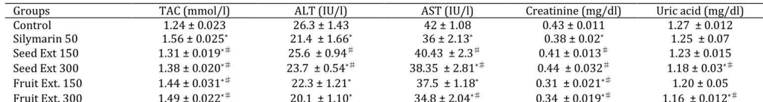
Table 1. (A-on normal rats). Effect of Pleiogynium solandri (Benth.) methanol water (70%) extracts of seed and pericarp on total
antioxidant capacity, liver enzymes marker, and kidney function in serum of normal rats
One-way ANOVA, significant at P-value ≤5.50; * Significantly different from control group; ♯ Compared to silymarin treatment group; TAC:
total antioxidant capacity; ALT: alanine aminotransferase; AST: aspartate aminotransferase; CCL4: carbon tetra chloride
Elution with water: methanol (70:30) afforded the presence of flavonoids. The fraction was applied to subcolumn on Sephadex LH-20 using methanol as eluent Fractions (50 ml each) were collected and separately concentrated to a small volume. All fractions were screened by PC (Whatmann No. 1) using n-butanol-acetic acid-water (4:1:5,v/v/v) and acetic acid :water(15:85,v/v) as solvent systems; where similar fraction were pooled and the solvents were, separately, evaporated under reduced pressure.
(Fractions 2-6) afforded one compound which gave dark purple color under UV light changing to yellow on exposure to ammonia vapor and yellow color with AlCl3. It was purified on Sephadex LH-20 column using methanol as eluent to give compound 2.
Elution with water: methanol (40:60) afforded the presence of flavonoids. The fraction was applied to subcolumn on Sephadex LH-20; using methanol as eluent fractions (50 ml each) were collected and separately concentrated to a small volume. All fractions were screened by PC (Whatmann No. 1) using n-butanol-acetic acid-water (4:1:5,v/v/v) and acetic acid: water(15:85,v/v) as solvent systems; where similar fraction were pooled and the solvents
were, separately, evaporated under reduced
pressure.
(Fractions 4-8) afforded one compound which gave yellow color under UV light, on exposure to ammonia vapor and with AlCl3. It was purified on Sephadex LH-20 column using methanol as eluent to give compound 3.
Elution with water: methanol (30:70) afforded one compound which gave pink color with vanillin sulfuric acid reagent. It was purified on Sephadex LH-20 column using methanol as eluent to give compound 4.
Results
LD50 determination revealed that the investigated
extracts were nontoxic up to 5g/kg which was the maximum soluble dose and also it is the maximum dose can be given according to the typical protocol for an acute study with rats (16).
TAC showed significant increase in all treatment groups with two studied extracts compared with the control values. The two dose levels of the seeds and pericarp extracts resulted in significant reduction of TAC value when compared with silymarin treated group. Liver enzymes (ALT and AST) significantly decreased in all treatment groups except in group with lower dose of seed extract that did not differ with control value. As well as, ALT and AST significantly decreased in groups treated with silymarin while compared with seed extract (at both doses) (Table 1). Creatinine serum concentration was decreased significantly in groups treated with the two doses of pericarp extract comparing with the control or silymarin groups while creatinine value did not changed in groups treated with seed extract. Uric acid level was not significantly changed compared with control in groups treated with seed extract (150 mg/kg) and silymarin, while in other treated groups uric acid values significantly decreased in comparison to control and silymarin treatment groups (Table 1).
Table 2 demonstrates that CCL4 treatment caused
significant depletion in TAC serum content. The lower dose of seed extract (150 mg/kg) increased TAC level in CCL4 hepatic damaged rats compared with CCL4-treatment alone; while the higher dose of seed extract had similar effect on TAC with that of
silymarin-treatment group. However, pericarp
Table 2. (B-On injured rat). Hepatoportective effect of Pleiogynium solandri (Benth.) methanol water (70%) extracts of seed and pericarp
on total antioxidant capacity, liver enzymes marker, and kidney function in serum of carbon tetrachloride-induced hepatic damage rats
Groups TAC (mmol/l) ALT (IU/l) AST (IU/l) Creatinine (mg/dl) Uric acid (mg/dl)
CCL4 0. 89 ± 0.014 46.5 ± 1.12 88.2 ± 2.3 1.32 ± 0.07 2.43 ± 0.04
Silymarin 50+CCL4 1.21 ± 0.018* 25.8 ± 1.33* 67.3 ± 0.94* 0.76 ±0.06* 1.72 ± 0.05*
Seed Ext 150+CCL4 0.95 ± 0.024*♯ 36.6 ± 1.09*♯ 74.8 ± 1.12*♯ 0.89 ± 0.04*♯ 2.1 ± 0.03♯
Seed Ext 300+ CCL4 1.20 ± 0.017* 32.1 ± 1.34*♯ 70.2 ± 2.00*♯ 0.85 ±0.05*♯ 1.97 ± 0.05*
Fruit Ext. 150+CCL4 1.31 ± 0.012*♯ 37.2 ± 1.21*♯ 65.2 ± 1.62* 0.74 ± 0.03 1.67 ± 0.05*
Fruit Ext. 300+CCL4 1.39 ± 0.022*♯ 33.6 ± 0.87*♯ 61.6 ± 1.33*♯ 0.78 ± 0.04 1.60 ± 0.06*
One-way ANOVA test, significant at P-value ≤5.50; * compared to CCL4- treatment group; ♯ compared to Silymarin treatment group; TAC: total antioxidant capacity; ALT: alanine aminotransferase; AST: aspartate aminotransferase; CCL4: carbon tetra chloride
Groups TAC (mmol/l) ALT (IU/l) AST (IU/l) Creatinine (mg/dl) Uric acid (mg/dl)
Control 1.24 ± 0.023 26.3 ± 1.43 42 ± 1.08 0.43 ± 0.011 1.27 ± 0.012
Silymarin 50 1.56 ± 0.025* 21.4 ± 1.66* 36 ± 2.13* 0.38 ± 0.02* 1.25 ± 0.07
Seed Ext 150 1.31 ± 0.019*♯ 25.6 ± 0.94♯ 40.43 ± 2.3♯ 0.41 ± 0.013♯ 1.23 ± 0.015
Seed Ext 300 1.38 ± 0.020*♯ 23.7 ± 0.54*♯ 38.35 ± 2.81*♯ 0.44 ± 0.032♯ 1.18 ± 0.03*♯
Fruit Ext. 150 1.44 ± 0.031*♯ 22.3 ± 1.21* 37.5 ± 1.18* 0.31 ± 0.021*♯ 1.20 ± 0.05
Iran J Basic Med Sci, Vol. 18, No. 2, Feb 2015
168
Table 3. Analgesic effect of Pleiogynium solandri (Benth.) methanol water (70%) extracts of seed and pericarp compared to control and
paracetamol- treated groups on hot plate- induced pain in rats
Groups 30 min 60 min 120 min 180 min 240 min
Control 5.81 ± 0.22 5.62 ± 0.33 5.53 ± 0.17 5.48 ± 0.14 5.40 ± 0.34
Paracetamol 50 mg/kg 11.52 ± 0.25* 10.22 ± 0.45* 8.42 ± 0.66* 7.81 ±0.24* 7.44 ± 0.21*
Seed Ext 150 mg/kg 5.92 ± 0.32♯ 6.42 ± 0.54♯ 7.16 ± 0.22*♯ 7.62 ± 0.30* 7.55 ± 0.43*
Seed Ext 300 mg/kg 6.73 ± 0.33*♯ 7.50 ± 0.24*♯ 7.80 ± 0.36*♯ 8.21 ±0.44* 7.78 ± 0.21* Fruit Ext. 150 mg/kg 6.55 ± 0.27♯ 7.81 ± 0.23*♯ 8.11 ± 0.41* 7.86 ± 0.54* 7.31 ± 0.18* Fruit Ext. 300 mg/kg 7.35 ± 0.22*♯ 7.93 ± 0.34* 8.45 ± 0.50* 8.90 ± 0.29*♯ 8.15 ± 0.31*
One-way ANOVA, P-value ≤5.50; * Significantly different from control group; # Significantly different from paracetamol group
extract showed significant increase in TAC value more than silymarin effect in a dose dependent manner in hepatotoxic groups. Liver enzymes values significantly elevated in CCL4-toxicated rats than the other CCL4 toxicated treated groups. All tested extracts significantly inhibited the elevation of ALT
and AST caused by CCL4 in a dose dependent
manner. Serum creatinine and uric acid significantly increased by CCL4 intoxication. Pericarp extract was significantly different from silymarin treatment group in decreasing creatinine value. Although seed extract treatment significantly reduced creatinine value, it was still lower than the reduction due to silymarin treatment (Table 2).
CCL4-treatment group showed significant
elevation in uric acid, however silymarin pretreating significantly decreased uric acid elevation caused by
CCL4. The lower dose of seed extract did not change
the uric acid value when compared with CCL4 alone.
The higher dose of seed extract and the two dose levels of pericarp extract reduced uric acid value in a
dose dependent manner compared with CCL4 or
silymarin. Both seed and pericarp extracts at their two dose levels showed significant increase in the reaction time compared with the control group (Table 3).
Table 4 shows the effect of different tested materials on rat paw edema versus indomethacine (stander drug). Pericarp extract exerted its powerful anti-inflammatory effect after 120 and 180 min of treatment with 300 and 150 mg/kg doses, respectively. However, seed extract had lower anti-inflammatory effect in a dose dependent manner.
Isolated compounds
Compound 1 was isolated as yellow powder, 15 mg, single spot; Rf values = 32 and 44 in solvent
systems: n-butanol:acetic acid:water (4:1:5 v/v/v) and 15% acetic acid, respectively. It gave dark purple color under UV changing to yellow on exposure to
ammonia vapor and yellow color with AlCl3.
UV spectral data of compound 1
λ max nm (MeOH): 258, 359 flavonol, (NaOMe): 272, 410 (free OH at 4), (AlCl3): 275, 433 (free OH on ring A and B), (AlCl3/HCl): 271, 400 (free OH at 5 and ortho OH at ring B), (NaOAc): 271, 393 (free OH at 7), (NaOAc/H3BO3): 262, 387 (ortho OH at ring B).
1H-NMR spectrum (DMSO-d6)
7.5 (1 H, d, J = 2.1 Hz, H-2), 7.49 (1 H, dd, J = 2.1, 8.4 Hz, H-6), 6.7 (1 H, d, J= 8.4 Hz, 5), 6.4 (1 H, d, J= 2.1 H-8), 6.2 (1 H, d, J= 2.1 H-6), 5.2 (1 H, d,J =7.2 Hz, H-1), 4.3 (1H, d,J = 1.5 Hz, H-1'), 1.1 (3 H, d, J = 6.6 Hz, Me).
Thus compound 1 is identified as quercetin 3-O- -rhamnosyl (1´´´→6´´)- -glucoside (Rutin)
Compound 2 was isolated as yellow amorphous powder (4 mg) with Rfvalues 0.58 (BAW) and 0.66 (15% HOAc) on PC. It appeared as deep purple spot under UV light changed to yellow with ammonia
vapor indicated that compound 2 is flavonol with
with free 5- and 4'-OH. Complete acid hydrolysis yielded quercetin as an aglycone and rhamnose as the sugar moiety in which both of them were co-chromatographed with authentic samples.
UV spectral data of compound 2
λ max nm (MeOH): 256, 350 (flavonol), (NaOMe): 270, 393 (free OH at 4), (AlCl3): 276, 430 (free OH on ring A and B), (AlCl3/HCl): 272, 401 (free OH at 5 and ortho OH at ring B), (NaOAc): 272, 372 (free OH at 7), (NaOAc/H3BO3): 260, 367 (ortho OH at ring B).
Table 4. Anti-inflammatory effect of Pleiogynium solandri (Benth.) methanol water (70%) extracts of seed and pericarp on rat paw edema,
compared to control and indomethacine- treated groups
Groups 30 min 60 min 120 min 180 min 240 min
Control 0.381±0.013 0.378 ±0.010 0.370±0.009 0.373±0.004 0.365±0.008
Indomethacine 5 mg/kg 0.192±0.006* 0.182±0.004* 0.180±0.008* 0.272±0.011* 0.310±0.005*♯
Seed Ext 150 mg/kg 0.241±0.014*♯ 0.230±0.016*♯ 0.226±0.003*♯ 0.219±0.11*♯ 0.221±0.007*♯
Seed Ext 300 mg/kg 0.220±0.005*♯ 0.216±0.006*♯ 0.205±0.008* 0.198±0.001*♯ 0.208±0.006*♯
Fruit Ext. 150 mg/kg 0.231±0.007*♯ 0.211±0.012*♯ 0.200±0.014*♯ 0.196±0.005*♯ 0.188±0.007*♯
Fruit Ext. 300 mg/kg 0.202±0.007* 0.194±0.003*♯ 0.186±0.011* 0.176±0.004*♯ 0.170±0.006*♯
Iran J Basic Med Sci, Vol. 18, No. 2, Feb 2015 169
1H-NMR spectrum (DMSO-d6)
7.26 (2H, m, H-2and H-6), 6.83 (1H, d, J = 9 Hz, 5), 6.4 (1H, d, J = 2.5, H-8), 6.14 (1H, d, J = 2.5, H-6), 5.2 (1H, d,J = 2 Hz, H-1), 0.78 (3H, d, J = 6Hz, Me).
Thus compound 2 is identified as quercetin 3-O
-–rhamnoside.
Compound 3 was isolated as yellow powder (5 mg) with Rf values 0.91 (BAW) and 0.12 (15% HOAc) on PC and these values were within the range of flavonoid aglycone. It gave yellow spot under UV light changed to yellow fluorescence with ammonia vapor indicated that compound P3 is flavonol with free 3- and 5-OH groups.
UV spectral data of compound 3
λ max nm (MeOH): 260, 368 (flavonol), (NaOMe): 272, 406 (free OH at 4), (AlCl3): 272, 446 (free OH on ring A and B), (AlCl3/HCl): 266, 430 (free OH at 3,5,and ortho OH at ring B), (NaOAc): 272, 404 (free OH at 7), (NaOAc/H3BO3): 260, 384 (ortho OH at ring B)
1H-NMR spectrum (MeOD)
7.74 (1 H, d, J = 2 Hz, H-2), 7.55 (1 H, d, J = 2.1, 8.4 Hz, H-6), 6.92 (1H, d, J=Hz, 5), 6.4 (1 H, d, J = 2, H-8), 6.15 (1 H, d, J= 2, H-6).
Thus compound 3 is identified as quercetin. Compound 4 isolated as white crystals (15 mg) with Rf0.6 (in 30% methanol:chloroform) give pink color with vanillin sulphuric acid reagent.
1H-NMR spectral data of compound 4
δ4.56 [H-2, d, J(H-2, H-3a) 7.8 Hz], 4.00 (H-3, m), 2.54 [H-4a, dd, J(H-4a, H-3a) 8.50 Hz, J(H-4a, H-4e) 16.10 Hz], 2.90 [H-4e, dd, 4e, H-3a) 5.50 Hz, J(H-4e, H-4a) 16.10 Hz], 5.87 [H-6, d, J(H-6, H-8) 2.3 Hz], 6.01 [H-8, d, J(H-8, H-6) 2.3 Hz], 6.89 [H-2′, d, J(H-2′, H-6′) 1.95 Hz], 6.79 [H-5′, d, J(H-5′, H-6′) 8.07 Hz], 6.73 [H-6′, dd, J(H-6′, H-2′) 1.94 Hz, J(H-6′, H-5′) 8.19 Hz], and 8.00 (phenolic protons, m).
13C-NMR spectral data of compound 4
Carbon atoms showed peaks at δ 27.7 (C-4), 66.3
(C-3), 80.9 (C-2), 93.9 (C-6), 95.1 (C-8), 114.5 (C-2 َ), 115.1 (C-5َ), 18.4 (C-6َ).Other aromatic carbons showed peaks at δ of 99.1, 130.6, 144.6, 144.8, 155.3, 156.1, and 156.4.
Thus compound 4is identified as catechin.
Discussion
Recently, the ethanolic extract of the leaves showed significant hypoglycemic, antioxidant and anti-inflammatory properties (6). The fruits have been reported to have antioxidant activity (7). Reviewing the current literature, nothing was traced concerning the biological activities of seeds and pericarp of this plant.
In this study, antioxidant, hepatoprotective, renal function protective, analgesic and anti-inflammatory
effects of both seeds and pericarp of P. solandri have been studied. The phenolic compounds isolated from the methanol extract of pericarp were responsible for bioactivity.
According to the results, the two dose levels of the seeds and pericarp extracts resulted significant reduction in TAC value when compared with silymarin treated group. All treatment groups had hepatoprotective effect in both normal and hepatotoxic groups except the lower dose of seed extract that did not differ than control value. The two doses of pericarp extract had a powerful renal function protective effects comparing with the control or silymarin groups while seeds extracts have lower effect than that of pericarp (Tables 1, 2).
Both seed and pericarp extracts with their two dose levels showed powerful analgesic effect by increasing the reaction time compared with the control group (Table 3). Table 4 showed the effect of different tested materials on rat paw edema versus indomethacine (stander drug). Pericarp extract exerted its powerful anti-inflammatory effect at two estimated doses. However, seed extract had lower anti-inflammatory effect in a dose dependent manner.
These biological activities are due to the chemical composition of the extract since catechin has been reported to have antioxidant activity (18-20).
Isolated compounds
UV spectral data of compound 1 in MeOH with few drops of NaOMe showed a bathochromic shift of 50 nm relative to band I in MeOH with increase in intensity; which indicated the presence of free 4'-OH. The bathochromic shift of 16 nm in band II in the presence of NaOAc indicated the presence of a free 7-OH. Bathochromic shift of 42 nm in band Ia in the presence of AlCl3/HCl relative to band I in methanol indicated the presence of free 5-OH. The presence of
orthodihydroxyl pattern at ring–B was confirmed
due to 21 nm bathochromic shift in band I with NaOAc/H3BO3 relative to band I in MeOH.
1H-NMR spectrum of compound 1 was in
consistence with the presence of quercetin, glucose, and rhamnose. The spectrum showed a broad signal at d 12.7 for 5-OH. The aromatic protons of B-ring appeared as multiplet at d 7.55 assigned to H2'-6' and doublet for H-5' at d 6.85 with J= 8.5 Hz due to
orthocoupling with H-6'. Two doublets of ring A at d
Iran J Basic Med Sci, Vol. 18, No. 2, Feb 2015
170
glycone, glucose, and rhamnose as the sugar moieties in which both of them were co-chromatographed with authentic samples.
Thus compound 1 is identified as quercetin 3-O- -rhamnosyl (1´´´→6´´)- -glucoside (Rutin) pre-viously isolated from P. solandri leaves (5).
UV spectral data of compound 2 in MeOH with few drops of NaOMe showed a bathochromic shift of 47 nm relative to band I in MeOH with the increase in intensity confirming the presence of free 4'-hydroxyl group. The bathochromic shift of 13 nm in band II in the presence of NaOAc suggests the presence of a free 7-OH group. Bathochromic shift of 47 nm in band Ia in the presence of AlCl3/HCl relative to band I in methanol indicates the presence of free 5-OH group. The presence of orthodihydroxyl pattern at ring–B was confirmed by 27 nm bathochromic shift in band I with NaOAc/H3BO3 relative to band I in MeOH.
1H-NMR spectrum of compound 2 showed the
consistence of the presence of quercetin and rhamnose where the aromatic protons of the B-ring appeared as multiplet at d 7.26 assigned to H-2' and
H-6', and doublet for H-5' at d 6.83 with J= 9 Hz due
to ortho-coupling with H-6'. Two doublets of ring A at d 6.49 and d 6.14 with J= 2.5 Hz due to meta-coupling were assigned for H-8 and H-6, respectively. Doublet signal of the anomeric proton of the rhamnose moiety at d 5.2 with J= 2 Hz indicated the
-configuration of rhamnose moiety and a doublet of
methyl group of rhamnose at 0.78 with J= 6 Hz. Complete acid hydrolysis yielded quercetin as aglycone, and rhamnose as the sugar moiety in which both of them were co-chromatographed with authentic samples. Thus compound 2 is identified as
quercetin 3-O- -rhamnoside which was previously
isolated from P. solandri leaves (5).
UV spectral data of compound 3 in MeOH with few drops of NaOMe showed a bathochromic shift of 39 nm relative to band I in MeOH with increase in intensity confirming the presence of free 4'-OH group. The Bathochromic shift of 7 nm in band II in the presence of NaOAc confirms a free 7-OH group. Bathochromic shift of 57 nm of band Ia in the presence of AlCl3/HCl relative to band I in methanol indicating the presence of free 5-OH group. The presence of ortho-dihydroxyl pattern at ring–B was confirmed from 16 nm bathochromic shift in band I with NaOAc/H3BO3 relative to band I in MeOH.
1H-NMR spectrum of compound 3 showed a
broad signal at d 12.5 ppm for 5-OH. The aromatic protons of the B-ring appeared as doublet at d 7.74 assigned to H-2' with= 2 Hz due to meta-coupling with the proton H-6' at d 7.55 and a doublet forH-5'
at d 6.92 with J=8 Hz due to ortho-coupling with
H-6'. The two doublets of ring A at d 6.42 and d 6.15 with J= 2 Hz due to meta-coupling were assigned to H-8 and H-6,respectively (21). Thus compound 3 is
identified as quercetin previously isolated from P. solandri leaves (5).
1H-NMR spectrum of compound 4 showed peaks
at δ4.56 as doublet for one proton H-2, at δ 4.00 for
one proton H-3 as multiple, at δ 2.54 for one proton
H-4a, at δ 2.90 for one protonH-4e, at δ 5.87 as doublet for proton H-6,with meta-coupling with H-8, at δ 6.01 for proton H-8 as doublet with meta-coupling with H-6, at δ 6.89 for proton H-2′ as doublet with meta-coupling with H-6′, at δ 6.79 for proton H-5′ as doublet with ortho-coupling with H-6′, at δ 6.73 for proton H-6′ as doublet of doublet
with meta-coupling with H-2′ and ortho-coupling
with H-5′ and 8.00 (phenolic protons, m).
13C-NMR spectrum of compound 4 showed peaks
at δ 27.7 for carbon (C-4), at δ 66.3 for carbon (C-3), at δ 80.9for carbon (C-2),atδ 93.9 for carbon(C-6), atδ 95.1 for carbon(C-8), atδ 114.5 for carbon (C-2), at δ 115.1 for carbon(C-5َ ), at δ 18.4 for carbon (C-6َ ) and other aromatic carbons showed peaks at δ of 99.1, 130.6, 144.6, 144.8, 155.3, 156.1 and 156.4.
Thus compound 4is identified as catechin, which
was isolated, for the first time in this article from the plant.
Conclusion
This study showed that methanol extract of pericarp of P. solandri is more powerful than that the extract of the seed regarding its antioxidant, hepatoprotective, renal function protective, analgesic, and anti-inflammatory effects. The phenolic compounds isolated from the methanol extract of pericarp are responsible for its bioactivity.
Acknowledgment
This study was financially supported by National Research Center. The results described in this paper were part of PhD thesis of Gehan Fawzy Abdel Raoof entitled “Phytochemical and Biological Evaluation of
Pleiogynium solandri (Benth.) Engl. (Family:
Anacardiaceae) growing in Egypt ".
References
1. Bailey LH. The Standard Cyclopedia of Horticulture. Vol. III. New York: The MacMillan Company; 1953.p. 2713.
2. Jessup LW. Pleiogynium. Vol 25. Flora of Australia: 1985.
3. Winton AL, Winton KB. The Structure and Composition of Foods. Vol. II. New York: John Wiley and Sons, INC; 1935.p.728.
4. Everett TH. The New York Botanical Garden Illustrated Encyclopedia of Horticulture. Vol. 8. Carland Publishing Inc; New York and London:1981.p.2721. 5. El-Fiki NM, Ahmed FI. Phytochemical study of
Pleiogynium solandri (Benth.) Engl J Pharm Sci1999; 24:38-50.
6. Al Sayed E, Martiskainen O, Sinkkonen J, Pihiaja K,
Iran J Basic Med Sci, Vol. 18, No. 2, Feb 2015 171
bioactivitiy of pliogynium timorense (Anacardiaceae).
Nat Prod Commun 2010; 5:545-550.
7.Netzel M, Netzel G, Tian Q, Schwartz S, Konczak I. Native
Australian Fruits- A novel Source of Antioxidants for Food. Innov Food Sci Emerg Technol 2007; 8:339-346.
8. Karber,Quantal Responses. Calculation of ED50. In:
Turner RA. editor. Screening Methods in
Pharmacology. New York and London: Acadmic Press; 1931.p.63,64.
9. Cocchetto DM, Bjornsson TD. Methods for vascular access and collection of body fluids from the laboratory rat. J Pharm Sci 1983; 72:465-492.
10. Koracevic D, Koracevic G, Djordjevic V, Andrejevic S, Cosic V. Method for the measurement of antioxidant activity in human fluids. J Clin Pathol 2001; 54:356-361. 11. Reitman S, Frankel S. Colorimetric method for aspartate and alanine transferases. Am J Clin Pathol
1957; 28:56–63.
12. Barham D, Trinder P. An improved colour reagent for the determination of blood glucose by the oxidase system. Analyst 1972; 97:142-145.
13. Bartles H, Bohmer M, Heirli C. Serum creatinine determination without protein precipitation. Clin Chem Acta 1972; 37:193-197.
14. Roszkowski A, Rooks WH, Tonolonis AJ, Miller LM.
Anti-inflammatory and analgesic properties of
Naproxien. J Pharmacol Exp Therap 1971; 179:1.
15. Winter GA, Rislfy EA, Nuss GW. Cargeenin induced oedema in hind paw of rats as assay for
antiinflammatory drugs. Proc Soc Exp Biol Med 1962; 111:121.
16. Sharma JN, Samud AM, Asmawi MZ .Comparison between plethysmometer and micrometer methods to measure acute paw oedema for screening anti-inflammatory activity in mice. Inflammopharmacology
2004; 12; 1:89–94.
17. Semler DE. The rat "Toxicology" In: Gad SC, Chengelis CP.editors. Animal Models in Toxicology.
Marcel Dekker. Inc. NewYork. Basel, Hong
Kong:1992.p.39.
18. Sang S, Tian S, Wang H, Stark RE, Rosen RT, Yang CS, et al. Chemical studies of the antioxidantmechanism of tea catechins. Radical reaction products of epicatechin with peroxyl radicals. Bioorg Med Chem 2003; 11:3371-3378.
19. Sano M, Yoshida R, Degawa M, Miyase T, Yoshino K. Determination of peroxyl radical scavenging activity of flavonoids and plant extracts using an automatic potentiometric titrator. J Agric Food Chem 2003;
51:2912–2916.
20. Santos-Buelga C, Scalbert A. Proanthocyanidins and tannin-like compounds-nature, occurrence, dietary intake and effects on nutrition and health. J Sci Food
Agric 2000; 80:1094–1117.
21. Mabry TJ, Markham KR, Thomas MB. The Systematic Identification of Flavonoids. New York, Heidelberg, Berlin: