1
Arquivos Brasileiros de Cardiologia - Volume 84, Nº 3, Março 2005Volume 84, Nº 3, Março 2005Volume 84, Nº 3, Março 2005Volume 84, Nº 3, Março 2005Volume 84, Nº 3, Março 2005
Imagem
Multiple Simultaneous Embolisms of Right
and Left Coronary Arteries
Moacir Fernandes Godoy, Thiago Augustus Portes, Paulo Leandro Alves Bernardo,
Flávio Correa Pivatelli
São José do Rio Preto, SP - Brazil
Faculdade de Medicina de São José do Rio Preto - Famerp
Mailing address: Thiago Augustus Portes - Rua José Picerni 419/33 Cep 15091-200 - São José do Rio Preto, SP, Brazil
E-mail: [email protected]
Received for publication: 08/11/2004 Accepted for publication: 09/29/2004 English version by Stela Maris Costalonga
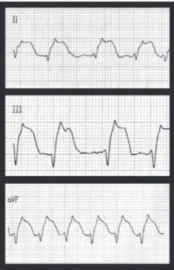
The patient is a 39-year-old female smoker presenting with typical clinical findings of acute myocardial infarction, who has had a mechanic valvular prosthesis in the aortic and mitral position for 2 years, being under irregular use of oral anticoagulants (INR= 1.1). The electrocardiogram showed a 9-mm elevation in the ST segment in the II, III and aVf leads (fig. 1). The CK serum level reached 6240 IU/L (normal < 145 IU/L) and that of CKMB rea-ched 236 IU/L (normal < 10 IU/L) in 12 hours. Primary angioplasty was indicated due to persistence of the clinical and electrocardio-graphic findings, despite the use of vasodilating agents, antiplatelet therapy and general measures.
The coronary angiography identified images of multiple obs-tructions in the distal branches of the left and right coronary arteries, with angiographic characteristics of thrombi (fig. 2). The transthoracic echocardiographic study did not show any atrial or ventricular intracavitary thrombus, any vegetation that would suggest infectious endocarditis, or any dysfunction of the metallic prosthesis. The ejection fraction was 0.18. An angiography dated from 2 years back had evidenced coronary arteries free from obs-tructive disease. The patient was admitted into the intensive care unit and submitted to thrombolytic therapy with streptokinase (1,500,000 IU, intravenously, in 30 min), and did not meet the reperfusion criteria. Tachyarrhythmia, chest pain and hemodynamic instability disappeared only after 10 days of intensive clinical treat-ment with dobutamine, heparin, metoprolol, diuretics, ACE inhi-bitor, opioids, and acetylsalicylic acid. The scintigraphic study evidenced dilation of the cardiac chambers, septal contractile dysfunction, and anterior and posterior contractile dysfunction of the left ventricular walls, with ejection fraction of 0.17. The patient refused to undergo control coronary arteriography. Once adequate anticoagulation was obtained (INR=3.2), she was dis-charged from the hospital. On her first follow-up visit 8 weeks later, the patient was asymptomatic.
Acute myocardial infarction with normal coronary arteries is a rare syndrome whose etiology and pathophysiology remain undefined in most cases. Coronary spasm and thromboembolism are involved.
Fig. 1 - Emergency electrocardiogram: elevation in the ST segment in the II, III, and aVF leads.
Its prevalence in angiographic, endosonographic, and histopatho-logic studies of the coronary arteries of infarcted patients has ranged from 1 to 7%. The mean age of patients experiencing acute myocardial infarction with normal coronary arteries is 40 years; among infarcted patients under 30 years of age, it may represent 16-35% of the cases 1.
2
Arquivos Brasileiros de Cardiologia - Volume 84, Nº 3, Março 2005Volume 84, Nº 3, Março 2005Volume 84, Nº 3, Março 2005Volume 84, Nº 3, Março 2005Volume 84, Nº 3, Março 2005 Multiple Simultaneous Embolisms of Right and Left Coronary Arteries
1. Tun A, Khan IA, Tampa FL, Omaha NE. Myocardial infarction with normal coronary arteries: The Pathologic and Clinical Perspectives. Angiology 2001; 52:299-304. 2. Lanza GM, Berman BJ, Taniuchi M. Multifocal coronary thromboembolism from
a left ventricular thrombus. N Engl J Med 1999; 341: 1083-84.
3. Takenaka T, Horimoto M, Igarashi K et al. Multiple coronary thromboemboli compli-cating valvular heart disease and atrial fibrillation. Am Heart J 1996; 131: 194-6.
References
4. Miranda IA, Esplieguero RA, Ruiz JC et al. Embolismo coronario múltiple en una mujer con factores de riesgo para enfermedad tromboembólica. Rev Esp Cardiol 2003; 56:318-320.
5. Loire R, Tabib A. Les embolies coronaires: a propos de 61 cas anatomo-cliniques. Arch Mal Coeur Vaiss 1985; 78:821-7.
disease, chronic atrial fibrillation, left ventricular thrombus, con-gestive heart failure, irregular use of anticoagulants, smoking, use of oral contraceptives, prothrombotic variant of factor II, and patent oval foramen 2-4. Takenaka et al 3 have attributed that rare
condition to the rupture of a thrombus passing through the left main coronary artery, with consequent obstruction of its distal branches. Consecutive coronary thromboembolism affecting diffe-rent coronary branches during a short period of time would be a less likely mechanism 5.
The risk factors for thromboembolism observed in our patient include smoking, metallic cardiac valvular prosthesis, and inade-quate use of anticoagulants. The following did not occur: pain and cyanosis of the extremities, petechiae, muscle pain, paresthesia of the limbs, paleness or absence of pulse, abdominal pain with gastrointestinal bleeding, flank pain and hematuria, or any other evidence of systemic embolism. The patient did not have neurologic alterations, sensorial or motor deficits, convulsions, dysphasia, or anopsia. The emboli reaching the coronary circulation may have originated from fragile thrombi possibly formed in the aortic pros-thesis close to the ostium of the coronary arteries, or, more unli-kely, in the mitral prosthesis. This is the fourth report of a distal multiple embolic pattern found on coronary angiography, and the second simultaneously affecting the left and right coronary arteries.