Assessment of first molars sagittal and rotational
position in Class II, division 1 malocclusion
Paulo Estevão Scanavini1, Renata Pilli Jóias2, Maria Helena Ferreira Vasconcelos3,
Marco Antonio Scanavini4, Luiz Renato Paranhos5
How to cite this article: Scanavini PE, Jóias RP, Vasconcelos MHF, Scana-vini MA, Paranhos LR. Evaluation of the first molars sagittal and rotational po-sition in Class II, division 1, malocclusion. Dental Press J Orthod. 2013 Nov-Dec;18(6):106-11.
Submitted: September 13, 2011 - Revised and accepted: December 27, 2011
Contact address: Paulo Estevão Scanavini Rua Antônio Pereira de Camargo, 129 – Centro
CEP: 13.170-030 – Sumaré/SP – E-mail: [email protected]
1 MSc in Orthodontics, UMESP. Professor, Specialization Course in
Orthodontics, APCD.
2 PhD Student, Oral Biopathology, UNESP. 3 PhD in Orthodontics, FOB/USP. Professor, UMESP. 4 PhD in Orthodontics, USP. Professor, UMESP.
5 Post-Doc in Dentistry, UNICAMP. Adjunct Professor, Federal University
of Sergipe.
» The authors report no commercial, proprietary or financial interest in the prod-ucts or companies described in this article.
Objective:This study assessed the anterior-posterior positioning of the upper and lower first molars, and the degree of rotation of the upper first molars in individuals with Class II, division 1, malocclusion. Methods: Asymmetry I, an accurate device, was used to assess sixty sets of dental casts from 27 females and 33 males, aged between 12 and 21 years old, with bilateral Class II, division 1. The sagittal position of the molars was determined by positioning the casts onto the device, con-sidering the midpalatal suture as a symmetry reference, and then measuring the distance between the mesial marginal ridge of the most distal molar and the mesial marginal ridge of its counterpart. With regard to the degree of rotation of the upper molar, the distance between landmarks on the mesial marginal ridge was measured. Chi-square test with a 5% significance level was used to verify the variation in molars position. Student’s t test at 5% significance was used for statistical analysis. Results: A great number of lower molars mesially positioned was registered, and the comparison between the right and left sides also demonstrated a higher number of mesially positioned molars on the right side of both arches. The average rotation of the molars was found to be 0.76 mm and 0.93 mm for the right and left sides, respectively. Conclusion: No statistically significant difference was detected between the mean values of molars mesialization regardless of the side and arch. Molars rotation, measured in millimeters, represented ¼ of Class II.
Keywords:Molar tooth. Angle Class II malocclusion. Orthodontics.
Objetivo:esse estudo avaliou o posicionamento anteroposterior dos primeiros molares superiores (1º MS) e inferiores, e o grau de rotação dos 1º MS, em indivíduos com má oclusão de Classe II, divisão 1. Métodos: mensuraram-se, em aparelho de precisão Assimetria I, 60 pares de modelos, de 27 indivíduos do sexo feminino e 33 do masculino, entre 12 e 21 anos de idade, com má oclusão de Classe II, divisão 1. Utilizando a sutura palatina mediana como referência de eixo de simetria, os modelos foram posicionados no aparelho para mensuração da distância entre a crista marginal mesial do molar mais distal e a crista marginal mesial do molar do lado oposto, a fim de verificar o posicionamento sagital dos molares. Em relação à giroversão, mediu-se a distância entre pontos na crista marginal mesial. O teste qui-quadrado a 5% foi utilizado para veri-ficar a variação de posicionamento dos molares, por arcos e por lado. O teste t de Student a 5% foi utilizado para comparar
esses valores. Resultados: houve maior número de molares inferiores mesializados e, comparando os lados, maior número de molares mesializados no lado direito em ambas as arcadas. As rotações médias dos molares foram de 0,76mm do lado direito e 0,93mm do esquerdo. Conclusão: não houve diferença estatisticamente significativa entre os valores médios das mesializações dos molares quanto a lado ou arco. Quando observada isoladamente, a rotação dos molares, quantificada em milímetros, representou uma situação de ¼ de Classe II.
INTRODUCTION
Dental arch symmetry and dimension are of great interest for orthodontists when making diagnosis and
treatment planning.1,2
Molar positioning in the anterior-posterior direc-tion determines the sagittal classificadirec-tion of
malocclu-sion3,4 and can be easily detected in cases of Class II
subdivision. However, molar positioning should also be considered in cases of bilateral malocclusion, since a molar more mesially positioned on one side, even if
not clinically2 apparent, can influence diagnosis,
treat-ment planning and,5-8 especially occlusal stability.
The asymmetric position may determine impor-tant aspects regarding the orthodontic mechanics that will be used, such as the correction of rotations, or
distalization methods.9-12 These procedures may be
applied with different intensities in each to obtain an arch with symmetrical positioning of the posterior
teeth.13 With the purpose of carrying out a detailed
assessment of teeth positioning, the dental cast analy-sis is an important tool due to its practicality,
reliabil-ity and reproducibilreliabil-ity.1,14-20
Thus, this study aimed at assessing the positioning of contralateral molars in the maxillary and mandibu-lar arches in the anterior-posterior direction, as well as to examine the degree of rotation of the upper molars in individuals with Class II, division 1 malocclusion.
MATERIAL AND METHODS
This research was previously approved by the Institu-tional Review Board of the Metodista University of São
Paulo (UMESP), under protocol number 0210135. The sample of this study was obtained from the files of the Postgraduate Program of the aforementioned University. Sixty sets of plaster models from 27 fe-males and 33 fe-males, aged between 12 and 21 years, with Class II, division 1 malocclusion, were selected.
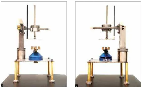
Measurements were made directly in the study mod-els by using Asymmetry I, a device developed in the Postgraduate Program Department to enable the visual-ization of the sagittal positioning of molars as well as the existence of possible rotations. This device consists of a structure similar to a parallelometer, containing: A base for positioning the model, a transparent horizontal acrylic plate with a millimeter ruler, and a rod which enables the identification of the molars positioning (Figs 1A and B).
The midpalatal suture was used as a symmetry
ref-erence.1,2 It was delimited in the maxillary arch by
demarcating landmarks over the mid-palatal suture from the incisive papilla until the most posterior
visi-ble landmark.19,20 The symmetry axis was obtained by
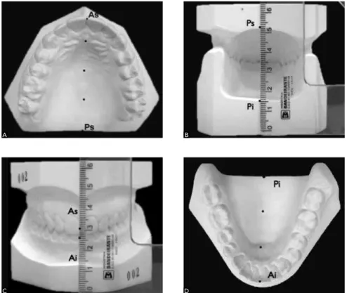
connecting these landmarks. Then, it was extended anteriorly up to the incisal edge of the maxillary inci-sor to determine the landmark As (anterior-superi-or). Conversely, it was posteriorly extended up to the posterior surface of the maxillary model to determine Ps (posterior-superior) (Fig 2A).
The midline projection, obtained in the maxil-lary arch, was used when the midpalatal suture was
transferred to the lower model.15,16 The upper
mid-line was transferred to the lower model by using the reference landmarks As and Ps.
Figure 1 - Measuring device - Asymmetry I -, frontal view (A); posterior view (B).
The models, properly trimmed, were placed in oc-clusion, so that the posterior surfaces matched in the same plane. The landmark Ps of the upper model was transferred to the lower model by means of a set tri-angle ruler positioned perpendicularly to the base of the lower model, thereby determining the landmark Pi (posterior-inferior) on the lower model (Fig 2B).
With the models still in occlusion and with the set triangle ruler equally placed anteriorly to the models matching with the landmark As of the upper model, the landmark Ai (anterior-inferior) was demarcated in the mandibular model (Fig 2C).
Obtaining and connecting Ai and Pi landmarks enabled the lower midline to be determined (Fig 2D).
The models were then placed onto the base of the parallelometer which was fixed to the base of the
Asymmetry I device (Figs 3A and B). When
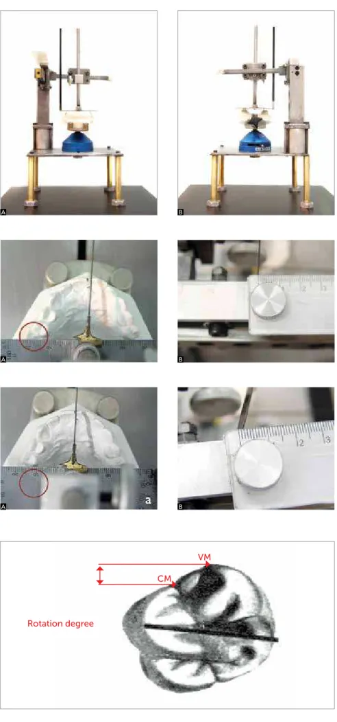
posi-tioning the models, the occlusal surface of the teeth should be parallel to the horizontal plane, and the protractor pointer positioned over the midpalatal su-ture. The reference used for assessing the molar posi-tioning was the mesial marginal ridge. Subsequently, the distance between the mesial marginal ridge posi-tioned more distally and the mesial marginal ridge of the opposite molar was measured in the longitudinal direction, as shown in Figures 4 and 5.
In order to verify the possible association of variation in molar positioning, concerning the arch and side of the
molars mesially positioned, chi-square test (c2) was used.
Student’s t test was employed to compare these values. In all tests, a significance level of 5% was adopted.
In addition to assessing the position of the molars in the anterior-posterior direction, the degree of rota-tion of the upper molars was also assessed, as follows: the distance between the most mesial portion of the mesial marginal ridge toward the mesiodistal sulcus (point CM), and the apex of the mesiobuccal cusp, at its most mesial portion, was measured. One hori-zontal line was projected from the CM landmark, and another from the VM landmark, both parallel. Thus, the distance between these lines was measured, in millimeters, indicating the rotation of the molars in the mesiodistal direction (Fig 6).
RESULTS
Table 1 shows the distribution of the individuals comprising the sample, considering the side of the molar mesially positioned and the dental arch.
The variation in the mean value of molar mesializa-tion was found to be 0.05 mm for the maxillary arch, with mean values of 1.55 mm and 1.50 mm for the left and right sides, respectively. For the mandibular arch, this
Figure 2 - A)determination of the upper midline (landmarks As and Ps); B) transference of the Ps to the lower model - obtaining PI; C) transfer-ence of the As landmark to the lower model – obtaining Ai; D) determination of the lower mid-line (landmarks Ai and Pi).
A
C
B
Figure 3 -Models positioned for measurement: A) front view, B) back view.
Figure 4 - A) Model positioned evidencing the more mesial position of the maxillary right first molar, in relation to left-side counterpart; B ) rul-er recording the position of the right maxillary first molar at zero position.
Figure 5 - A)Device positioned on the mesial marginal ridge of the right maxillary first molar B); ruler registering the most mesial position of the right maxillary first molar by 1.5 mm, in rela-tion to left-side counterpart.
Figure 6 -method used to assess the rotation of the first maxillary molars.
A
A
A
B
B
B
Rotation degree
VM
Mesialization
Arch
Maxillary Mandibular
n % 27 45,0
Right molar 21 35.0 27 45.0
Without mesialization 29 48.3 17 28.3
Left molar 10 16.7 16 26.7
Total 60 100.0 60 100.0
Table 1 - Distribution of the individuals comprising the sample considering the side of the molar mesially positioned and arch.
χ2 = 5.27; p = 0.072 (non-significant difference).
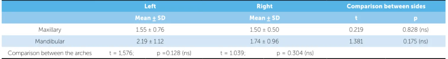
Table 2 - Mean value and standard deviation of molar mesialization with regard to the arch and side, and t test values for the respective comparisons.
ns – non-significant difference.
Left Right Comparison between sides
Mean ± SD Mean ± SD t p
Maxillary 1.55 ± 0.76 1.50 ± 0.50 0.219 0.828 (ns)
Mandibular 2.19 ± 1.12 1.74 ± 0.96 1.381 0.175 (ns)
Comparison between the arches t = 1,576; p =0.128 (ns) t = 1.039; p = 0.304 (ns) variation was 0.45 mm, with mean values of 2.19 mm and
1.74 mm for the left and right sides, respectively. Table 2 shows the mean values and respective standard deviation of molar mesialization with regard to the arch and side.
The t test indicated no significant difference in the
re-spective comparisons.
With regard to the rotation of the upper molars, mean rotations of 0.76 ± 0.37 mm and 0.93 ± 0.53 mm were observed for the right and left molars, respectively, and a mean rotation of 0.85 ± 0.45 mm was observed between both sides.
DISCUSSION
The models used herein were assessed by a device exclusively developed for the dental arch asymmetry analyses. It was carefully designed not only to allow the models to be positioned on a flat surface, but also to observe the inclination of the occlusal plane. Ad-ditionally, it was used a small-caliber rod (0.5 mm) to
avoid improper interferences on the measurements.14-17
In order to achieve more accurate and standard-ized measures, the models were placed onto a paral-lelometer base fixed to the base of the device,
consid-ering the midpalatal suture as a symmetry reference.1,2
The occlusal surfaces of the teeth on the models were leveled to the horizontal plane by means of a leveling board. Two-millimeter rulers were adapted, one on
the protractor base, and the other on the upper part of the device as a way to measure the positioning and degree of rotation of the molars. The part of the de-vice where the protractor with the pointer was fixed could move in transverse and longitudinal directions, through a set of rollers, causing the measurements to be easily and agilely obtained, thus, permitting higher reliability and standardization — once the models re-mained static after been positioned, and only the pro-tractor with the pointers could move.
The analysis of the results demonstrated that the com-parison between maxillary and mandibular arches showed a great number of lower molars more mesially positioned,
corroborating the literature,15,21,22 while the comparison
between the sides showed a higher number of molars me-sially positioned on the right side of both arches.
The mean values regarding variation in molar me-sialization suggest that the sample of this study showed greater variation in molar positioning in the mandibu-lar arch, which leads to a high incidence of asymmetry. This fact occurs because the mesialization of a molar in relation to its counterpart reveals an asymmetric posi-tioning of these teeth, which can also indicate asymme-try in the respective dental arch. Although numerical differences were observed with regard to the sagittal positioning of these molars, our results pointed out that the mean values and standard deviation of molar mesi-alization have no significant difference when compared between the arches, neither when compared to the side of the molar mesially positioned. These findings are in
disagreement with those of other authors15,16,21 who
as-sessed dental asymmetry in individuals with the same malocclusion and found significant differences between the maxillary and mandibular arches.
Asymmetric positioning of molars can be attrib-uted to genetic, postural, chewing and harmful
hab-its.1,6,7,21 A correct diagnosis and adequate treatment
obtain-ing a stable occlusion at retention and post-retention stages. In addition, achieving a symmetrical posi-tioning of molars at an early stage of the orthodontic
treatment favors the success of such a treatment.6,7,8
With regard to factors that possibly influence the asymmetric positioning of molars, the rotation of max-illary molars was assessed. This rotation may indicate an increased length of the maxillary dental arch, besides being responsible for the Class II relationship of molars. Accordingly, it is justified the need to assess the rotation of the first maxillary molars, once the degree of rota-tion may determine the level of Class II malocclusion. The correction of rotation of the first maxillary molars can even transform a Class II molar relationship into a Class I molar relationship, depending on the degree of rotation of the first maxillary molars.
This study aimed at assessing the rotation of molars in millimeters, i.e., foreseeing the space that would be gained in the dental arch so that the molars would be driven to the ideal position. The mean values of
rotation of the molars were found to be 0.76 mm and 0.93 mm for the right and left molars, respectively.
In general, the literature quantifies Class II into ¼ or ½ Class II, or full Class II. Taking into account that the average mesiodistal width of an upper
mo-lar is 10.41 mm23 and that the average rotation of the
molars was 0.84 mm, it was possible to understand that the molar rotation observed herein, when singly considered, would correspond to ¼ of Class II.
CONCLUSION
In conclusion, the results obtained by the meth-odology applied showed that:
» No significant difference was detected between the mean values and standard deviation of the molars mesialization when compared per side (right and left) and per arch (maxillary and mandibular);
» When singly observed, the molar rotation, quan-tified in millimeters, accounted for ¼ of Class II.
1. Lundstrom A. Some asymmetries of dental arches, jaws, and skull, and their etiological significance. Am J Orthod. 1961;47(2):81-106.
2. Lear C. Symmetry analyses of the palate and maxillary dental arch. Angle Orthod. 1968;38(1):56-62.
3. Angle EH. Classification of malocclusion. Dent Cosmo. 1899;41(3):248-64. 4. Andrews LF. The six keys to normal occlusion. Am J Orthod. 1972;
62(3):296-309.
5. Nery PCB, Barbosa JA. Rotação de primeiros molares superiores na oclusão normal e má oclusão de Classe II divisão 1 de Angle. Rev Dental Press Ortod Ortop Facial. 2003;8(5):101-12.
6. Burstone CJ. Diagnosis and treatment planning of patients with asymmetries. Semin Orthod. 1998;4(3):153-64.
7. Shroff B, Siegel SM. Treatment of patients with asymmetries using asymmetric mechanics. Semin Orthod. 1998;4(3):165-79.
8. Paranhos LR, Andrews WA, Jóias RP, Bérzin F, Daruge Júnior E, Triviño T. Dental arch morphology in normal occlusions. Braz J Oral Sci. 2011; 10(1):65-8.
9. Moscardini MS. Estudo comparativo da eficiência do aparelho extrabucal e da barra transpalatina como meios de ancoragem durante a fase de retração. Rev Dental Press Ortod Ortop Facial. 2007;12(2):86-95.
10. Shimizu RH, Ambrosio RA, Shimizu IA, Godoy-Bezerra J, Ribeiro JS, Staszak KR. Princípios biomecânicos do aparelho extrabucal. Rev Dental Press Ortod Ortop Facial. 2004;9(6):122-56.
11. Dahlquist A. The effect of a transpalatal arch for the correction of first molar rotation. Eur J Orthod. 1996;18(3):257-67.
REFERENCES 12. Choi YJ, Lee JS, Cha JY, Park YC. Total distalization of the maxillary arch in a
patient with skeletal Class II malocclusion. Am J Orthod Dentofacial Orthop. 2011;139(6):823-33.
13. Quaglio LC, Freitas KMS, Freitas MR, Janson G, Henriques JFC. Stability and relapse of maxillary anterior crowding treatment in Class I and Class II division 1 malocclusions. Am J Orthod Dentofacial Orthop. 2011;139(6):768-74. 14. Wertz RA. Diagnosis and treatment planning of unilateral Class II
malocclusions. Angle Orthod. 1975;45(2):85-94.
15. Araújo TM. Skeletal and dental arch asymmetries in individuals with normal dental occlusion. Int J Adult Orthod Orthog Surg. 1994;9(2):111-8. 16. Araújo TM. Skeletal and dental arch asymmetries in Class II division 1
malocclusion. J Clin Pediatr Dent. 1994;18(3):181-5.
17. Korkhaus G. A new orthodontic symmetrograph. Int J Orthod Oral Surg Radiol. 1930;16(6):665-8.
18. Mucha JN, Bolognese AM. Análise de modelos em Ortodontia. Rev Bras Odontol. 1985;42(1):28-44.
19. Maurice TJ, Kula K. Dental arch asymmetry in the mixed dentition. Angle Orthod. 1998;68(1):37-44.
20. Alavi DG. Facial and dental arch asymmetries in Class II subdivision malocclusion. Am J Orthod Dentofacial Orthop. 1988;93(1):38-46. 21. Janson GR. Assimetria dentária e suas complicações no tratamento
ortodôntico: Apresentação de um caso clínico. Ortodontia. 1995;28(3):68-73. 22. Rose JM. Mandibular skeletal and dental asymmetry in Class II malocclusions.
Am J Orthod Dentofacial Orthop. 1994;105(5):489-95.