Decreased Eccentric Exercise-Induced
Macrophage Infiltration in Skeletal Muscle
after Supplementation with a Class of
Ginseng-Derived Steroids
Szu-Hsien Yu1,2, Chih-Yang Huang3,4, Shin-Da Lee3,4, Ming-Fen Hsu1, Ray-Yau Wang5, Chung-Lan Kao3., Chia-Hua Kuo1*.
1.Laboratory of Exercise Biochemistry, University of Taipei, Taipei City, Taiwan, Republic of China,2. Department of Leisure Industry and Health Promotion, National Ilan University, Yilan County, Taiwan, Republic of China,3.Institute of Basic Medical Science, China Medical University, Taichung City, Taiwan, Republic of China,4.Department of Health and Biotechnology, Asia University, Taichung City, Taiwan, Republic of China, 5.Department of Physical Therapy and Assistive Technology, National Yang-Ming University, Taipei City, Taiwan, Republic of China
.These authors contributed equally to this work.
Abstract
Dammarane steroids (DS) are a class of chemical compounds present in Panax ginseng. Here, we evaluated the effect of 10 weeks of DS supplementation on inflammatory modulation in the soleus muscle following eccentric exercise (EE)-induced muscle damage (downhill running). Eighty rats were randomized into 4 groups of DS supplementation (saline, 20, 60, 120 mg/kg body weight).
Inflammatory markers were measured at rest and again 1 h after EE. At rest, NFkB signaling, TNF-alpha and IL-6 mRNAs, 3-nitrotyrosine, glutathione peroxidase, and GCS (glutamylcysteine synthetase) levels were significantly elevated in the skeletal muscle of DS-treated rats in a dose-dependent manner. Additionally, there were no detectable increases in the number of necrotic muscle fibers or CD68+ M1
macrophages. However, muscle strength, centronucleation, IL-10 mRNA
expression, and the number of CD163+M2 macrophages increased significantly
over controls with DS treatment in rat soleus muscle. Under EE-challenged conditions, significant increases in muscle fiber necrosis, CD68+ M1 macrophage
distribution, and 3-nitrotyrosine were absent in rats that received low and medium doses (20 and 60 mg/kg) of DS treatment, suggesting that DS possess anti-inflammatory action protecting against a muscle-damaging challenge. However, this protective activity was diminished when a high dose of DS (120 mg/kg) was administered, suggesting that DS possess hormetic properties. In conclusion, our study provides new evidence suggesting that DS is an ergogenic component of
OPEN ACCESS
Citation:Yu S-H, Huang C-Y, Lee S-D, Hsu M-F, Wang R-Y, et al. (2014) Decreased Eccentric Exercise-Induced Macrophage Infiltration in Skeletal Muscle after Supplementation with a Class of Ginseng-Derived Steroids. PLoS ONE 9(12): e114649. doi:10.1371/journal.pone.0114649
Editor:Hsin-Chih Lai, Chang-Gung University, Taiwan
Received:August 28, 2014
Accepted:November 12, 2014
Published:December 11, 2014
Copyright:ß2014 Yu et al. This is an open-access article distributed under the terms of the Creative Commons Attribution License, which permits unrestricted use, distribution, and repro-duction in any medium, provided the original author and source are credited.
Data Availability:The authors confirm that all data underlying the findings are fully available without restriction. All relevant data are within the paper.
Funding:This study was supported in part by National Science Council of Taiwan, Pegasus Pharmaceuticals (101-2622-H-154-001-CC2), http://www.ppginco.com/eng/aboutus/index.html, and the University of Taipei, Taiwan,www.utaipei. edu.tw. The funders had no role in study design, data collection and analysis, manuscript prepara-tion, or decision to publish.
ginseng that potentiate inflammation at baseline but that produce anti-inflammatory effects on skeletal muscle following muscle-damaging exercise. Furthermore, high doses should be avoided in formulating ginseng-based products.
Introduction
Inflammation, an innate immune response, plays a key role in eliminating unhealthy cells generated by various adverse conditions [1]; this process is essential for the regeneration of new muscle fibers after damage [2]. At the cellular level, NFkB activation in damaged skeletal muscle facilitates an inflammatory response, which increases interleukin-1beta (IL-1b), tumor necrosis factor alpha (TNF-a), interleukin-6 (IL-6), inducible nitric oxide synthase (iNOS), and cyclooxygenase-2 (COX-2) [3]. This response facilitates further clearance of damaged fibers by communicating with surrounding mononuclear cells. Two phenotypes of mononuclear cells, M1 (CD68+ population) and M2 (CD163+ population) macrophages, are known to orchestrate this reconstructive program in damaged skeletal muscle. During the early phase, M1 macrophages are recruited to damaged muscle fibers, and free radicals are released to lyse cells in inflamed tissue [4]. This process makes room for tissue repopulation. Changes in redox state after the burst of oxidative free radicals, followed by increased antioxidant production, appears to be essential for the recruitment of stem cells to the damaged site for cell regeneration within a controlled period of time [5]. During the late phase, M1 macrophages are replaced by M2 macrophages to drive a protracted, tissue-specific differentiation. Release of interleukin-10 (IL-10) from M2 macrophages is associated with inhibition of M1 macrophages to end the first destructive phase [4,6].
Inflammation plays a significant part in the remodeling and adaptation of skeletal muscle in response to physical challenge [7]. In young healthy animals, local anti-inflammatory treatment delays skeletal muscle fiber differentiation after an over-load lesion [8], blocks stem cell proliferation in EE-challenged muscle in humans [9], slows hypertrophy in a rat synergist ablation model [10,11], and decreases recovery of force development in muscle injury [12]. These previous findings suggest that inflammatory modulation plays a crucial role in muscle adaptation against physical challenges.
Dammarane steroids (DS), a class of steroids present in many ginseng species, have been shown to have an anti-inflammatory activity [13,14]. However, other studies have reported an increase in cell death after DS treatment in cultured cells [15,16], suggesting that DS possess pro-inflammatory properties [17]. The underlying mechanisms behind this paradox are not clearly known. Ginseng has been used for thousands of years as a popular supplement to enhance stamina and coping capacity against physical fatigue. However, most studies have not
confounder and may be responsible for the inconsistent results observed among studies [18,19]. The steroid profile of ginseng varies with species and cultivating season [20,21]. In this study, the effects of the ginseng-derived steroids DS on inflammation and strength were examined in rat skeletal muscle after 10 weeks of oral supplementation. Eccentric exercise is known to cause fiber injury and elicit an inflammatory response in contracted skeletal muscle. Inflammation-associated markers were measured in skeletal muscle at rest and 1 h following an acute bout of downhill running, which consists of eccentric muscle contraction, in rats that received various doses of DS supplementation.
Materials and Methods
Dammarane steroids (DS)
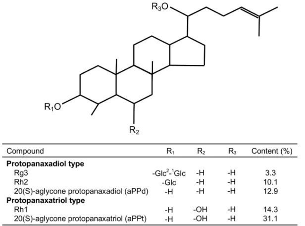
The chemical structures and content of DS (ChemSpider ID: 7827637) are shown in Fig. 1, and they were obtained from Pegasus Inc. (Vancouver, Canada). DS (20 mg) was dissolved in 10 ml methanol as test solution and purified by high performance liquid chromatography (HPLC) on a C-18 Silica-based reversed phase HPLC column (Agilent ODS-C18, 5 mm, 4.66150 mm). The mobile phase was a 2:4:2:1 mixture of chloroform, ethyl acetate, methanol and water, and the separation conditions were as follows: 120 ml mobile phase was injected for each separation, the flow rate was 1 ml/min, and was eluted with a retention time of 120 min. The DS was confirmed by comparison of the physical and spectral data using above process with reference standard solution containing 1.5 mg of Rh1 (C36H64O9), Rg3 (C30H54O3), Rh2 (C36H64O8), 20(S)-aglycone protopanaxadiol (aPPd) (C30H54O3) and 20(S)-aglycone protopanaxatriol (aPPt) (C30H54O4). The content of DS powder contained Rh1 (14.3%), Rg3 (3.3%), Rh2 (10.1%), aPPd (12.9%) and aPPt (31.1%).
Animals
Experimental design
A 262 factorial design (supplemental dosage and exercise challenge) was used in this study. Following a 7-day acclimatization to the housing environment at University of Taipei, the 80 rats were randomized by weight into 4 groups. Each group was assigned one of 4 daily DS doses: saline (Control, N520) and 20 (DS20, N520), 60 (DS60, N520), or 120 mg/kg BW (DS120, N520). DS were dissolved in saline by vortexing immediately before oral intubation. Equal volumes of DS and saline were delivered daily by gastric gavage to DS-treated and control rats, respectively. To determine the effect of DS supplementation on muscle strength and baseline inflammation, DS supplemented groups were compared with controls in sedentary animals (N540) after 10 weeks of DS treatment. To determine the effect of DS supplementation on eccentric exercise-induced muscle damage and inflammatory responses, rats at each dosage were equally split into sedentary (Sedentary, N510) and eccentric exercise (EE) groups (N510).
The eccentric exercise protocol was modified from previously published methods using downhill running [22]. One week prior to experiments, all animals were acclimated to running on a rat treadmill at 10 m/min for 10 min per day. Rats in the EE group were challenged with an acute bout of intermittent downhill
Fig. 1. Chemical structure of dammarane steroids fromPanax ginseng.
exercise at a 16
˚
decline. Exercises consisted of 18 sessions (5 min/session) separated by 2 min of rest at 16 m/min for a total of 90 min. All rats in EE groups completed the entire exercise session. Two hour after exercise, rats wereanaesthetized with intraperitoneal injection of chloral hydrate (400 mg/kg BW). Soleus muscles were excised from both legs and frozen immediately with liquid nitrogen and then stored at 280
˚
C for further analysis.Histology and Immunohistochemistry (IHC)
Histology and immunohistochemistry staining were carried out by a pathologist at the Taipei Institute of Pathology (Taipei, Taiwan). Muscle tissues were fixed in formalin and embedded in paraffin. Muscle samples were then sliced to produce 2-mm-thick sections. Sections were stained with hematoxylin and eosin (H&E) to observe tissue histology, and immunohistochemistry was performed to evaluate macrophage invasion and centronucleation as a muscle regeneration marker and to confirm H&E staining [23]. Fixed muscle sections were incubated with mouse antibodies against rat CD68 (dilution 1:250) (abcam, Cambridge, UK) and rabbit CD163 (dilution 1:450) (epitomic, San Diego, CA, USA) and then visualized using mouse and rabbit specific HRP/DAB (ABC) detection IHC kit (abcam,
Cambridge, UK). Image analysis software Image J (NIH, Bethesda, MD, USA) was used to count the number of CD68- and CD163-positive cells per 100 muscle fibers as described previously [24].
Western Blotting Analysis
Soleus muscle (,100 mg) was homogenized in 1 mL of HES buffer containing 20 mM Hepes Aldrich, St Louis, MO, USA), 1 mM EDTA (Sigma-Aldrich, St Louis, MO, USA), and 250 mM sucrose (Sigma-(Sigma-Aldrich, St Louis, MO, USA). Homogenates were centrifuged at 10,000 g for 10 min at 4
˚
C, and the supernatant was collected for Western blotting analysis. Protein (50 mg) was separated by SDS-PAGE (10–12% acrylamide) and transferred to PVDFmembranes (PALL Life Science, Ann Arbor, MI, USA) by standard wet transfer methods. After 1 h blocking in PBS containing 5% skim milk, membranes were incubated with primary antibody overnight at 4
˚
C. Primary antibodies against nitrotyrosine were purchased from Millipore (Bedford, MA, USA) (dilution 1:1000); antibodies against iNOS and eNOS were purchased from BD(Santa Cruz, CA, USA). Antibody-bound protein was detected using a peroxidase-conjugated mouse secondary antibody (Sigma, St Louis, MO, USA) or anti-rabbit IgG (Cell Signaling Technology, Beverly, MA, USA) and enhanced
chemiluminescent HRP substrate (PerkinElmer Life and Analytical Sciences, Shelton, CT, USA). To verify equal protein loading, GAPDH was used as an internal control. Western blot bands were quantified using a ChemiDoc XRS+
System (BioRad, Hercules, CA, USA).
Quantitative Polymerase Chain Reaction (qPCR)
Total RNA was isolated using Qiagen RNeasy Fibrous Tissue Mini Kit (Qiagen, Valencia, CA, USA). Reverse transcription of RNA was carried out using iScriptTM cDNA Synthesis Kit (BioRad, Hercules, CA, USA). The following primer and probe sequences were used for amplification of target genes: COX-2 (NM_017232) forward primer: CAGTCTCTCATCTGCAATA; reverse primer: AGGGTTAATGTCATCTAGTC; probe: TCCCTTTGCCTCTTTCAATGTGC. iNOS (NM_012611) forward primer: TGAGGATTACTTCTTCCAG; reverse primer: TGCTCCATAGGAAAAGAC; probe:
CACCGAAGATATCCTCATGATA-ACGT. TNF-a (NM_012675) forward primer: GAGTCATTGCTCTGTGAG;
reverse primer: CTCTGAGGAGTAGACGATA; probe: CTGGCGTGTTCAT-CCGTTCTCT. IL-6 (NM_012589) forward primer: GAGCAATACTGAAAC-CCTA; reverse primer: GATGGTCTTGGTCCTTAG; probe:
ACTCCTTCTGTG-ACTCTAACTTCTCCA. IL-1b (NM_031512) forward primer:
CCAAGCACCTT-CTTTTCC; reverse primer: GTTGGCTTATGTTCTGTCC; probe: CCGTCCTCT-GTGACTCGTGG. TaqMan probes contained a FAM reporter at the 5’-end and a TAMRA quencher at the 3’-end. The RT-qPCR conditions have been previously described [25]. Gene expression was normalized to 18S rRNA.
Wire suspension test
Rat muscle strength was measured by a wire suspension test [26,27] conducted one night after the final DS treatment. A 12 gauge, 50 cm plastic wire was suspended between two platforms 80 cm above a foam cushioned surface. At the beginning of the test, forepaws were placed on the wire and the time (seconds) to fall was measured in two trials with a 5-minute separation. Muscle strength was defined as maximum time the rat held on to the wire.
Fiber typing
Staining methods to determine muscle fiber type have been described previously [25]. Briefly, 8 mm thick frozen cross-section of each soleus muscle were sliced at
Statistical analysis
One-way analysis of variance (ANOVA) was conducted to compare the mean of all variables among groups, with the exception of fiber typing analysis. The Duncan post hoc test, which holds the probability value of a type I error to 5 percent for each test, was employed to distinguish differences between pairs of groups. Two-way ANOVA was used to determine the interactive effect of dosage and exercise challenge. Changes in fiber type were analyzed using the non-parametric chi-square method. Variable values of each group are presented as the mean ¡standard error (SE). A level ofP,0.05 was set for statistical significance of difference for all tests.
Results
To evaluate the immune response to EE in skeletal muscle after downhill running, we examined leukocyte invasion and the number of necrotic muscle fibers. H&E staining and immunohistochemical staining for CD68+ (M1 macrophages) was performed in rat soleus following 10 weeks of DS supplementation at various doses (Fig. 2). Necrotic muscle fibers and CD68-positive cells were identified by cells invasion (Fig. 2A) and brown color (Fig. 2B), respectively. In non-exercised rats, there was no increase in necrotic muscle fibers (Fig. 2A) or CD68+ M1 macrophages (Fig. 2B) in skeletal muscle with DS treatment. However, levels of 3-nitrotyrosine, an oxidative damage marker, increased linearly with dosage (Fig. 2C). Following EE challenge, the number of necrotic muscle fibers increased significantly above the non-exercise control, along with leukocyte and CD68+M1 macrophage infiltration. In rats treated with low and medium doses of DS, such increases were not observed with EE challenge (Figs. 2A and 2B). In saline control rats, 3-nitrotyrosine levels in EE-challenged muscle were significantly greater than in non-exercised muscle. However, this increase was not observed at all doses of DS treatment (Fig. 2C).
muscle of DS-treated rats than in those of saline control rats; however, levels were lower than those of EE-challenged saline control rats (Figs. 3F–H).
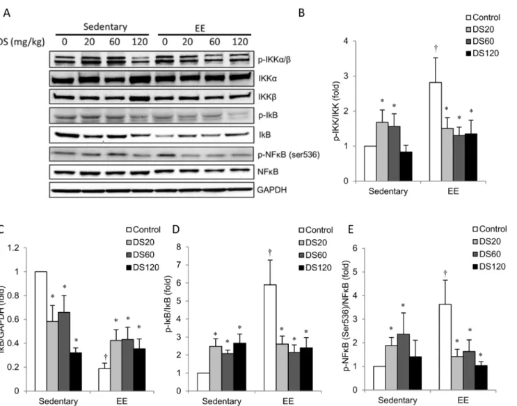
The NFkB signaling data are shown in Fig. 4. Representative radioactivity is displayed inFig. 4Ausing GAPDH as an internal standard. Among saline control rats, p-IKK/IKK (Fig. 4B), p-IkB/IkB (Fig. 4D) and p-NFkB/NFkB (Fig. 4E) levels in muscle of the EE-challenged group were greater than those in the non-exercise control group. IkB was changed in the opposite direction. These changes were attenuated in DS-treated rats at all doses (Fig. 4C). Among non-exercise rats, the 20 mg/kg and 60 mg/kg doses of DS resulted in a significant increase in p-IKK/IKK (Fig. 4B) and p-NFkB/NFkB (Fig. 4E) above saline control levels in muscle. Conversely, DS treatment significantly decreased IkB expression below saline control levels in a dose-dependent manner (Fig. 4C). p-IkB/IkB levels in muscle for all DS-treated rats were significantly greater than those in saline control rats (Fig. 4D).
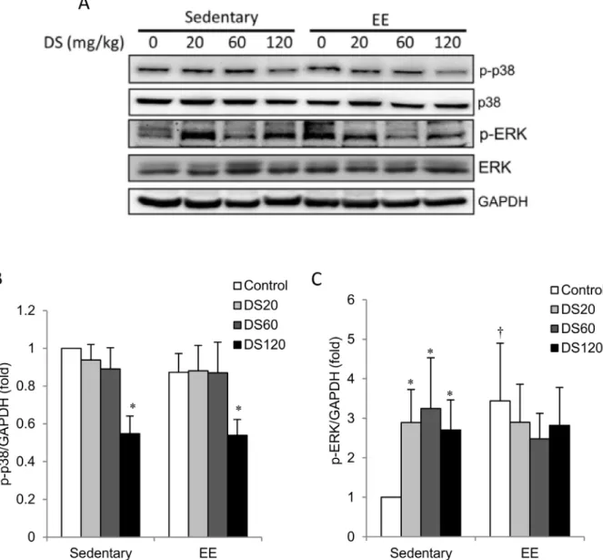
P38 mitogen-activated protein kinases (MAPK) are generally activated by stress-induced inflammation. Here, we found no difference in phosphorylation of p38 MAPK (phospho-p38) between sedentary and EE-challenged muscles (Figs. 5A and 5B). DS treatment at 120 mg/kg resulted in reduced muscle phospho-p38 levels relative to saline controls in both sedentary and EE-challenged rats; however, no apparent change was found at low and medium doses. In contrast, phosphorylation of ERK (phospho-ERK) increased significantly above non-exercise controls in the muscle of EE-challenged rats (Figs. 5A and 5C). All doses of DS supplementation and exercise had similar effects on phospho-ERK levels. No additive effect of DS and exercise on phospho-ERK was observed.
Antioxidant enzymes, including MnSOD, Cu/ZnSOD, GCS and GPx, were evaluated in muscle as shown in Fig. 6. Muscle MnSOD and Cu/ZnSOD were unaffected by EE-challenge (Figs. 6A–6C). MnSOD protein levels in both sedentary and EE-challenged rats were significantly decreased below saline control levels with 120 mg/kg DS treatment (Figs. 6A and 6B). No significant effects of DS on Cu/ZnSOD protein levels were observed (Figs. 6A and 6C). In non-exercise control rats, muscle GCS protein levels were increased with DS treatment in a dose-dependent manner relative to the saline control (Figs. 6A and 6D). Among saline-control rats, EE-challenge resulted in a significant increase in GCS protein levels over sedentary control levels. However, this increase was attenuated in a dose-dependent manner by DS treatment. Only low a dose of DS treatment resulted in increased muscle GPx (Fig. 6E). No effect on GPx was observed with EE-challenge.
Fig. 2. Muscle injury, macrophage invasion and oxidative stress in soleus muscle.(A) Representative histochemical analysis of muscle sections from soleus muscle with H&E staining. Arrowheads indicate immune cell invasion. Scoring is shown on the right. (B) Representative immunohistochemical staining of CD68-positive cells (brown color) in a soleus muscle section. Nucleolus was labeled with eosin staining (blue color). Original magnification was 400x, and scoring of CD68-positive cells is shown on the bottom. (C) Representative western blot showing levels of nitrotyrosine extracted from soleus muscle. Bars represent the relative quantification of nitrated protein normalized to GAPDH. In (A–C), and data are presented as the mean¡SEM.{p,0.05 compared with the sedentary group. *p,0.05 compared with the control group of sedentary or EE. Scale bar550mm.
Muscle strength was evaluated using a wire suspension test (Fig. 7A).
Suspension time increased by 40–50% in all doses of DS treatment compared to control rats. Regenerative muscle fibers and CD163-positive cells were identified by centronucleation (Fig. 7A) and brown color (Fig. 7B), respectively.
Histological (Fig. 7B) and immunohistochemical (Fig. 7C) analyses show that 10
Fig. 3. Inflammatory gene and cytokine expression in soleus muscle.(A–E) Bars represent the relative quantification of COX-2, iNOS, TNF-a, IL-6, and IL-1bmRNA expression levels normalized to 18S rRNA. (F) Representative western blot showing protein levels of iNOS and eNOS extracted from soleus muscle. (G–H) Bars represent the relative protein quantification of iNOS (G) and eNOS (H) normalized to GAPDH. In (A–E, G–H), and data are presented as the mean¡SEM.{p,0.05 compared with the sedentary group. *p,0.05 compared with the control group of sedentary or EE.
doi:10.1371/journal.pone.0114649.g003
Fig. 4. NFkB pathway.(A) Representative western blot showing protein levels of total and phosphorylated IKKa, IkBaand NFkB extracted from soleus
muscle. (B–E) Bars represent relative protein quantification of p-IKK/IKK, p-IkBa/IkBa, total IkBaand p-NFkB/NFkB normalized to GAPDH. In (B–E), and data are presented as the mean¡SEM.{p,0.05 compared with the sedentary group. *p,0.05 compared with the control group of sedentary or EE.
weeks of DS supplementation increased centronucleation and the number of CD163+ M2 macrophages in muscle above saline control level, indicating increased muscle fiber regeneration. Consistent with these data, IL-10 mRNA expression was elevated in DS-treated rats relative to saline controls (Fig. 7D). No effect of DS supplementation on muscle fiber type composition was observed (Fig. 7E).
Fig. 5. MAPK phosphorylation.(A) Representative western blot showing protein levels of total and phosphorylated p38 and ERK extracted from soleus muscle. (B–C) Bars represent the relative protein quantification of p-p38/p38 and p-ERK/ERK normalized to GAPDH. In (B–C), data are presented as the mean¡SEM.{p,0.05 compared with the sedentary group. *p,0.05 compared with the control group of sedentary or EE.
Discussion
The major findings of the study are (1) the ginseng-derived steroids DS can potentiate inflammatory signaling of skeletal muscle in a dose-dependent manner, as evidenced by increases in nitrotyrosine levels, NFkB signaling, and gene expression of pro-inflammatory mediators, without apparent muscle fiber injury or CD68+M1 macrophage invasion in skeletal muscle; (2) muscle strength can be increased by long-term DS supplementation at all doses tested; and (3) muscle damage, nitrotyrosine levels, and CD68+ M1 macrophage infiltration induced by EE can be minimized at low and medium DS doses, but this protective effect is diminished at a maximal dose. The reverse U-shape dose-dependent outcome suggests that DS have hormetic properties.
Fig. 6. Antioxidant enzymes in soleus muscle.(A) Representative western blot showing protein levels of MnSOD, Cu/ZnSOD, GCS and GPx1 extracted from soleus muscle. (B–E) Bars represent the relative protein quantification of MnSOD (B), Cu/ZnSOD (C), GCS (D) and GPx1 (E) normalized to GAPDH. In (B–E), data are presented as the mean¡SEM.{p,0.05 compared with the sedentary group. *p,0.05 compared with the control group of sedentary or EE.
Given that muscle fiber composition and muscle mass were not changed, the increased muscle strength in DS-treated rats appears to be associated with increased muscle fiber renewal. DS have been reported to stimulate cell
regeneration in skin cells [28], neurons [29], and b cells [30]. Here, we supply additional evidence that increases in muscle strength with DS supplementation are associated with muscle regeneration, evidenced by increased centronucleation. Inflammation is required for muscle fiber turnover in rodents and humans. Pro-inflammatory interventions, such as delivery of myotoxic agents [31] or glucose [32], have been shown to increase muscle regeneration and improve muscle strength. In this study, increased IL-10 mRNA expression and CD163+ M2 macrophage localization in skeletal muscle with DS supplementation are consistent with the observed increase in centronucleation. IL-10 is highly
expressed in M2 macrophages, which are known to promote muscle regeneration [33,34]. Taken together, increased cell turnover is expected to reduce average cell age in skeletal muscle, which may underlie the increased resilience of skeletal muscle after long-term DS supplementation.
Though DS supplementation appears to potentiate inflammatory signaling in skeletal muscle, injured fibers and EE-induced oxidative damage were absent in rats with long-term, low-dose DS supplementation. This is consistent with reduced M1 macrophage infiltration, a primary source of NO production during inflammation [4]. M1 macrophage infiltration into tissues typically occurs with cell injury, particularly after traumatic challenge or during cell aging. In early phases of inflammation, phagocytic CD68+M1 macrophages invade and lyse existing injured muscle fibers, followed by a second phase of accumulation of non-phagocytic CD163+ M2 macrophages to renew the muscle tissue [7]. Long-term DS supplementation may shorten the length of time required for this muscle regeneration program to produce a younger and healthier muscle fiber
population. In previous studies, Panax ginseng extract supplementation has been shown to reduce nitric oxide [34] and muscle damage [35] levels in untrained exercise. Our results suggest that DS may be the active component of ginseng that contributes to the putative ergogenic effect reported in previous studies.
In the present study, our data show that DS supplementation significantly increased oxidative stress at rest. Free radicals are a required mediator for perpetuating inflammatory responses and stem cell recruitment [5]. Oxidative stress generated during inflammation amplifies the inflammatory responses mediated by activation of NFkB and MAPK signaling [4] to increase gene expression of pro-inflammatory mediators, e.g., iNOS, eNOS, COX-2 and cytokines [3]. However, EE-induced NFkB/MAPK signaling, M1 macrophage infiltration, and gene expression of inflammatory mediators were attenuated in the skeletal muscle of DS-treated rats. Thus, early renewal of muscle fiber
scoring of CD163-positive cells is shown on the right. (D) Bars represent the relative quantification of IL-10 mRNA expression levels normalized to 18S rRNA. (E) Bars represent the percentage of muscle fiber types in the control and 20 mg/kg DS, 60 mg/kg DS, and 120 mg/kg DS groups. In (A–E), data are presented as the mean¡SEM. *p,0.05 compared with the control group. Scale bar550mm.
populations by potentiating inflammatory signaling with long-term DS supple-mentation may explain the protective effect of DS against a muscle-damaging exercise. Whether or not the inflammatory potentiation effects of increasing DS dosage can produce adverse outcome in humans demands further clinical investigation.
The NFkB/MAPK signaling system is also known to enhance expression of antioxidant enzymes [36]. The duration and degree of free radical surge during inflammatory processes can be controlled by alterations in antioxidant levels. The reduction in EE-induced oxidative damage in muscle of DS-treated rats may be explained, in part, by increased expression of antioxidant enzymes, such as GPx and GCS. This is consistent with previous in vitro data reporting that ginseng treatment upregulated GPx activity in the soleus muscle of rats [36] and GCS activity in pheochromocytoma PC12 cells [37]. Our data show increased
antioxidant enzyme expression and NFkB/MAPK signaling in the muscle of DS-treated rats at low and medium doses; this may explain the attenuated oxidative stress in muscle following EE challenge. However, increased GPx levels were attenuated at a high dose of DS. Additionally, decreased MnSOD levels were observed only with a high dose of DS supplementation; this indicates that too much DS may undermine its protective action. Similar outcomes have also been reported in hyperglycemia-associated chronic inflammation [38]. These results describe the important caveat that different dosages and durations of ginseng use can produce contradictory outcomes in vivo.
Panax ginseng is a popular herbal medicine used worldwide to enhance stamina and coping capacity against physical fatigue. To date, the active ginsenoside component (glycosylated steroids) that contributes to its alleged ergogenic benefit is unknown. Due to its structural similarity to many human steroid hormones, the steroid component of ginseng has been a target of research. However, the steroid profile of ginseng varies with species type and season [20]; this may be a major confounder that contributes to the conflicting results reported in previous ginseng studies [20,21]. Using ginseng-derived steroids is one way to standardize ginseng and provide a more reliable pharmacological outcomes. The results of this study provide a new perspective and suggest that modulating inflammation with ginseng-derived steroids can influence skeletal muscle performance.
Acknowledgments
We thank Ming-Chieh Wu and Mallikarjuna Korivi for their technical assistance.
Author Contributions
Conceived and designed the experiments: SHY CHK. Performed the experiments: SHY MFH. Analyzed the data: SHY CYH SDL CHK. Contributed reagents/ materials/analysis tools: CYH SDL RYW CLK. Wrote the paper: SHY CLK CHK.
References
1. Ungaro R, Fukata M, Hsu D, Hernandez Y, Breglio K, et al. (2009) A novel Toll-like receptor 4 antagonist antibody ameliorates inflammation but impairs mucosal healing in murine colitis.
Am J Physiol 296: G1167–G1179.
2. Tidball J, Wehling-Henricks M(2007) Macrophages promote muscle membrane repair and muscle fibre growth and regeneration during modified muscle loading in mice in vivo. J Physiol 578: 327–336.
3. Ji LL(2008) Modulation of skeletal muscle antioxidant defense by exercise: role of redox signaling. Free Radic Biol Med 44: 142–152.
4. Tidball JG, Villalta SA(2010) Regulatory interactions between muscle and the immune system during muscle regeneration. Am J Physiol 298: R1173–R1187.
5. Vezzoli M, Castellani P, Corna G, Castiglioni A, Bosurgi L, et al.(2011) High-mobility group box 1 release and redox regulation accompany regeneration and remodeling of skeletal muscle. Antioxid Redox Signal 15: 2161–2174.
6. McLennan I(1993) Resident macrophages (ED2-and ED3-positive) do not phagocytose degenerating rat skeletal muscle fibres. Cell Tissue Res 272: 193–196.
7. St Pierre BA, Tidball JG(1994) Differential response of macrophage subpopulations to soleus muscle reloading after rat hindlimb suspension. J Appl Physiol 77: 290–297.
8. Monda M, Vicidomini C, Viggiano A, Sampaolo S, Di Iorio G, et al.(2009) Inhibition of prostaglandin synthesis reduces the induction of MyoD expression in rat soleus muscle. J Muscle Res Cell Motil 30: 139–144.
9. Mikkelsen UR, Langberg H, Helmark IC, Skovgaard D, Andersen LL, et al. (2009) Local NSAID infusion inhibits satellite cell proliferation in human skeletal muscle after eccentric exercise. J Appl Physiol 107: 1600–1611.
10. Novak ML, Billich W, Smith SM, Sukhija KB, McLoughlin TJ, et al.(2009) COX-2 inhibitor reduces skeletal muscle hypertrophy in mice. Am J Physiol 296: R1132–R1139.
11. Soltow QA, Betters JL, Sellman JE, Lira VA, Long JH, et al.(2006) Ibuprofen inhibits skeletal muscle hypertrophy in rats. Med Sci Sports Exerc 38: 840–846.
12. Mishra DK, Fride´n J, Schmitz MC, Lieber RL(1995) Anti-inflammatory medication after muscle injury. A treatment resulting in short-term improvement but subsequent loss of muscle function. J Bone Joint Surg Am 77: 1510–1519.
13. Scholz D, Baumann K, Grassberger M, Wolff-Winiski B, Rihs G, et al. (2004) Synthesis of dammarane-type triterpenoids with anti-inflammatory activity in vivo. Bioorg Med Chem Lett 14: 2983– 2986.
14. Song SB, Tung NH, Quang TH, Ngan NT, Kim KE, et al.(2012) Inhibition of TNF-a-mediated NF-kB Transcriptional Activity in HepG2 Cells by Dammarane-type Saponins from Panax ginseng Leaves. J Ginseng Res 36: 146–152.
16. Park IH, Piao LZ, Kwon SW, Lee YJ, Cho SY, et al. (2002) Cytotoxic dammarane glycosides from processed ginseng. Chem Pharm Bull (Tokyo) 50: 538–540.
17. Wallach D, Kang TB, Kovalenko A(2014) Concepts of tissue injury and cell death in inflammation: a historical perspective. Nat Rev Immunol 14: 51–59.
18. Bahrke MS, Morgan WR(2000) Evaluation of the ergogenic properties of ginseng: an update. Sports Med 29: 113–133.
19. Bucci LR(2000) Selected herbals and human exercise performance. Am J Clin Nutri 72: 624s–636s.
20. Sievenpiper JL, Arnason JT, Leiter LA, Vuksan V (2003) Variable effects of American ginseng: a batch of American ginseng (Panax quinquefolius L.) with a depressed ginsenoside profile does not affect postprandial glycemia. Eur J Clin Nutri 57: 243–248.
21. Sievenpiper JL, Arnason JT, Leiter LA, Vuksan V(2004) Decreasing, null and increasing effects of eight popular types of ginseng on acute postprandial glycemic indices in healthy humans: the role of ginsenosides. J Am Coll Nutr 23: 248–258.
22. Lima-Cabello E, Cuevas MJ, Garatachea N, Baldini M, Almar M, et al. (2010) Eccentric exercise induces nitric oxide synthase expression through nuclear factor-kB modulation in rat skeletal muscle. J Appl Physiol 108: 575–583.
23. Sato K, Li Y, Foster W, Fukushima K, Badlani N, et al.(2003) Improvement of muscle healing through enhancement of muscle regeneration and prevention of fibrosis. Muscle & Nerve 28: 365–372.
24. Przybyla B, Gurley C, Harvey JF, Bearden E, Kortebein P, et al.(2006) Aging alters macrophage properties in human skeletal muscle both at rest and in response to acute resistance exercise. Exp Gerontol 41: 320–327.
25. Chen CY, Tsai YL, Kao CL, Lee SD, Wu MC, et al.(2010) Effect of mild intermittent hypoxia on glucose tolerance, muscle morphology and AMPK-PGC-1alpha signaling. Chin J Physiol 53: 62–71.
26. Adori C, Zelena D, Tı´ma´r J, Gyarmati Z, Domokos A, et al. (2010) Intermittent prenatal MDMA exposure alters physiological but not mood related parameters in adult rat offspring. Behav Brain Res 206: 299–309.
27. Johnson SA, Fournier NM, Kalynchuk LE (2006) Effect of different doses of corticosterone on depression-like behavior and HPA axis responses to a novel stressor. Behav Brain Res 168: 280–288.
28. Kim W-K, Song S-Y, Oh WK, Kaewsuwan S, Tran TL, et al. (2013) Wound-healing effect of ginsenoside Rd from leaves of Panax ginseng via cyclic AMP-dependent protein kinase pathway. Eur J Pharmacol 702: 285–293.
29. Zheng G, Cheng W, Wang Y, Wang X, Zhao S, et al. (2011) Ginseng total saponins enhance neurogenesis after focal cerebral ischemia. J Ethnopharmacol 133: 724–728.
30. Wang Y, Wang H, Liu Y, Li C, Qi P, et al. (2012) Antihyperglycemic effect of ginsenoside Rh2 by inducing isletb-cell regeneration in mice. Horm Metab Res 44: 33–40.
31. Howell J, Walker KR, Creed KE, Dunton E, Davies L, et al.(2013) Phosphorylase re-expression, increase in the force of contraction and decreased fatigue following notexin-induced muscle damage and regeneration in the ovine model of McArdle disease. Neuromuscul Disord 24: 167–177.
32. Jensen KT, Rabago DP, Best TM, Patterson JJ, Vanderby R(2008) Early inflammatory response of knee ligaments to prolotherapy in a rat model. J Orthop Res 26: 816–823.
33. Deng B, Wehling-Henricks M, Villalta SA, Wang Y, Tidball JG (2012) IL-10 triggers changes in macrophage phenotype that promote muscle growth and regeneration. J Immunol 189: 3669–3680.
34. Cabral dOAC, Perez AC, Prieto JG, Duarte ID, Alvarez AI (2005) Protection of Panax ginseng in injured muscles after eccentric exercise. J Ethnopharmacol 97: 211–214.
35. Cabral dOAC, Perez AC, Merino G, Prieto JG, Alvarez AI(2001) Protective effects of Panax ginseng on muscle injury and inflammation after eccentric exercise. Comp Biochem Physiol C Toxicol Pharmacol 130: 369–377.
37. Park SH, Jang JH, Chen CY, Na HK, Surh YJ(2010) A formulated red ginseng extract rescues PC12 cells from PCB-induced oxidative cell death through Nrf2-mediated upregulation of heme oxygenase-1 and glutamate cysteine ligase. Toxicology 278: 131–139.