FREDERICO AUGUSTO DE ALCÂNTARA COSTA
Molecular epidemiology and development of vaccines against emerging pathogens
for Brazilian fish farming:
Streptococcus dysgalactiae
and
Weissella ceti
Tese apresentada ao Programa de Pós Graduação em
Ciência Animal da Escola de Veterinária da Universidade
Federal de Minas Gerais como requisito parcial para
obtenção do grau de Doutor em Ciência Animal.
Área de concentração:
Medicina Veterinária Preventiva
Orientador:
Prof. Henrique César Pereira Figueiredo
Co-orientadores:
Prof. Rômulo Cerqueira Leite
Prof. Carlos Augusto Gomes Leal
Belo Horizonte
5
Ao amigo Lamartine (in memorian), pelo seu exemplo de simplicidade.
DEDICO
6
AGRADECIMENTOS
À Universidade Federal de Minas Gerais e à Escola de Veterinária por
oferecerem o Programa de Pós-Graduação em Ciência Animal, concedendo-me a
oportunidade de realização do doutorado.
Aos meus pais, Nelson e Liege, a vocês minha eterna gratidão, por junto a Deus
me proporcionarem as oportunidades dessa vida. Aos meus irmãos Fabrício e Fabíola
pelo amor e apoio. Aos meus avós, Carlos Acácio e Maria Escolástica, por estarem
sempre presentes. À Bárbara pelo carinho, compreensão e companheirismo em todos os
momentos, apesar da distância. E aos “amigos” que sempre me intuem e me amparam.
7
SUMMARY
ABSTRACT ... 11
RESUMO ... 11
1.
CHAPTER 1 –
INTRODUCTION ... 12
2.
CHAPTER 2 –
LITERATURE REVIEW ... 13
2.1
Brazilian aquaculture ... 13
2.2
Emerging pathogens for fish farming ... 13
2.2.1
Streptococcus dysgalactiae ...
14
2.2.2
Weissella ceti ...
16
2.3
Molecular epidemiology ... 17
2.4
Fish vaccines ... 19
2.5
REFERENCES ... 21
3.
CHAPTER 3
–
Genotyping of
Streptococcus dysgalactiae
strains isolated
from Nile tilapia,
Oreochromis niloticus
(L.)
...
28
4.
CHAPTER 4
–
Efficacy of an oil adjuvanted vaccine against
Streptococcus
dysgalactiae
for Nile tilapia,
Oreochromis niloticus
(L.)
...
38
5.
CHAPTER 5
–
Characterization of
Weissella ceti
infections in Brazilian
rainbow trout farms and development of an oil adjuvanted vaccine
...
46
6.
CHAPTER 6
–
CONCLUSIONS
...
59
8
LIST OF TABLES
CHAPTER 3
Table 3.1 Streptococcus dysgalactiae strains isolated from Nile tilapia used in this study ... 29
CHAPTER 4
Table 4.1 Results of mortality and presence of lesions observed in different experimentally challenged groups ... 41
Table 4.2 Mortality, relative percent survival (RPS) and presence of subcutaneous abscess in caudal peduncle of diseased fish after immunization with different Streptococcus dysgalactiae
vaccines in Nile tilapia ... 42
CHAPTER 5
Table 5.1 Mortality, RPS and bacterial reisolation from surviving fish after vaccination with different Weissella ceti vaccines in rainbow trout ... 53
9
LIST OF FIGURES
CHAPTER 3
Figure 3.1 Phylogenetic neighbor-joining tree based on sodA gene sequences showing the phylogenetic relationships of Brazilian isolates, amberjack isolates, and reference strains of S. dysgalactiae subsp. dysgalactiae ATCC 43078, S. dysgalactiae subsp. equisimilis ATCC 35666,
S. equi subsp. equi ATCC 33398, and Streptococcus agalactiae ATCC 12403. Bootstrap percentages (based on 1000 replications) are shown at branch points ... 32
Figure 3.2 PFGE SmaI macrorestriction profile analysis of 21 Brazilian strains of Streptococcus dysgalactiae isolated from diseased Nile tilapia, using Dice’s coefficient and clustered using the UPGMA approach ... 33
Figure 3.3 Typing results of 21 Brazilian strains of S. dysgalactiae obtained using ERIC-PCR. The dendogram was constructed using Dice’s coefficient and the UPGMA approach ... 34
CHAPTER 4
Figure 4.1 Nile tilapia experimentally infected by intramuscular injection with Streptococcus dysgalactiae showing subcutaneous abscesses ... 41
Figure 4.2 Survival rates after intramuscular challenge in different groups of fish vaccinated with solution containing adjuvanted vaccine, bacterin, or PBS. Nile tilapia challenged at 30 d.p.v. (A) and 60 d.p.v. (B) ... 43
CHAPTER 5
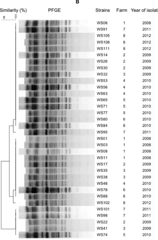
Figure 5.1 Cluster analysis of Weissella ceti strains using ERIC-PCR (A), and PFGE (B). The dendrograms were constructed using Dice’s coefficient and the UPGMA approach. W. ceti
isolates were obtained from eight different Brazilian farms between 2008 and 2012 ... 52
10
ABBREVIATION LIST
bp
base pair
BSFGE
Biased sinusoidal gel electrophoresis
CFU
Colony forming units
d.p.v.
days post-vaccination
ERIC
Enterobacterial repetitive intergenic consensus
g
gram
GCSD
Group C
Streptococcus dysgalactiae
HDL
High-density lipoprotein
IP
Intraperitoneal
L
Litre
MLST
Multi-locus sequence typing
MRS
Man, Rogosa and Sharpe
PBS
Phosphate buffered saline
PCR
Polymerase chain reaction
PFGE
Pulsed field gel electrophoresis
REP
Repetitive extragenic palindromic
RPS
Relative percent survival
SOF
Serum opacity factor
THA
Todd Hewitt agar
11
ABSTRACT
Streptococcus dysgalactiae and Weissella ceti have been considered emergent pathogens for Brazilian Nile tilapia (Oreochromis niloticus) and rainbow trout (Onchorhynchus mykiss) farming, respectively. Both diseases cause great economic losses in fish farms. Disease prevention by vaccination is a fundamental method for pathogen control in aquaculture. The aims of this study were to evaluate the genetic diversity of S. dysgalactiae and W. ceti strains isolated from distinct geographic origins and to develop efficient vaccines against these pathogens. Different methods (REP-PCR, ERIC-PCR and PFGE) were used for the genetically characterization of strains of both bacteria, and sodA gene sequencing was also tested for genotyping of S. dysgalactiae isolates. Aqueous-based whole cell killed bacterin and oil-adjuvanted vaccine were tested. The PFGE was the best genotyping method for S. dysgalactiae
and detected three different genetic profiles among the evaluated strains. The genetic variability of patterns was clearly associated with the geographic origin of isolates. According to the methods used, Brazilian W. ceti isolates was found to be highly homogeneous with a clonal population. Vaccines formulated with whole cell bacterin emulsified in oil adjuvant provided significant protection against S. dysgalactiae (RPS = 92,5) and W. ceti (RPS = 92,0) infections in Nile tilapia and rainbow trout, respectively. The present results provide scientific information for development of adequate methods to control two emerging infectious diseases that threaten Brazilian fish farming industry.
Keywords: Genotyping; vaccine; aquaculture.
RESUMO
As bactérias Streptococcus dysgalactiae e Weissella ceti são consideradas patógenos emergentes para a piscicultura nacional, causando perdas significativas nas cadeias produtivas de tilápia do Nilo (Oreochromis niloticus) e truta arco-íris (Onchorhynchus mykiss), respectivamente. Os objetivos do presente estudo foram avaliar a diversidade genética de amostras de S. dysgalactiae
e W. ceti provenientes de regiões geográficas diversas e desenvolver vacinas eficazes na proteção contra esses patógenos. Diferentes métodos (REP-PCR, ERIC-PCR e PFGE) foram utilizados para caracterização genética das bactérias. A análise do sequenciamento do gene sodA
foi utilizada somente para a genotipagem de amostras de S. dysgalactiae. Duas diferentes vacinas foram testadas: bacterina e bacterina emulsificada com adjuvante Montanide. A técnica de PFGE foi a mais eficaz para a genotipagem dos isolados de S. dysgalactiae, detectando três perfis genéticos diferentes. Os padrões genéticos estão associados com a origem geográfica dos isolados. De acordo com os métodos testados, as amostras brasileiras de W. ceti apresentam alta similaridade genética, sendo consideradas clonalmente relacionadas. As vacinas formuladas a partir de células inteiras inativadas (bacterina) emulsificadas em adjuvante oleoso conferiram proteção frente as infecções por S. dysgalactiae (RPS = 92,5) e W. ceti (RPS = 92,0) em tilápia do Nilo e truta arco-íris, respectivamente. Os resultados do presente projeto fornecem informações científicas e ferramentas fundamentais para o controle de duas doenças infecciosas emergentes da piscicultura nacional.
Palavras-chaves: Genotipagem; vacina; aquicultura.
1 – CHAPTER 1
INTRODUCTION
Global aquaculture production has continued to growth over the last years, while the
capture production has levelled off.
Therefore, the contribution of aquaculture to world food fish production for human consumption is growing each year. In Brazil, the scenario is not different: driven by population growth, rising demand for fish food and a stabilization of production from capture fisheries, the practice of farming aquatic animals becomes an interesting economic sector of the agribusiness.
The rapid growth of aquaculture in Brazil, mainly fish farming, has been associated with culture in high density of fish stocks and animals exposed to environmental stress. The consequence has been the emergence and spread of an increasing number of infectious diseases. In this sense, the Laboratory of Aquatic Animals Diseases (AQUAVET/EV-UFMG) is performing, since 2002, the diagnostic and research of the main infectious pathogens of the Brazilian aquaculture.
In the last years, two emerging diseases have been diagnosed causing economic impact to
fish farming in many countries.
Streptococcus dysgalactiae and Weissella ceti are causing substantial losses among farms of Nile tilapia (Oreochromis niloticus) and rainbow trout (Onchorhynchus mykiss), respectively.
Streptococcal infections have become an increasing problem in cultured fish around the world. In Brazil disease caused by bacteria from the genus Streptococcus, mainly S. agalactiae, is one of the major threats to Nile tilapia farming, the major fish
species produced in the country.
Streptococcus dysgalactiae was first described as a pathogen of fish during outbreaks in marine fish cultured (amberjack and yellowtail) in Japan. Recently a disease characterized by moderate mortality and presence of abscesses, in musculature of Nile tilapia in Brazil, is attributed to S. dysgalactiae infection.
Weissella ceti infections were first described in fish in 2009. Chinese researchers accompanied outbreaks in a rainbow trout farm and the disease was attributed to W. ceti. In the same period, our group isolated strains of this pathogen from diseased rainbow trout from farms located in different Brazilian states (São Paulo, Minas Gerais and Rio de Janeiro). Outbreaks were characterized by high mortality rates and the main clinical signs in diseased fish were lethargy, anorexia, melanosis, exophthalmia, ascites, and haemorrhages in the mouth, tongue, eyes, and fins. The disease affected fingerling, juvenile, and adult fish, and occurred during the summer, when water temperature usually is above 17 ºC.
The main method used to control infectious diseases in fish farming is the oral antibiotic therapy. However, this approach is not always effective to control infectious fish diseases because anorexia is present as the first clinical sign of illness. Therefore, disease prevention by vaccination is on
economic, environmental and ethical
grounds the most adequate method for pathogen control currently available to the aquaculture industry worldwide.
The development of efficient vaccines to prevent Streptococcus dysgalactiae and Weissella ceti infections in Nile tilapia and rainbow trout, respectively, is one of the main points to control these diseases. To design new vaccines, the identification of molecular features of strains is essential to understand whether a genetic variation implies in an antigenic diversity. Hence, studies on molecular characterization of field isolates of S. dysgalactiae and W. ceti occurring in Brazil is important to implement efficient control management practices. Furthermore, the genotyping of strains is also essential to understand pathogens distribution and determine if epidemiologically related isolates are genetically related strains pose a clonal structure or not.
isolates from different national regions are essential to evaluate the effectiveness of this immunoprophylactic method. Furthermore, there are no studies about the relationship among genetic variability, origin of isolates and vaccine protection against different strains for both bacteria.
Objectives
The aims of this study were to evaluate the genetic diversity of S. dysgalactiae and W.
ceti strains isolated from different
geographic origins in Brazil; and to develop vaccines to protect against these pathogens infections in Nile tilapia and rainbow trout farms, respectively.
2 – CHAPTER 2
LITERATURE REVIEW
2.1
Brazilian aquaculture
Fish and fishery products are among the most traded food commodities worldwide, with developing countries leading the bulk of world exports. While capture fisheries production remains stable, aquaculture production continues to expand. Aquaculture is one of the fastest-growing animal food-producing sectors and, in the next decade, total production from both capture and aquaculture will exceed that of beef, pork or poultry. In 2011, the total world of fisheries production was 154 million tonnes of which aquaculture contributed with 63,9 million tonnes or about 41,3% of the total world production (FAO, 2012).
Brazil is one of the countries with the highest growth in aquaculture worldwide, and with a further potential to growth due to a coastline of 8,500 km, and 12% of the world’s freshwater reserves. Freshwater reservoirs are mainly representing by 10 million hectares of water, which are associated with hydroelectric production (BRASIL, 2012; FAO, 2012).
The average annual growth rate for Brazilian aquaculture production from 2009 to 2011 was 51,2% per cent. The total aquaculture production, in 2011, was 628.704 tonnes of which freshwater fish farming accounts for
86,6% (544.490 tonnes) of total. From 2010 to 2011 the freshwater fish production increased significantly, with a growth rate of 38%. However, the Brazilian domestic consumption of fish increased in the last years, and the national fisheries production is still not enough to supply this market. In 2011, Brazil exported 42 million tonnes (US$ 271 million) and imported 349 million tonnes of fish (US$ 1.262 million), with a
trade deficit of US$ 991 million,
approximately (BRASIL, 2012).
The main fish species cultured in Brazil is the Nile tilapia (Oreochromis niloticus). About a half of freshwater fish production comes from Nile tilapia farms. It is an omnivorous tropical fish species with optimal temperature ranging from 28 to 35ºC. Nile tilapia is considered suitable for culture because the high tolerance to adverse conditions, relative fast growth and the well stablished protocols for breeding (Guerrero, 1985). In 2011, the total production of Nile tilapia in Brazil was 253.824 tonnes (BRASIL, 2012).
Another exotic fish species cultured in Brazil is the rainbow trout (Onchorhynchus mykiss). It is a species of salmonid, native to the Pacific drainages of North America, with optimal temperature ranging from 9 to 15ºC. The rainbow trout is a hardy fish that is easy to spawn and fast growing. The species has a high demand of dissolved oxygen in the water, and the lowest concentration survival is 3 ppm (Hardy et al., 2000). The rainbow trout production in Brazil is restricted to some regions (cold and hilly areas of South and Southeast), due its requirement for a cold water. The total production of rainbow trout in Brazil was 3.277 tonnes in 2011
(BRASIL, 2012). Despite this small
production, rainbow trout has a high market value compared to other cultured fish species.
2.2 Emerging pathogens for fish
farming
fish diseases has been driven by anthropogenic influences associated with growing of global aquaculture. Farming of aquatic animals commonly involves a stressful environment, the use of artificial feeds, and culture in high stocking densities. This has provided opportunities for exposure to pathogens and conditions that can compromise the fish immune system, and also facilitate pathogen replication and transmission. In addition, the growth in aquaculture and increasing international trade has resulted in the rapid movement of aquatic animals, with associated risks of the transmission of pathogens (Noga, 1996; Walker and Winton, 2010).
Apparent changes in the prevalence and distribution of fish diseases can also be explained by the improved surveillance as a result of the development of more sensitive molecular diagnostic methods and greatly growth in aquatic animal health researches (Eiras, 2008).
By concept an emerging disease can be defined as: a new or previously unknown disease; a known disease appearing for first time in a new species (expanding host range); a known disease in a new location (expanding geographic range); and/or new presentation (different signs) of a known disease due changes in the pathogen, mainly higher virulence (Walker, 2004).
The impacts of emerging pathogens and the extent of diseases spread are often exacerbated by some problems that are typically encountered including: delay in developing tools for the confirmatory diagnosis of disease; poor knowledge of the current or potential host range; inadequate knowledge of the present geographic range; no understanding of critical epidemiological factors; and poor understanding of genetic differences among strains and relationships to the epidemiological factors of disease (Walker and Winton, 2010). Therefore, knowledge of emerging fish diseases is of great relevance, both from a scientific as well as an applied point of view. Detailed knowledge of the emerging pathogens, their interactions with fish as well as the main responses of the host is a pre-requisite for
prevention and treatment of fish diseases (Eiras, 2008).
2.2.1
Streptococcus dysgalactiae
The taxonomic of the genus Streptococci was first described by Rosenbach, 1884 (Hardie, 1986), and recently reviewed by Facklam (2002). Lancefield (1933) reported
specific technique that is used to
demonstrate specific carbohydrate “group” antigens.
Streptococcosis was first described as a fish disease during an outbreak in a rainbow trout (Oncorhynchus mykiss) farm in Japan, in 1956 (Hoshina et al., 1958). Since that, Streptococossis is used as a generic term for some septicaemia infections in fish, caused by Gram-positive cocci. Four genera are associated with diseases in aquatic animals: Streptococcus, Lactococcus, Vagococcus
and Carnobacterium (Mata et al., 2004). In the last years Streptococcosis has increased in importance, with outbreaks occurring in numerous fish species and other aquatic animals (Agnew and Barnes, 2007; Romalde
et al., 2008; Netto et al., 2011).
Streptococcosis outbreaks have been
considered one of the major barriers to the development of world fish farming (Ghittino
et al., 2003; Austin and Austin, 2007). Unofficial data estimate that annual losses arising from Streptococcosis outbreaks exceed the value of US$ 150 million (Romalde et al., 2008). In Brazil, three species of Streptococcus were described as aetiological agents causing septicaemia in Nile tilapia (Oreochromis niloticus):
Streptococcus agalactiae, Streptococcus dysgalactiae and Streptococcus iniae
(Figueiredo et al., 2012a; Godoy et al.,
2013; Mian et al., 2009; Netto et al., 2011; Salvador et al., 2003, 2005). Streptococcus agalactiae infections are probably the most prevalent, and have been reported in farms located in several states of the country (Mian
et al., 2009).
Streptococcus dysgalactiae is divided into two subspecies: S. dysgalactiae subsp.
2011). This division was based on electrophoresis of cell wall proteins and physiological tests proposed by Vandamme
et al. (1996). Therefore, the name S. dysgalactiae subsp. dysgalactiae was proposed for strains of animal origin that belong to Lancefield Group C Streptococcus
(GCS) and Group L (GLS).
Lancefield group C Streptococcus
dysgalactiae (GCSD) is mainly associated with endometritis, subclinical or clinical mastitis, subcutaneous cellulitis, and toxic shock-like syndrome in bovine (Seno and Azuma, 1983; Aarestrup and Jensen, 1996). The same pathogen is reported as a cause of
bacteremia, meningoencephalitis and
mastitis in sheep (Scott, 2000; Chenier et al.,
2008).
The first report of GCSD in fish occurred in 2002 in Japan. The bacterium was isolated from two cultured marine fish species, amberjack (Seriola dumerili) and yellowtail (Seriola quinqueradiata), which were displaying necrotic lesions of the caudal peduncle (Nomoto et al., 2004). Since that,
S. dysgalactiae has been associated with severe necrosis lesions in musculature and mortalities of marine fish species as kingfish (Seriola lalandi), grey mullet (Mugil cephalus), basket mullet (Liza alata), soiny mullet (Liza haematocheila), amur sturgeon (Acipenser schrenckii), and cobia (Rachycentron canadum) (Nomoto et al.,
2006; Abdelsalam et al., 2009; Yang and Li, 2009; Qi et al., 2013).
GCSD has been identified as fish specific pathogen based on 16S rDNA sequence analysis, sodA and tuf genes sequence
analysis, biased sinusoidal field gel
electrophoresis and Lancefield grouping. Some biochemical characteristics of the fish isolates were different from those of the typical reference of mammalian strains S. dysgalactiae subsp. dysgalactiae and S. dysgalactiae subsp. equisimilis (Nomoto et al., 2008; Nishiki et al., 2010).
In 2007, Netto et al. (2011) investigated a disease outbreak in a Nile tilapia farm located in Ceará State, Brazil. Diseased fish showed clinical signs of septicaemia and subcutaneous abscesses in the caudal region.
From diseased Nile tilapia it was
successfully isolated Streptococcus
dysgalactiae strains and it was described the first infection of GCSD in freshwater fish. In the same study, the disease was reproduced in experimental infections using the same host. However, they were unable to reproduce the typical clinical sign of subcutaneous abscesses of S. dysgalactiae. In addition, Netto et al. (2011) used PFGE to genotype GCSD strains and the analysis showed that isolates belonged to a single pulsotype. This result could be explained by the restricted geographical (a single outbreak in one farm) and contemporary origin of the isolates analyzed.
The search for a competent vaccine against
S. dysgalactiae is hampered by the short knowledge about its pathogenesis and
virulence determinants. The virulence
factors of GCSD are mainly based on its cell
surface properties such as high
hemagglutination and hydrophobic
properties, which determine the main
adhesion and invasive pathogenic
mechanism of the pathogen. This has been confirmed by Abdelsalam et al. (2009), who have indicated that GCSD isolates were able to adhere to and invade fish epithelial cell line (EPC: Epithelial papiloma of Carp and
CHSE-214: Chinook salmon embryo)
cultured in vitro. Besides, the pyrogenic, exotoxin G, a superantigen, and streptolysin S genes are regarded as the most important virulence traits, with cultures recovered from diseased fish harboring the streptolysin S structural gene, sagA (Abdesalam et al., 2010).
Suzuki et al. (2011) demonstrated that some
Streptococcus dysgalactiae subsp.
dysgalactiae strains carry a number of prophage proteins that are considered putative virulence genes in Streptococcus pyogenes, including hyaluronidase and streptodornase type D, both of which are
documented as Streptococcus virulence
genes (Davies et al., 2007).
Serum opacity factor (SOF) was detected in
opacification domain, a putative fibronectin-binding domain, and an LPXTG Gram-positive anchor motif. The structure of SOF-FD is similar to that of the SOF in
Streptococcus pyogenes (Nishiki et al.,
2011; Nishiki et al., 2012). The substrate of SOF is a high-density lipoprotein (HDL), which has anti inflammation activity. The SOF binds to HDL and then disrupts its structure, which may contribute to the virulence of streptococci. S. dysgalactiae
strains isolated from fish have serum opacification activity and sof-FD genes with highly homologous sequences (Nishiki et al., 2011).
2.2.2
Weissella ceti
The genera Weissella, Leuconostoc, and
Oenococcus together constitute a genetic branch of lactic acid bacteria (Chelo et al., 2007). The genus Weissella contains species that are Gram-positive, catalase-negative, heterofermentative, non-motile, coccoid or rod-shaped organisms (Collins et al., 1993; De Bruyne et al., 2008). Microorganisms of the genus Weissella have been isolated from a variety of habitats such as soil, fresh vegetables and fermented foods or meat products (Schillinger and Lucke, 1987; Bjorkroth et al., 2002; Magnusson et al.,
2002; De Bruyne et al., 2010; Padonou et al., 2010).
Some species of Weissella have been
identified in intestinal contents of healthy humans, dogs, chickens and ducks (Kurzak
et al., 1998; Walter et al., 2001; Beasley et al., 2006; Chelo et al., 2007; Wise and Siragusa, 2007; Sirirat et al., 2008).
Weissella confusa has been isolated from intestines of healthy fish farmed Asian seabass (Lates calcarifer) (Cai et al., 1998; Rengpipat et al., 2008). In addition,
Weissella confusa have also been associated with cases of bacteremia in humans and primates as well as otitis in dogs (Olano et al., 2001; Bjorkroth et al., 2002; Flaherty et al., 2003; Vela et al., 2003; Svec et al., 2007).
Liu et al. (2009) provided the first description of Weissella sp. infection in a cultivated fish species. They documented sequential outbreaks of the pathogen in one
commercial rainbow trout (Oncorhynchus mykiss) facility in China. The diseased fish exhibited hemorrhage in eyes, anal region and intestine, and petechiae in liver. During the epidemic outbreak, a loss of 40% was suffered. Weissella sp. strains were isolated from kidney, liver and/or brain of four adult rainbow trout with typical clinical signs (Liu
et al., 2009).
Figueiredo et al. (2012b) described the occurrence of outbreaks of hemorrhagic septicaemia caused by Weissella sp. in three commercial rainbow trout farms in Brazil, from 2008 to 2009. During outbreaks was observed that the first clinical sign of disease is anorexia, followed by: exophthalmia, ascites, and hemorrhage in the mouth, tongue, and eyes. High mortality rates (50-80%) occurred during outbreaks and disease affected different age groups as fingerlings, juveniles, adults, and broodstock.
In addition, Figueiredo et al. (2012b) investigated the potential routes of infection of Weissella sp. in rainbow trout. The disease was successfully reproduced in the laboratory by intraperitoneal injection,
immersion, and cohabitation between
diseased and healthy fish. These data indicated that bacteria can actively infect healthy fish and are able to proliferate, survive, and be transmitted under normal conditions. Figueiredo et al. (2012b) also evaluated the resistance of the pathogen to five antibiotics. The results showed that all 77 Weissella sp. strains tested were resistant to sulfonamide, and based in normalized resistance interepretation (NRI) analysis, one, two, and three isolates were classified as non-wild type (NWT) for erythromycin,
oxytetracycline, and norfloxacin,
respectively.
The origin of the bacteria Weissella sp.
associated with these described fish
outbreaks is unknown. During an
investigation into the microbiota of beaked whales
(
Mesoplodon bidens)
, Vela et al.genius Weissella, named Weissella ceti sp.
nov. (Vela et al., 2011). 16S rRNA
sequences analysis of W. ceti isolated from whales compared with the sequences of the Brazilian and Chinese isolates showed a high genetic similarity (>99%), suggesting that all strains belonged to the same species.
The third report of a Weissella ceti causing infection and loss in rainbow trout farms occurred in the United States (Welch and Good, 2013). The disease signs and bacteria isolated in this outbreak were very similar to those observed in the Brazilian and Chinese cases. Affected fish exhibited as clinical signs: dark skin coloration, bilateral exophthalmia, corneal opacity and ocular hemorrhage.
Elevated temperature is often associated with severe fish disease outbreaks involving mesophillic bacterial pathogens, and this is likely due to higher temperature of water induced increases in microorganism growth rate and/or causes physiological responses to thermal stress in fish. All outbreaks described for Weissella ceti in rainbow trout
coinciding with seasonal high water
temperatures, usually above 17 ºC. This suggests that one of the main predisposing
factors associated with Weissella ceti
infections is the elevation of water temperature.
Ladner et al. (2013) presented the first genome sequence of Weissella ceti, using a strain from United States. Results of comparative analysis of this genome demonstrated several putative virulence factors, which do not have homologous encoded in any of the other sequenced
Weissella genomes. These included five collagen adhesins, a platelet-associated adhesion, and a mucus-binding protein (Ladner et al., 2013).
Immunoprophylaxis generally has a positive effect in fish farming by reducing the economic losses caused by disease. In some countries, commercial vaccines are available to protect cultured rainbow trout against
some pathogens, including Lactococcus
garviae and Yersinia ruckeri (Vendrell et al.,
2006). These vaccines are usually
intraperitoneally injected and the requisite
anesthetization and handling associated with this process causes significant stress. Thus, it is advantageous, when possible, mix vaccines into multivalent formulations that can be delivered together, minimizing the impact of fish stress by handling, which would be caused by multiple injections. Welch and Good (2013) developed an aqueous-based whole-cell killed bacterin vaccine able to protect rainbow trout from
Weissella ceti infection in laboratory trials. The authors also proposed a bivalent formulation that conferred a level of protection against W. ceti or Y. ruckeri
challenge that was equivalent to that
conferred by vaccination with each
component.
The occurrence of several outbreaks of
Weissella ceti on three continents over a relatively short period of time, suggests that this pathogen is a significant and rapidly emerging concern for the farmed rainbow trout industry.
2.3
Molecular epidemiology
The science of “Molecular epidemiology” has emerged in 1970s when the term was first coined in relation to the study of
influenza virus (Kilbourne, 1973).
Molecular epidemiology utilizes molecular biology to define the distribution of disease in a population and based on integration of traditional epidemiological approaches with molecular results to identify the etiological determinants of this distribution (Snow, 2011). Understanding pathogen distribution and relatedness is essential for determining the epidemiology of emerging infectious diseases of aquaculture, supporting in design of efficient pathogen control methods (Singh
et al., 2006).
Over the years, approaches to the
epidemiological analysis of infectious
disease have undergone a remarkable
evolutionary transition moving from
to transfer some genetic elements in a process of acquisition and recombination of
foreign DNA. These mobile genetic
elements are segments of DNA that encode enzymes and other proteins that mediate the movement of DNA within genomes or among bacterial cells (Wellington et al.,
2013). This process is essential to understand some genetic variability during molecular epidemiologic studies.
The search for a clearer comparison of genomic relatedness between bacterial isolates from diseases has involved four generations of molecular approaches. First generation, plasmid analysis, was replaced by a second-generation with the use of restriction enzymes and probes. This was followed by third-generation pulsed field gel electrophoresis (PFGE) and PCR-based methods with movement now to fourth-generation DNA sequence-based approaches (Goering, 1998, 2000). In this context, it is interesting to note that while far from the most current molecular technique, PFGE still has a relative valuable method of genomic analysis and comparison. This is especially true in the clinical setting where for some organisms it can be used for assessing isolate interrelationships (Goering, 2010).
The chromosome is the most fundamental component of identity of the cell and therefore represents a preferred measure for
assessing strain interrelatedness.
Chromosomal DNA can be digest with restriction enzymes, resulting in a series of fragments of different sizes that form different patterns when analyzed by agarose gel electrophoresis (Singh et al., 2006). Enzymes used to cleave DNA often
recognize numerous sites within the
bacterial chromosome, resulting in too many band fragments to efficiently and accurately compare. However, the resulting DNA fragments are too large to be separated by conventional agarose gel electrophoresis. A number of alternative methods, generally classified as PFGE, are capable of separating these large DNA fragments (Chang and Chui, 1998). In conventional agarose gel electrophoresis, DNA molecules that are more than 40 to 50 kbp in size fail to migrate efficiently. By periodically changing
the direction of the electrical field in which the DNA is separated, PFGE allows the separation of DNA molecules of over 1,000 kbp in length (Carle et al., 1986).
To interpret DNA fragment patterns generated by PFGE and transform them into epidemiologically useful information for typing pathogens, is essential understand how to compare PFGE patterns and how random genetic events can alter these
patterns. Ideally, the PFGE isolates
representing an outbreak will be
indistinguishable from each other and
distinctly different from those of
epidemiologically unrelated strains.
Alternatively, random genetic events can occur, such as point mutations or insertions and deletions of DNA that can alter the restriction profile obtained during the course of an outbreak (Hall, 1994).
Because only a small portion of the organism’s genetic component is undergoing analysis, isolates that give identical results are classified as “indistinguishable”, not “identical”. Guidelines proposed by Tenover
et al. (1995) are often used for the
interpretation of PFGE. With these
guidelines, a banding pattern difference of three fragments could have occurred due to a single genetic event and thus these isolates are classified as highly related. Differences of four to six restriction fragments are likely due to two genetic events, and differences of greater than seven restriction fragments are due to three or more genetic events. Isolates that differ by three fragments in PFGE analysis may represent epidemiologically related sub-types of the same strain.
Conversely, isolates differing in the
positions of more than three restriction
fragments may represent lower
epidemiologic relation.
Commercially available programs to
analysis PFGE and other molecular results have been developed for computer-assisted analysis (CAA) such as BioNumerics and GelCompar (Applied Maths,
Sint-Martens-Latem) and Diversity Database
Fingerprinting Software (Bio-Rad
Laboratories, Hercules, Ca) (Goering, 2010).
and highly discriminatory typing methods available. However, the process of PFGE is time consuming, mainly because of the need to extract DNA without subjecting it to forces that would cause disruption by shearing. This delay can be a serious problem when epidemiological data are needed urgently (Singletone, 2000). A further disadvantage of PFGE, compared with other molecular methods, is the susceptibility to endogenous nucleases, which can degrade target DNA (Marshall et al., 1999).
In this point, rapid and inexpensive
PCR-based typing techniques, such as
enterobacterial repetitive intergenic
consensus - PCR (ERIC-PCR) and repetitive element sequence - PCR (REP-PCR) can be used to screen for genetic relatedness among strains. In many bacterial species there are repetitive DNA sequences spread throughout the genome. PCR primers can be designed for these elements to amplify the genomic DNA between repetitive elements when two of these elements are in relatively close proximity (Versalovic et al., 1991).
The regions located between the repeated elements often vary in size due to difference among separate strains, and thus fragments of different sizes will be amplified, creating
unique profiles following gel
electrophoresis. These different banding patterns are compared one to another to genotype the organisms (Georghiou et al.,
1994). Repetitive-element PCR systems have been developed for a variety of pathogens (Hulton et al. 1991; Shutt et al., 2005).
In recent years, the rapidly expanding number of sequenced microbial genomes has served as a catalyst for the development of a variety of molecular typing approaches that
focus on either single or multiple
chromosomal loci. The better example is the multilocus sequence typing (MLST) that are considered as the reference tools for investigating genetic and epidemiological
relationships for some pathogens
(Haguenoer et al. 2011; Nakib et al. 2011). Sequence-based molecular epidemiology is attractive in offering the promise of reproducible typing profiles that are highly
amenable to standardization, uniform
interpretation, and database cataloging, since they are based on simple quaternary data (A, T, G, and C) (Kemp et al., 2005).
The development of next generation
sequencing technology promises a
revolution in the ability to generate sequence data and thus information of potential epidemiological relevance. The process of
disease involves many genetic
polymorphisms, across the genome, which is now being aggressively explored. Therefore, the analyses performed to choose specific
genome regions that can provide
assessments consistent with those predicted with full-length genome sequences, would be preferable to serve as phylogenetic markers for molecular epidemiological studies of pathogens (Wellington et al., 2013).
However, descriptive molecular data alone is inadequate in tracing pathogen spread, especially when variation is limited and evolution does not occur in a regular way.
The development of methods to
systematically record and share such epidemiologically important information thus represents a major challenge for fish health professionals in making the best future use of molecular data in supporting fish health surveillance and disease control. The implementation of the best available
molecular epidemiological information
would offers great support to surveillance programs in further developing a healthy and sustainable aquaculture industry, which is necessary to satisfy an increasing world demand for cultured fish products (Snow, 2011).
2.4
Fish vaccines
Antibiotic therapy can be used for the
treatment of bacterial diseases in
The goal of successful vaccination is the capacity to induce the immune system of fish to develop immunological memory. A first contact with an antigen usually induces relatively short-lived effector cell (activated Th, Treg, cytotoxic T cells or plasma cells). However, there are also long-lived memory cells among the progeny of the original non-primed lymphocytes. These memory cells retain the capacity to be stimulated by the same antigen. The height of the secondary response is dependent on the amount of priming antigen (Willem and Nakao, 2013).
Several differences between the secondary responses of mammals and fish have been found. One distinction is that the ratio between the secondary and the primary response is much higher in mammals than in fish. In fish B-cells memory is probably due to an increase in the antigen-sensitive
precursor pool without any of the
accompanying characteristics observed in mammals (such as a switch in isotype) (Hikima et al., 2011; Willem and Nakao, 2013).
Fish can be immunized by three routes:
injection preferably intraperitoneally;
immersion, dipping the fish in a diluted vaccine solution; or oral administration of the vaccine. Although these methods have different advantages and disadvantages with respect to the level of protection, side effects, practicality and cost-efficiency, it is widely accepted that injection route give the higher protection to be used as the primary route of fish immunization in commercial production (Taffala et al., 2013). There is still a limited understanding of the mechanisms involved in antigen uptake and presentation after immersion and oral vaccination (Nakanishi and Ototake, 1997).
Oral vaccination usually evokes only minimal immune responses in the host. It is not easy to explain this phenomenon. It was showed that the 2nd gut segment plays a key role in antigen transport and antigen processing by macrophages (Rombout and Van Den Berg, 1989). Numerous lymphoid cells are also present in this part of the gut, playing a role in mucosal responses (Rombout et al., 1989a). Repeated oral administration of bacterial antigen resulted
in presence of antibodies in skin mucus and bile, but not in serum (Rombout et al., 1989b).
For oral vaccination, research has been focused on protecting the antigens from digestion and decomposition during passage through the stomach and anterior part of the
gut (Quentel and Vigneulle, 1997).
Promising results have been obtained using encapsulation of antigens in alginate or polylactic glycolic acid microparticles (Sinyakov et al., 2006). From the economic standpoint, oral vaccination is the ideal route to be employed in a vaccination program, which requires one or more booster immunizations.
Four types of vaccines are tested for a vaccine formulation for fish: bacterin, live
attenuated, purified subunits of the
pathogen, and DNA vaccines. Most of bacterial vaccines used in aquaculture have been inactivated vaccines obtained from a broth culture of a specific strain(s) subjected
to subsequent formalin inactivation
(Newman, 1993; Toranzo et al., 1997). The best results are obtained with those bacterins that include both bacterial cells and extracellular products. Whereas with some vaccines acceptable levels of protection are
achieved with aqueous formulations
administered by injection or immersion. For other bacterins, such as those devised for salmonids against Aeromonas salmonicida
subsp. salmonicida, an acceptable level of protection can only be achieved by immunization with oil-adjuvanted bacterins delivered by injection.
The live attenuated vaccines should
DNA vaccines consist of a bacterial plasmid with a strong promoter, the gene of interest,
and a polyadenylation/transcriptional
termination sequence. This is one of the most promising fish vaccine preparations
against viral diseases (delivered
intramuscularly). They consisted of naked plasmid DNA that will result in gene expression of viral proteins in the muscle tissue of the vaccinated fish. Overall, the inactivated, subunit or whole virus vaccines in general confer lower level of protection compared to DNA and live, attenuated vaccines (Evensen and Leong, 2013).
A subunit vaccine is produced by MSD Animal Health against infectious pancreatic necrosis (IPN) and is intended for use in salmonids. The vaccine consists of the major IPNV capsid protein (VP2) produced in bacteria (Escherichia coli), and is injected intraperitoneally in Atlantic salmon prior to transfer to seawater. There are also subunit, oral vaccines against IPN, infectious salmon
anemia (ISA) and salmon rickettsia
syndrome (SRS) for use in salmonids in Chile (produced by Centrovet, Chile) (Evensen and Leong, 2013).
Many vaccines based upon recombinant antigens or killed pathogens are not very effective in fish as such. In most cases the use of adjuvants is necessary to stimulate the response of innate and acquired immunity and increase vaccine efficacy. Adjuvants (from Latin adjuvare meaning “to help”) have traditionally been defined as helper substances that increase the magnitude of an adaptive response to a vaccine (potency), or ability to prevent infection and death (efficacy). However, nowadays scientists have acknowledged that adjuvants may become more important as a group of structurally heterogeneous compounds able to modulate the intrinsic immunogenicity of an antigen (Guy, 2007).
Traditional adjuvants such as mineral oils are routinely used in different commercial bacterial vaccines available for fish; however, important side effects may occur with this type of adjuvants. A search for
alternative molecules or certain
combinations of them as adjuvants is desirable in order to increase animal welfare
without reducing protection levels.
Especially, combinations that may target specific cell responses and thus a specific pathogen, with no or minor side effects, should be explored. Despite this, the oil adjuvants currently used are quite friendlier with respect to side effects compared with the oil adjuvants previously used (Taffala et al., 2013).
The development of effective vaccines should be approached by combining the search for protective antigens together with the application of specific, and targeting, adjuvants that increase the immunogenicity with a desired immune response. (Taffala et al., 2013). Many aspects of fish immunology are still unknown and we are far from close
to understanding on which immune
mechanisms the protection against many of these pathogens resides (Secombes, 2008). Better understanding of the immune system of different fish species is essential to achieve progress in fish vaccinology.
2.5 – REFERENCES
AARESTRUP, F.M.; JENSEN, N.E. Genotypic and phenotypic diversity of
Streptococcus dysgalactiae strains isolated from clinical and subclinical cases of bovine mastitis. Vet. Microbiol., v.53, p.315–23, 1996.
ABDELSALAM, M.; CHEN, S.C.; YOSHIDA, T. Surface properties of
Streptococcus dysgalactiae strains isolated from marine fish. Bull. Eur. Assoc. Fish Pathol., v.29, p.15–23, 2009.
ABDELSALAM, M.; CHEN, S., YOSHIDA, T. Phenotypic and genetic characterizations of Streptococcus
dysgalactiae strains isolated from fish collected in Japan and other Asian countries.
FEMS Microb. Lett., v. 302, n. 1, p. 32-38, 2010.
AGNEW, W.; BARNES, A. C.
Streptococcus iniae: an aquatic pathogen of global veterinary significance and
challenging candidate for reliable
AUSTIN, B.; AUSTIN, D. A. Bacterial fish pathogens: diseases of farmed and wild fish. 4th ed. Chichester: Springer, 2007. 552 p.
BEASLEY, S.S.; MANNINEN, T.J.; SARIS, P.E. Lactic acid bacteria isolated from canine faeces. J. Appl. Microbiol., v.101, p.131–138, 2006.
BENMANSOUR, A.; KINKELIN, C. (Eds.), Fish Vaccinology, Dev. Biol. Stand., Karger, Basel, 1997. p. 279.
BJORKROTH, K. J.; SCHILLINGER, U.; GEISEN, R. et al. Taxonomic study of
Weissella confusa and description of
Weissella cibaria sp. nov., detected in food and clinical samples. Int. J. Syst. Evol. Microbiol., v.52, p.141–148, 2002.
BRASIL. Ministério da Pesca e Aquicultura. Boletim estatístico da pesca e aquicultura 2011. Brasília, 2012. 60 p.
CAI, Y; BENNO, Y.; NAKASE, T. et al.
Specific probiotic characterization of
Weissella hellenica DS-12 isolated from flounder intestine. J. Gen. Appl. Microbiol., v.44, p.311–316, 1998.
CARLE, G. F.; FRANK, M.; OLSON, M.V. Electrophoretic separations of large DNA molecules by periodic inversion of the electric field. Science, v. 232, n.4746, p. 65-68, 1986.
CHANG, N.; L. CHUI. A standardized protocol for the rapid prep- aration of bacterial DNA for pulsed-field gel electrophoresis. Diagn. Microbiol. Infect. Dis., v.31, p.275–279, 1998.
CHELO, I.M.; ZE-ZE, L.; TENREITO, R. Congruence of evolutionary relationships
inside the Leuconostoc–Oenococcus–
Weissella clade assessed by phylogenetic analysis of the 16S rRNA gene, dnaA, gyrB, rpoC and dnaK. Int. J. Syst. Evol. Microbiol.
v.57, p.276–286, 2007.
CHENIER, S.; LECLERE, M.; MESSIER, S. et al.Streptococcus dysgalactiae cellulitis and toxic shock like syndrome in a brown Swiss cow. J. Vet. Diagn. Invest., v.20, p.99–103, 2008.
COLLINS, M.D.; SAMELIS, J.;
METAXOPOULOS, J. et al. Taxonomic
studies on some leuconostoc-like organisms from fermented sau- sages: description of a new genus Weissella for the Leuconostoc paramesenteroides group of species. J. Appl. Bacteriol., v.75, p.595–603, 1993.
DAVIES, M.R.; McMILLAN, D.J.; BEIKO, R.G. et al. Virulence profiling of
Streptococcus dysgalactiae subspecies
equisimilis isolated from infected humans reveals 2 distinct genetic lineages that do not segregate with their phenotypes or
propensity to cause diseases. Clin. Infect. Dis., v.44, p.1442–1454, 2007.
DE BRUYNE, K.; CAMU, N.; LEFEBVRE, K. et al. Weissella ghanensis sp. nov., isolated from a Ghanaian cocoa
fermentation. Int. J. Syst. Evol. Microbiol.
v.58, p.2721–2725, 2008.
DE BRUYNE, K.; CAMU, N.; DE VUYST, L. et al. Weissella fabaria sp. nov., from a Ghanaian cocoa fermentation. Int. J. Syst. Evol. Microbiol., v.60, p.1999–2005, 2010.
EIRAS, J.C.; SEGNER, H.; WAHLI, T. et
al. Fish diseases. Science Publishers, Enfield, Jersey, Plymouth, USA. 2008. Vol. 1 and 2; 1312 pp.
EVENSEN, O.; LEONG, J.C. DNA vaccines against viral diseases of farmed fish. Fish Shellfish Immunol., p.1-8, 2013.
FACKLAM, R. What happened to the streptococci: overview of taxonomic and. nomenclature changes. Clin. Microbiol. Rev., v.15, p.613–30, 2002.
FIGUEIREDO, H.C.P.; NETTO, L.N.; LEAL, C.A.G. et al. Streptococcus iniae
outbreaks in Nile tilapia (Oreochromis niloticus L.) farms. Brazilian Journal of Microbiology, São Paulo, 2011. Braz. J. Microbiol., v.43, n.2, 2012a.
FIGUEIREDO, H.C.; COSTA, F.A.A.; LEAL, C.A.G. et al.Weissella sp. out- breaks in commercial rainbow trout
FLAHERTY, J.D.; LEVETT, P.N.; DEWHIRST, F.E. et al. Fatal case of endocarditis due to Weissella confusa. J. Clin. Microbiol., v.41, p.2237–2239, 2003.
FOOD AND AGRICULTURE ORGANIZATION OF THE UNITED NATIONS (FAO). The state of world fisheries and aquaculture. Rome, 2012. 196p.
GEORGHIOU, P. R.; DOGGET, A.M.;
KIELHOFNER, M.A. et al. Molecular
fingerprinting of Legionella species by repetitive element PCR. J. Clin. Microbiol., v.32, p.2989-2994, 1994.
GHITTINO, C.; LANTINI, M.; AGNETTI, F. et al. Emerging pathologies in
aquaculture: effects on production and food safety. Vet. Res. Commun., v. 27, n. 1, p. 471-479, 2003.
GOERING, R.V. The molecular
epidemiology of nosocomial infection: an overview of principles, application, and interpretation. In: Specter, S., Bendi- nelli, M., Friedman, H. (Eds.), Rapid Detection of Infectious Agents. Plenum Press, New York, 1998, pp. 131-157.
GOERING, R.V. The molecular
epidemiology of nosocomial infection: past, present, and future. Rev. Med. Microbiol. v.11, p.145–152, 2000.
GOERING, R. V. Pulsed field gel
electrophoresis: A review of application and interpretation in the molecular epidemiology of infectious disease. Inf. Gen. Evol., v. 10, p. 866–875, 2010.
GODOY, D.T.; CARVALHO-CASTRO; G.A.; LEAL, C.A.G. et al. Genetic diversity and new genotyping scheme for fish
pathogenic Streptococcus agalactiae. Lett. App. Microb., v. 57, p. 476-483, 2013.
GUERRERO, R.D. III. Tilapia farming in the Phillippines: Practices, problems and prospects. In:I.R. Smith, E.B. Torres and E.O. Tan, eds. Philippine Tilapia
Economics. ICLARM Conf. Proc.12. 1985 pp. 261.
GUY, B. The perfect mix: recent progress in adjuvant research. Nat. Rev. Microbiol., v.5, p.505-517, 2007.
HALL, L. M. C. Are point mutations or DNA rearrangements responsible for the restriction fragment length polymorphisms that are used to type bacteria? Microb., v.140, p.197–204, 1994.
HARDIE, J. M. Genus streptococcus Rosenbach 1884, 22 AL, pp. 1043-1071. In: P.H.A. Sneath, N.S. Mair, M.E. Sharpe and J.G. Holt, eds. Bergey’s Mannual of Systematic Bacteriology. Williams and Wilkins, Baltimare, 1986, vol. 2.
HARDY, R.W.; FORNSHELL, G.C.G.; BRANNON, E.L. Rainbow trout culture. In: R. Stickney (ed.) Fish Culture, John Wiley & Sons, New York, USA. 2000. pp. 716-722.
HAGUENOER, E.; BATY, G.; POURCEL, C. et al. A multi locus variable number of tandem repeat analysis (MLVA) scheme for Streptococcus agalactiae genotyping. BMC Microbiol., v.11, n. 171, p.1–13, 2011.
HIKIMA, J.; JUNG, T.S.; AOKI, T. Immunoglobulin genes and their transcriptional control in teleosts. Dev. Comp. Immunol., v.35, p.924-936, 2011.
HOSHINA, T.; SANO, T.; MORIMOTO, T.
Streptococcus pathogenic to fish. Journal of Tokyo University of Fisheries, v. 44, p. 57-68, 1958.
HULTON, C. S.; HIGGINS, C.F.; SHARP, P.M. ERIC sequences: a novel family of repetitive elements in the genomes of
Escherichia coli, Salmonella typhimurium
and other enterobacteria. Mol. Microbiol., v.5, p.825-834, 1991.
KEMP, R.; LEATHERBARROW, A.J.; WILLIAMS, N.J. et al. Prevalence and genetic diversity of Campylobacter spp. in environmental water samples from a 100-square-kilometer predominantly dairy farming area. Appl. Environ. Microbiol., v.71, p.1876–1882, 2005.
epidemiology of influenza. J. Infect. Dis., v.127, p.478-487, 1973.
KUMAR, S.; FILIPSKI, A. Molecular phylogeny reconstruction. Encyclopedia of Life Sciences (ELS) Chichester: John Wiley & Sons, Ltd; 2008.
KURZAK, P.; EHRMANN, M.A.; VOGEL, R.F. Diversity of lactic acid bacteria
associated with ducks. Syst. Appl. Microbiol.
v.21, p.588–592, 1998.
LADNER, J.T.; WELCH, J.T.; WHITEHOUSE, C.A. et al. Genome sequence of Weissella ceti NC36, an emerging pathogen of farmed rainbow trout in the United States. Genome Announc. 1(1): e00187-12. doi:10.1128/genomeA.00187-12. 2013.
LANCEFIELD, R. A serological
differentiation of human and other groups of hemolytic Streptococci. J. Exp. Med., v.59, p.571–91, 1933.
LIU, J.Y., LI, A.U.; JI, C. et al. First description of a novel Weissella species as an opportunistic pathogen for rainbow trout
Oncorhynchus mykiss (Walbaum) in China.
Vet. Microbiol., v.136, p.314–320, 2009.
OLANO, A.; CHUA, J.; SCHROEDER, S.
et al.Weissella confusa (Basonym:
Lactobacillus confusus) bacteremia: a case report. J. Clin. Microbiol., v.39, p.1604– 1607, 2001.
MAGNUSSON, J.; JONSSON, H; SCHNURER, J. et al. Weissella soli sp. nov., a lactic acid bacterium isolated from soil. Int. J. Syst. Evol. Microbiol., v.52, p.831–834, 2002.
MARSHALL, S.; CLARK, C.G.; WANG, G. et al. Comparision of molecular methods for typing Vibrio parahaemolyticus. J. Clin. Microbiol., v. 37, p. 2473-2478, 1999.
MATA, A. I.; GIBELLO, A.;
CASAMAYOR, A. et al. Multiplex PCR
assay for detection of bacterial pathogens associated with warm-water streptococcosis in fish. App. Env. Microb., v. 70, n. 5, p. 3183-3187, 2004.
MIAN, G. F.; GODOY, D.T.; LEAL, C.A.G. et al. Aspects of the natural history and virulence of S. agalactiae infection in Nile tilapia. Vet. Microb., v. 136, n. 2, p. 180-183, 2009.
NAKANISHI, T.; OTOTAKE, M. Antigen uptake and immune responses after
immersion vaccination. Dev. Biol. Stand., v.90, p.59-68, 1997.
NAKIB, M.; LONGO, M.; TAZI, A. et al.
Comparison of the Diversilab" system with multi-locus sequence typing and pulsed-field gel electrophoresis for the characterization of Streptococcus agalactiae invasive strains.
J. Microbiol. Methods, v.85, p.137–142, 2010.
NETTO, L. N.; LEAL, C. A. G.;
FIGUEIREDO, H. C. P. Streptococcus
dysgalactiaeas an agent of septicaemia in
Nile tilapia, Oreochromis niloticus(L.). J. Fish Dis.,v. 34, n. 9, p. 251-254, 2011.
NEWMAN, S.G. Bacterial vaccines of fish.
Ann. Rev. Fish Dis., v.3, p. 145-186, 1993.
NISHIKI, I.; MASAHIRO, N.; ITAMI, T. et
al. Homogeneity of Streptococcus
dysgalactiae from farmed amberjack Seriola dumerili in Japan. Fish Sci., v.76, p.661–8, 2010.
NISHIKI, I.; HORIKIRI, Y.; ITAMI, T. et al. Cloning and expression of serum opacity factor in fish pathogenic Streptococcus dysgalactiae and its application to
discriminate between fish and mammalian isolates. FEMS Microbiol. Lett., v.323, p.68–74, 2011.
NISHIKI, I.; MINAMI, T.; CHEN, S.C. et
al. Expression of the serum opacity factor gene and the variation in its upstream region in Streptococcus dysgalactiae isolates from fish. J. Gen. Appl. Microbiol., v.58, p.457– 63, 2012.
NOGA, E. J. Fish diseases: diagnosis and treatment. Saint Louis: Mosby-Year Book, 1996. 367 p.
Streptococcus dysgalactiae infection responsible for fish mortalities in Japan. J. Fish Dis., v. 27, n. 12, p. 679-686, 2004.
NOMOTO, R.; UNOSE, N.; SHIMAHARA, Y. et al. Characterization of group C
Streptococcus dysgalactiae isolated from farmed fish. J. Fish Dis., v.29, n. 11, p. 673-682, 2006.
NOMOTO, R.; KAGAWA, H.; YOSHIDA, T. Partial sequencing of sodA gene and its application to identification of Streptococcus dysgalactiae subsp. dysgalactiae isolated from farmed fish. Lett. App. Microb., v. 46, n. 1, p. 95-100, 2008.
PADONOU, S. W.; SCHILLINGER, U.; NIELSEN, D.S. et al.Weissella beninensis
sp. nov., a motile lactic acid bacterium from submerged cassava fermentations, and emended description of the genus Weissella.
Int. J. Syst. Evol. Microbiol., v.60, p.2193– 2198, 2010.
PASNIK, D. J. Antigenicity of Streptococcus agalactiaeextracellular products and
vaccine efficacy. Journal of Fish Diseases,
Oxford, v. 28, n. 4, p. 205-212, 2005.
PASNIK, D. J.; EVANS, J. J.; KLESIUS, P. H. Duration of protective antibodies and correlation with survival in Nile tilapia Oreochromis niloticusfollowing
Streptococcus agalactiaevaccination.
Diseases of Aquatic Organisms, v. 66, n. 1, p. 129-234, 2005.
PEREIRA, U. P. et al. Genotyping of
Streptococcus agalactiaestrains isolated from fish, human and cattle and their
virulence potential in Nile tilapia. Veterinary
Microbiology, v. 140, n. 1/2, p. 186-192, 2010.
QI, Z.T.; TIAN, J.Y.; ZHANG, Q.H. et al.
Susceptibility of Soiny Mullet (Liza
haematocheila) to Streptococcus dysgalactiae and physiological response to formalin inactivated S. dysgalactiae. Pak. Vet. J., v.33, n.2, p.234-237, 2013.
QUENTEL, C.; VIGNEULLE, B.(Eds.),
Fish Vaccinology, Dev. Biol. Stand., Karger, Basel, 1997, p.69.
RENGPIPAT, S.; RUEANGRUKLIKHIT, T.; PIYATIRATITIVORAKUL, S.
Evaluations of lactic acid bacteria as probiotics for juvenile seabass Lates calcarifer. Aquacult. Res., v.39, p.134–143, 2008.
RIJKERS, G.T.; TEUNISSEN, A.G.; VAN
OOSTEROM, R. et al. The immune system
of cyprinid fish. The immunosuppressive effect of the antibiotic oxytetracycline in carp (Cyprinus carpio L.). Aquac., v.19, p.177–189, 1980.
ROMALDE, J. L.; RAVELO, C.; VALDES, I. et al.Streptococcus phocae, an emerging pathogen for salmonid culture. Vet. Microb., v. 130, n. 2, p. 198-207, 2008.
ROMBOUT, J.H.W.M.; VAN DEN BERG, A.A. Immunological importance of the second gut segment of carp. I. Uptake and processing of antigens by epithelial cells and macrophages. J. Fish Biol., v.35, p.13–22, 1989.
ROMBOUT, J.H.W.M.; BOT, H.E.; TAVERNE-THIELE, J.J. Immunological importance of the second gut segment in carp II. Characterization of mucosal leucocytes. J. Fish Biol., v.35, p.167–178, 1989a.
ROMBOUT, J.H.W.M.; BOT, H.E.; TAVERNE-THIELE, J.J. Immunological importance of the second gut segment of carp III. Systemic and/or mucosal immune responses after immunization with soluble or particulate antigen. J. Fish Biol., v.35, p.179–186, 1989b.
SALVADOR, R.; MULLER, E.E.; LEONHARDT, J.H. et al. Isolation of
Streptococcus spp. from nile tilapia
(Oreochromis niloticus) and quality of water in hapas nets in North Region of Parana State, Brazil. Semina: Ciências Agrárias, v. 24, n. 1, p. 35-42, 2003.