J of Evolution of Med and Dent Sci/ eISSN- 2278-4802, pISSN- 2278-4748/ Vol. 4/ Issue 10/Feb 02, 2015 Page 1622
THE STUDY OF OUTCOME OF CHRONIC PYOGENIC LONG BONE
OSTEOMYELITIS TREATED BY ANTIBIOTIC IMPREGNATED BONE CEMENT
BEADS AND NAILS
Soumyajit Mondal1, Rajiv Roy2, Tapas Kumar Ghosh3, Rajeeb Banik4, Gautam Bhattacharya5
HOW TO CITE THIS ARTICLE:
Soumyajit Mondal, Rajiv Roy, Tapas Kumar Ghosh, Rajeeb Banik, Gautam Bhattacharya. The Study of Outcome of Chronic Pyogenic Long Bone Osteomyelitis Treated by Antibiotic Impregnated Bone Cement Beads and Nails . Journal of Evolution of Medical and Dental Sciences 2015; Vol. 4, Issue 10, February 02; Page: 1622-1627, DOI: 10.14260/jemds/2015/229
ABSTRACT: OBJECTIVE: In developing country the incidence of chronic pyogenic infection of long bone is high among children and adults. This entity is difficult to be managed by conventional systemic antibiotics alone. Many treatment methods for long bone osteomyelitis were attempted but unfortunately the eradication of chronic osteomyelitis remains a problem. Local antibiotic therapy has been introduced by arthroplasty surgeons and subsequently this method has been tried for treating chronic osteomyelitis. Treatment of chronic osteomyelitis using antibiotic-impregnated bone cement beads or nail after thorough debridement has become a good option of treatment. Therefore this prospective study has been designed to evaluate the effect of local antibiotic therapy in the form of bone cement beads or nail for eradication of chronic pyogenic long bone osteomyelitis. METHODS:
Thirty patients with chronic osteomyelitis of metaphyseal and diaphyseal area of long bones were studied prospectively about outcome of treatment. The diagnosis of chronic osteomyelitis was made on the basis of clinical and radiological features and confirmed by deep aspiration, staining and culture sensitivity of the aspirate. Patients suffering from fungal or tubercular osteomyelitis were excluded. Also the patients with small bones osteomyelitis or open injury more than Gustilo type 2 were excluded. Antibiotic impregnated polymethylmethacrylate beads or nails were implanted after thorough debridement and wound closed primarily. Two dose of intervenous antibiotic were used, one before and another after operation. Beads or nails were removed at the end of six weeks. Patients were followed up for an average period of two years. RESULT: Out of thirty patients in this study, twenty eight patients were cured completely and two patients had persistent discharge till the last follow up visit. No organism found in six cases. No systemic adverse reactions were seen.
CONCLUSION: The present study observes that approximately 93% patients were completely free from recurrence in two year follow-up. However, two patients with metaphyseal osteomylitis had persistence discharge probably due to inadequate removal of glycocalyx in the metaphyseal region where the bone tissue is spongy in nature.
KEYWORDS: Chronic osteomyelitis, pyogenic infection, long bone, thorough debridement, antibiotic impregnated polymethylmethacrylate beads and nails.
INTRODUCTION: Treatment of chronic osteomyelitis is an eternal problem due to cavity and dead bone formation, poor blood supply, ineffective systemic antibiotic therapy with antecedent toxicity and bacterial resistance mostly due to unique biofilm formation.1 Before 1970s, principle of
treatment of chronic osteomyelitis2 was not established because of lack of investigation facilities to
J of Evolution of Med and Dent Sci/ eISSN- 2278-4802, pISSN- 2278-4748/ Vol. 4/ Issue 10/Feb 02, 2015 Page 1623 In 1980s, after the invention of investigation facilities like CT and MRI, osteomyelitis cavity and sequestrum is being delineated easily and surgery toward removal of sequestrum and obliteration of cavities with vascular tissue come to replace previous methods.
In 1990, with the success in management of complication in arthroplasty with antibiotic impregnated bone cement, interest develops in using local antibiotic for the management of chronic osteomyelitis. Various studies since 1970, have shown that heat stable powder antibiotic impregnated in bone cement eluted for prolonged periods3, maintaining lethal concentration locally4
without associated hazard of systemic toxicity5 and also obliterate the dead space.
MATERIAL AND METHODS: Thirty patients suffering from diaphyseal and metaphyseal osteo-myelitis were recruited for this study. These patients were selected from a pool of fifty patients attended the OPD and emergency from 1/7/11 to 30/6/12. A detailed history, physical examination, haematological and radiological investigation were done for all these patients. Subsequently they were posted for deep aspiration for the detection of organism and its sensitivity to antibiotic.
Culture sensitive heat stable powder antibiotic6 impregnated bone cement beads or nails
were placed in the affected site after thorough debridement of all necrotic and infected tissue till the punctate bleeding appears. Several beads of 5-7 mm diameter were prepared and those beads were placed on 18-20 gauge wire to form a chain.7
Antibiotic mixed bone cement was placed in a 50 ml syringe and injected into T-95 chest tube of desired length. Before the cement set up enders/rush nail inserted into chest tube. Proximal end of the inserted nail was used as a handle. After cement hardening, the plastic tube was cut and then the nail was ready for insertion.
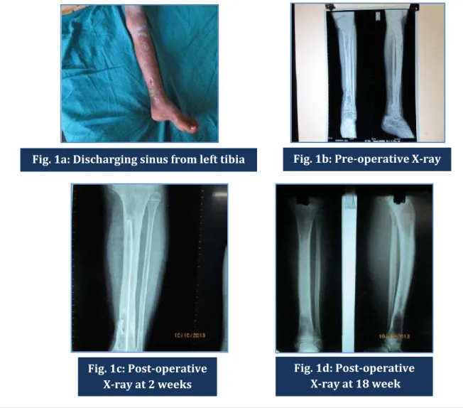
Sutures were removed after two weeks and antibiotic mixed bone cement beads or nails were removed after six weeks. Two doses of prophylactic intravenous antibiotic were used; one before, and another after the operation. All patients were followed up for a period of two years. The pictures of preoperative clinical condition of the wound, preoperative X-ray of the diseased bone, immediate postoperative X-ray, follow up X-ray and clinical condition at recovery of one patient are given in figures 1a to 1e.
RESULTS:
Sl. No
Age
(Yrs) Sex Aetiology
Clinical features
Bone
involve Site involve Organism
C/S Antibioti
c
Antib iotic given
Treatment response
1 14 F Haematogen ous
Discharging sinus
Left
femur Metaphysis S.aureus
Vancomy cin
Vanco
mycin Cured
2 10 F Haematogen ous
Discharging sinus
Left
femur Metaphysis S.aureus
Vancomy cin
Vanco
mycin Cured
3 11 F Haematogen ous
Discharging sinus
Right
tibia Metaphysis S.aureus
Vancomy cin
Vanco
mycin Cured
4 19 F
Post-operative
Discharging sinus
Left
femur Diaphysis S.aureus
Cefuroxi me
Cefur
oxime Cured
5 36 M
Post-operative
Discharging
sinus Left ulna Diaphysis No-growth Nil
Vanco
mycin Cured
6 42 F
Post-operative Quisent sinus
Right
humerus Diaphysis S.aureus
Vancomy cin
Vanco
mycin Cured
7 14 M Haematogen ous
Discharging
sinus Left tibia Metaphysis Steptococcus
Cefuroxi me
Cefur
J of Evolution of Med and Dent Sci/ eISSN- 2278-4802, pISSN- 2278-4748/ Vol. 4/ Issue 10/Feb 02, 2015 Page 1624
8 35 M
Post-operative
Discharging
sinus, Ulcer Left tibia Diaphysis Steptococcus
Cefuroxi me
Cefur
oxime Cured
9 7 F Haematogen ous
Discharging
sinus Left tibia Metaphysis S.aureus
Vancomy cin
Vanco
mycin Cured
10 50 M Post-operative
Discharging sinus
Right
femur Diaphysis S.aureus
Cefuroxi me
Cefur
oxime Cured
11 38 M
Post-traumatic Quisent sinus
Right
humerus Diaphysis No-growth Nil
Vanco
mycin Cured
12 18 M Post-traumatic
Discharging sinus
Right
tibia Metaphysis S.aureus
Cefuroxi me
Cefur
oxime Cured
13 14 M Post-traumatic
Discharging sinus
Right
femur Metaphysis S.aureus
Cefuroxi me
Cefur
oxime Cured
14 18 M Post-traumatic
Discharging sinus
Right
femur Metaphysis S.aureus
Cefuroxi me
Cefur
oxime Failure
15 15 F Haematogen
ous Quisent sinus
Right
humerus Metaphysis S.aureus
Vancomy cin
Vanco
mycin Cured
16 6 F Haematogen ous
Discharging sinus
Right
tibia Metaphysis S.aureus
Vancomy cin
Vanco
mycin Cured
17 40 F Post-operative
Discharging sinus
Left
femur Metaphysis S.aureus
Cefuroxi me
Cefur
oxime Cured
18 7 M Haematogen
ous Quisent sinus
Right
radius Diaphysis S.aureus
Vancomy cin
Vanco
mycin Cured
19 53 M Post-traumatic
Discharging sinus
Right
ulna Diaphysis No-growth Nil
Cefur
oxime Cured
20 56 M Post-operative
Discharging sinus
Right
radius Diaphysis Steptococcus
Vancomy cin
Vanco
mycin Cured
21 14 M Haematogen
ous Quisent sinus
Right
tibia Diaphysis S.aureus
Cefuroxi me
Cefur
oxime Cured
22 22 M Post-operative
Discharging sinus
Left
femur Diaphysis No-growth Nil
Vanco
mycin Cured
23 22 M Post-operative
Discharging sinus
Left
femur Metaphysis E.coli
Meropen am Mero pena m Cured
24 32 M Post-traumatic
Discharging sinus
Right
tibia Diaphysis No-growth Nil
Cefur
oxime Cured
25 24 M Post-traumatic
Discharging sinus
Right
tibia Metaphysis
S.aureus& Streptococcus
Vancomy cin
Vanco
mycin Failure
26 34 M Post-operative
Discharging
sinus Left tibia Metaphysis S.aureus
Cefuroxi me
Cefur
oxime Cured
27 48 M Post-operative
Discharging sinus
Right
femur Metaphysis No-growth Nil
Vanco
mycin Cured
28 18 M Post-traumatic
Discharging sinus
Right
humerus Diaphysis S.aureus
Vancomy cin
Vanco
mycin Cured
29 17 F Haematogen
ous Quisent sinus Left ulna Diaphysis S.aureus
Cefuroxi me
Cefur
oxime Cured
30 9 F Haematogen ous
Discharging sinus
Left
humerus Metaphysis S.aureus
Cefuroxi me
Cefur
oxime Cured
DISCUSSION: Treatment of chronic osteomyelitis remains a problem in orthopaedics owing to the unique microarchitecture of bone tissue, precarious blood supply compared to soft tissue and consequently less delivery of antibiotics to the affected site. Moreover, the use of systemic antibiotics for prolonged period leads to systemic toxicity.8
Saucerization as a mode of treatment for chronic osteomyelitis was plagued by complications like fracture and non-union.9 Some micro-organism form biofilm, which is a highly structured
J of Evolution of Med and Dent Sci/ eISSN- 2278-4802, pISSN- 2278-4748/ Vol. 4/ Issue 10/Feb 02, 2015 Page 1625 actually made of glycocalyx12 and acts as a multicellular organism. It invites several other organisms
which remained dormant at the site of infection and now multiply in that favourable environment. On the other hand, antibiotics administered via systemic route reach to infected site in a sublethal dose as biofilm prevent penetration of antibiotics.
Therefore it is now understood that the best way to treat chronic osteomyelitis is to prevent the development of chronic osteomyelitis. Dedicated management of open injury to prevent infection, strict aseptic and antiseptic technique, improvement in operating room environment and implant design is a few of these developments.
With the success in controlling infection with antibiotic mixed bone cement in arthroplasty,13
ideas developed regarding the use of local antibiotic therapy in chronic osteomyelitis.14 Local
antibiotic therapy maintains lethal concentration locally against bacteria without escaping to systemic flow.
The elution properties of various bone cement, their heat stability, pharmacodynamic and pharmacokinetic, local tissue toxicity and systemic adverse reaction due to local use were thoroughly studied in last few decades.
In the present study, out of thirty patients involving diaphyseal and metaphyseal osteomyelitis, twenty eight patients were completely free from recurrence in two year follow-up. Two patients had persistence discharge. These two patients had metaphyseal osteomylitis following open injury.
The result of the current observation is encouraging in most of the cases particularly in diaphyseal region where medullary glycocalyx was removed easily. However, the authors faced difficulties in removing glycocalyx adequately in the metaphyseal region where the bone tissue is spongy in nature.
ACKNOWLEDGEMENT: The authors wish to thank Dr. M. C. Mandal, Associate Professor in Anaesthesiology, N.B.M.C. for his contribution in editing the first draft before submission.
REFERENCES:
1. Stewart PS. Mechanisms of antibiotic resistance in bacterial biofilms. Int J Med Microbiol 2002; 292:107-13.
2. Clawson DK, Dunn AW. Management of common bacterial infections of bones and joints. J Bone Joint Surg Am 1967; 49: 164-82.
3. Bayston R, Milner RD. The sustained release of antimicrobial drugs from bone cement. An appraisal of laboratory investigations and their significance. J Bone Joint Surg Br 1982; 64: 460-4.
4. Stewart PS, Costerton JW. Antibiotic resistance of bacteria in biofilm. Lancet 2001; 358: 135-8. 5. Edin ML, Miclau T, Lester GE, Lindsey RW, Dahners LE. Effect of cefazolin and vancomycin on
osteoblasts in vitro. Clin Orthop Relat Res 1996; 333: 245-51.
6. Marks KE, Nelson CL and Lautenschlager EP. Antibiotic-impregnated acrylic bone cement. J Bone Jt Surg Am 1976; 58: 358-64.
J of Evolution of Med and Dent Sci/ eISSN- 2278-4802, pISSN- 2278-4748/ Vol. 4/ Issue 10/Feb 02, 2015 Page 1626 8. Waldvogel FA, Papageorgiou PS. Osteomyelitis: the past decade. N Eng J Med. 1980; 303:
360-70.
9. Weaver DS, Perry GH, Macchiarelli R, Bondioli L. A surgical amputation in 2nd century Rome.
Lancet. 2000; 356: 686.
10.Sauer K, Camper AK, Enrlich GD, Costerton JW, Davis DG. Pseudomonas aeruginosa displays multiple phenotypes during development as a biofilm. J. Bacteriol. 2002; 184: 1140-54.
11.An YH, Friedman RJ. Concise review of mechanisms of bacterial adhesion to biomaterial surfaces. J Biomed Mater Res 1998; 43: 338-48.
12.Olson ME, Garvia KL, Fey PD, Rupp ME. Adherence of Staphylocorcus epidermidis to biomaterials is augmented by PIA. Clin Orthop Relat Res 2006; 451: 21-4.
13.Buchholz HW, Engelbrecht H. Depot effects of various antibiotics mixed with Palacos resins. Chirurg 1970; 41:511-5.
14.Vecsei V, Starlinger M. Gentamicin-PMMA bead chains in the treatment of posttraumatic osseous and tissue infections. Arch Orthop Trauma Surg 1982; 99: 259-63.
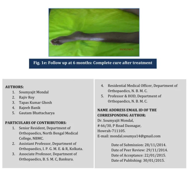
Figures 1a to 1e: Pictures showing the discharging sinus (1a), pre-operative X-ray (1b), Post-operative X-ray at 2 weeks (1c), at 18 weeks (1d) and healing at 6 months (1e) of left tibia of one girl aged 7 years.
Fig. 1a: Discharging sinus from left tibia Fig. 1b: Pre-operative X-ray
Fig. 1c: Post-operative X-ray at 2 weeks
J of Evolution of Med and Dent Sci/ eISSN- 2278-4802, pISSN- 2278-4748/ Vol. 4/ Issue 10/Feb 02, 2015 Page 1627
AUTHORS:
1. Soumyajit Mondal 2. Rajiv Roy
3. Tapas Kumar Ghosh 4. Rajeeb Banik
5. Gautam Bhattacharya
PARTICULARS OF CONTRIBUTORS:
1. Senior Resident, Department of Orthopaedics, North Bengal Medical College, NBMC.
2. Assistant Professor, Department of Orthopaedics, I. P. G. M. E. & R, Kolkata. 3. Associate Professor, Department of
Orthopaedics, B. S. M. C, Bankura.
4. Residential Medical Officer, Department of Orthopaedics, N. B. M. C.
5. Professor & HOD, Department of Orthopaedics, N. B. M. C.
NAME ADDRESS EMAIL ID OF THE CORRESPONDING AUTHOR:
Dr. Soumyajit Mondal, # 66/30, P Road Dasnagar, Howrah-711105.
E-mail: [email protected]
Date of Submission: 28/11/2014. Date of Peer Review: 29/11/2014. Date of Acceptance: 22/01/2015. Date of Publishing: 30/01/2015.