Heat Shock Protein 22 (Hsp22) Regulates
Oxidative Phosphorylation upon Its
Mitochondrial Translocation with the
Inducible Nitric Oxide Synthase in
Mammalian Heart
Eman Rashed1, Paulo Lizano1, Huacheng Dai1, Andrew Thomas2, Carolyn K. Suzuki3, Christophe Depre1, Hongyu Qiu1,4*
1Department of Cell Biology and Molecular Medicine, New Jersey Medical School, Rutgers, The State University of New Jersey, New Brunswick, New Jersey, United States of America,2Department of Pharmacology and Physiology, New Jersey Medical School, Rutgers, The State University of New Jersey, New Brunswick, New Jersey, United States of America,3Department of Microbiology, Biochemistry and Molecular Genetics, New Jersey Medical School, Rutgers, The State University of New Jersey, New Brunswick, New Jersey, United States of America,4Department of Basic Science, Division of Physiology, School of Medicine, Loma Linda University, Loma Linda, California, United States of America
Abstract
Objectives
Stress-inducible heat shock protein 22 (Hsp22) confers protection against ischemia through induction of the inducible isoform of nitric oxide synthase (iNOS). Hsp22 overexpressionin vivostimulates cardiac mitochondrial respiration, whereas Hsp22 deletionin vivo
signifi-cantly reduces respiration. We hypothesized that Hsp22-mediated regulation of mitochon-drial function is dependent upon its mitochonmitochon-drial translocation together with iNOS.
Methods and Results
Adenoviruses harboring either the full coding sequence of Hsp22 (Ad-WT-Hsp22) or a mu-tant lacking a N-terminal 20 amino acid putative mitochondrial localization sequence (Ad-N20-Hsp22) were generated, and infected in rat neonatal cardiomyocytes. Compared to β-Gal control, WT-Hsp22 accumulated in mitochondria by 2.5 fold (P<0.05) and increased
oxygen consumption rates by 2-fold (P<0.01). This latter effect was abolished upon
addi-tion of the selective iNOS inhibitor, 1400W. Ad-WT-Hsp22 significantly increased global iNOS expression by about 2.5-fold (P<0.01), and also increased iNOS mitochondrial
locali-zation by 4.5 fold vsβ-gal (P<0.05). Upon comparable overexpression, the N20-Hsp22
mutant did not show significant mitochondrial translocation or stimulation of mitochondrial respiration. Moreover, although N20-Hsp22 did increase global iNOS expression by 4.6-fold, it did not promote iNOS mitochondrial translocation.
OPEN ACCESS
Citation:Rashed E, Lizano P, Dai H, Thomas A, Suzuki CK, Depre C, et al. (2015) Heat Shock Protein 22 (Hsp22) Regulates Oxidative Phosphorylation upon Its Mitochondrial Translocation with the Inducible Nitric Oxide Synthase in Mammalian Heart. PLoS ONE 10(3): e0119537. doi:10.1371/journal. pone.0119537
Received:October 18, 2014
Accepted:January 18, 2015
Published:March 6, 2015
Copyright:© 2015 Rashed et al. This is an open access article distributed under the terms of the Creative Commons Attribution License, which permits unrestricted use, distribution, and reproduction in any medium, provided the original author and source are credited.
Data Availability Statement:All relevant data are within the paper and its supporting information files.
Funding:This work is partially supported by a grant 1R01 HL115195-01 (http://www.nih.gov/) from United States National Institute of Health, (H. Q.) and a grant 0835182N from American Heart Association (https:// research.americanheart.org) (H. Q.). The funders had no role in study design, data collection and analysis, decision to publish, or preparation of the manuscript.
Conclusion
Translocation of both Hsp22 and iNOS to the mitochondria is necessary for Hsp22-mediat-ed stimulation of oxidative phosphorylation.
Introduction
The heart depends on oxidative phosphorylation to supply the large amount of ATP required
for its continuous contractile activity[1]. Mitochondrial dysfunction due to decreased supply of
oxygen and substrates, such as that occurring during ischemia, leads to impaired ATP genera-tion and structural alteragenera-tions of the respiratory chain, eventually resulting in cell death by
both apoptosis and necrosis[2].
The heat shock protein 22/H11 Kinase (Hsp22) is a stress-inducible protein responsive to
various conditions of myocardial stress, including ischemia [3,4]. Cardiac-specific
overexpres-sion of Hsp22 in a transgenic (TG) mouse provides protection against myocardial ischemia
that is equally powerful to ischemic preconditioning [5] through the induction of the inducible
isoform of nitric oxide synthase (iNOS) [5], the effector of the second window of ischemic
pre-conditioning [6]. Accordingly, inhibition of iNOS abolishes the cardioprotection conferred by
Hsp22 [7].
We characterized previously the subcellular distribution of Hsp22 in the heart, and showed
that it is located in the mitochondrial, as well as in the nuclear and cytosolic fractions [8]. In
addition, it has been shown inDrosophilathat Hsp22 localization in mitochondria is due to a
translocation mechanism that depends on its N-terminal domain [9]. The biological function
of Hsp22 inside mammalian mitochondria remains unknown; however we showed in the TG
mouse model that increased Hsp22 expressionin vivostimulates mitochondrial oxidative
phosphorylation, whereas its deletion in a knockout model has the opposite effect [10]. In
addi-tion, the TG mouse is characterized by an inhibition of the mitochondrial pathway of apoptosis
[5]. These observations support the hypothesis that mitochondrial localization of Hsp22 might
promote both cardiac cell survival and oxidative metabolism. The main purpose of our study was to interrogate the physiological consequence of this mechanism on
mitochondrial respiration.
Although our previous studies showed that cardioprotection conferred by Hsp22 is iNOS-dependent, the role played by iNOS in the mitochondrial respiration is still relatively unknown. We also hypothesized that mitochondrial functions of Hsp22 could be mediated by iNOS. Therefore, in the present study, we tested whether mitochondrial localization of Hsp22 in mammalian heart promotes mitochondrial respiration, and whether this process involves iNOS.
Materials and Methods
Animal model
Three-month old male FVB wild type (WT) and Hsp22 transgenic (TG) mice were used. A
car-diac specific Hsp22 TG mouse was generated as described previously [11]. The Hsp22 protein
with the approval of the IACUC committee at New Jersey Medical School, Rutgers, The State University of New Jersey (IACUC# 11025 and #11024).
Generation of a mutant Hsp22 adenovirus
Two mutant adenoviruses were generated, in which the last 20 amino acids at either the N-ter-minus (Ad-N20-Hsp22) or the C-terN-ter-minus (Ad-C20-Hsp22) were deleted. A green fluorescent protein (GFP) coding sequence was fused at the C-terminus of each mutant. The sequences were ligated downstream from the CMV promoter of the AdEasy XL adenoviral vector system (Agilent, Santa Clara, CA), followed by homologous recombination with the adenoviral
back-bone plasmid [11]. The adenoviruses harboring the full length Hsp22 coding sequence
(Ad-WT-Hsp22) or theβ-galactosidase (β-Gal) control were described before [11,12]. The
recombi-nant adenoviruses were propagated in HEK 293 cells.
Culture of rat neonatal cardiac myocytes
Rat neonatal ventricular cardiac myocytes (RNCMs) were prepared from Sprague-Dawley rat
pups (Charles River Laboratories, Wilmington, MA) as described previously [11,13,14].
Neo-natal rats were sterilized with 70% ethanol and sacrificed by decapitation. Myocytes were dis-persed with 0.1% collagenase type IV (Worthington Biochem, Lakewood, NJ), 0.1% trypsin
(GIBCO, Grand Island, NY) and 15μg/mL DNase I (Sigma-Aldrich, St. Louis, MO). Cell
sus-pensions were applied on a discontinuous Percoll gradient, and the myocyte layer was
collect-ed. Cells were cultured in medium containing Dulbecco’s Modified Eagle Medium (DMEM)/
F12 (1:1) (Sigma-Aldrich, St. Louis, MO) for 24 hours. Myocytes were infected for 48 hours after 24 hours of serum-free starvation. Inhibition of iNOS was initiated 24 hours before the
collection of the cells upon addition of 100μM 1400W (Sigma-Aldrich, St. Louis, MO).
Protein extraction and subcellular fractions
Tissues were homogenized at 4°C in a buffer supplemented with protease and phosphatase
in-hibitors [5,15], and centrifuged at 12,000gfor 20 min. Proteins were denatured by boiling,
re-solved on SDS-PAGE gels and then transferred onto a nitrocellulose membrane. Primary antibodies were incubated overnight and after incubation with the secondary antibody, detec-tion was performed by chemiluminescence (Dupont/NEN, Boston, MA), and quantified by
densitometry. Subcellular fractions were prepared as described previously [15]. After an initial
spin at 100g, the nuclear fraction was pelleted at low-speed centrifugation (500g, 10 minutes).
The supernatant was further centrifuged (10,000g, 10 minutes) to pellet the mitochondrial
frac-tion. The resulting supernatant was ultracentrifuged (100,000g, 90 minutes) to obtain the
cyto-solic fraction (supernatant) and a microsomal fraction (pellet). Pellets were washed and re-suspended in 150 mM NaCl, 1% NP40, 0.5% deoxycholate, 0.1% SDS, 50 mM Tris (pH 8.0). Fraction purity was verified by western blotting, using glyceraldehyde 3-phosphate dehydroge-nase (GAPDH, cytosol), lamin A/C (nucleus), cytochrome oxidase IV (COX IV,
mitochon-dria), and plasma membrane calcium ATPase (membranes) as described previously [8].
Targeted proteins including Hsp22, Stat3 and iNOS were detected by western blotting as
de-scribed previously [8,10].
Mitochondrial sub-fractionation
Two techniques were used to sub-fractionate the mitochondria obtained by cell fractionation. First, isolated mitochondria were incubated with 2% digitonin for 20 minutes, and spun at
membrane (OM) together with the inter-membrane space (IMS) in the supernatant [9,16,17]. Second, mitochondria were incubated with 2% Nonidet P-40 for 15 minutes and spun at
18,000gfor 40 minutes to yield a soluble component including the matrix, IMS and an
insolu-ble fraction composed of mitochondrial inner (IM) and outer membranes (OM) [9]. The purity
of each mitochondrial sub-fraction was verified by western blotting using specific antibodies: COX IV (1:1000, rabbit polyclonal antibodies, Cell Signaling, Danvers, MA) for IM, voltage-de-pendent anion-selective channel protein 1 [VDAC] (1:1000, rabbit polyclonal antibodies, Cell Signaling, Danvers, MA) for OM, and Grp75 (1:500, Rabbit polyclonal antibody, Abcam, Cam-bridge, MA) for the matrix.
Oxygen consumption assay
Clark electrode. Mitochondrial oxygen consumption rates (OCR) were measured in intact
RNCMs with a Clark-type electrode as described previously [18,19]. Briefly, RNCMs infected
with Ad-β-Gal, Ad-WT-Hsp22 or Ad-N20-Hsp22 were collected and suspended in
extracellu-lar buffer as described[18] Glucose was used as a substrate. Oxygen concentration was
mea-sured at 30°C with a Clark-type electrode fitted to a 50μl water-jacketed reaction chamber.
OCR was determined after addition of 6μM oligomycin or 5μM carbonyl cyanide
4-(trifluoro-methoxy) phenylhydrazone (FCCP). The ratio of FCCP-stimulated to oligomycin-inhibited OCR in the myocytes was also calculated.
Immunofluorescence
Cardiac myocytes were cultured to confluence on a 4-chamber slide. The cells were washed
with PBS, fixed in methanol at−20°C for 10 min, blocked with 5% bovine serum albumin for
1h, and incubated with primary antibody. After washing, cells were incubated with a
fluoresce-in-labeled secondary antibody and mounted in a Vecto 4’-6-diamino-2-phenylindole (DAPI)
medium for observation at x40. Mitochondria were detected using the MitoTracker Red (Life Technologies, Carlsbad, CA).
Measurement of iNOS activity
Cell lysates were incubated in 20 mM Tris–HCl (pH 8.0), 2 mM NADPH, 2 mM L-arginine
and 10 mM FAD for 3 h at room temperature. NO production was measured from nitrite levels
based on the Griess reaction [20] (Nitric Oxide Assay Kit, Oxford Biomedical Research,
Ox-ford, UK). The absorbance values were determined at 540 nm using a microtiter plate reader. Nitrate was reduced to nitrite by incubation with 0.1 U/ml nitrate reductase, 0.1 mM NADPH and 5 mM FAD. The reaction was stopped by the addition of 10 U/ml lactate dehydrogenase and 10 mM pyruvate. Sodium nitrite was used as a standard, and lysis buffer as a blank. Nitrite value of the control test (without NADPH/ L-arginine) was subtracted from the experimental
values [21,22].
Apoptosis assays
Apoptosis was induced upon incubation of RCNMs with 1μM chelerythrine (Sigma Aldrich,
St. Louis, MO) for 4 hours and was measured, both by enzyme-linked colorimetric assay and
Statistical analysis
Results are the mean ± SEM for the number of samples indicated in the Figure legends. A one-way ANOVA was used and Student-Newman-Keuls post hoc correction was applied for
multi-group comparison. A value of P<0.05 was considered significant.
Results
Hsp22 is predominantly located to the mitochondrial inner membrane
We previously showed that Hsp22 is expressed in mitochondria from mouse heart [8]. To
elu-cidate the potential role of Hsp22 in the mitochondria, we first characterized its sub-mitochon-drial localization using two different fractionation techniques described in the Methods. As
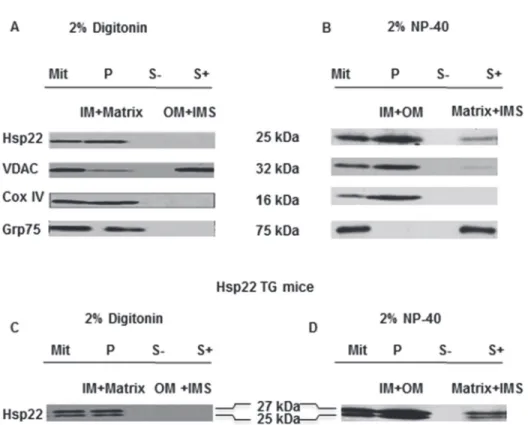
shown inFig. 1 A and B, the digitonin digestion method showed that Hsp22 was only detected
in the pellet sub-fraction containing the IM and matrix, but not in the fraction containing the OM and IMS. The NP-40 digestion showed that Hsp22 was located primarily within the mem-brane fraction, whereas only a residual amount was found in the matrix and IMS. Taken
Fig 1. Sub-mitochondrial localization of Hsp22 in mouse heart.Mitochondrial sub-fractionation was performed on WT (A and B) and TG mouse hearts (C and D) after treatment with digitonin (A and C) and NP-40 (B and D). In each panel, from left to right: Mit: untreated total mitochondria (positive control); P: pellet obtained from detergent-treated mitochondria; S-: supernatant obtained after centrifugation of untreated mitochondria (negative control); S+: supernatant obtained after treatment. In panels A and C, digitonin treatment shows that Hsp22 is distributed in the pellet (P) containing the IM and matrix but not in the supernatant (S+) containing the OM and IMS. In panels B and D, NP-40 treatment shows that Hsp22 is predominantly located in the pellet (P) containing the IM and OM, and to a low extent in the supernatant (S+) containing the matrix and IMS. Markers include VDAC (OM), COX IV (IM), and Grp75 (matrix). The figure shows one representative example of n = 4 per group.
together, these results demonstrate that, in the mammalian heart, mitochondrial Hsp22 is pre-dominantly associated with the IM. We repeated this experiment in the TG model of
cardiac-specific Hsp22 overexpression. The results shown inFig. 1 C and Dare consistent with the
data obtained in the WT mouse, indicating that over-expression of the protein does not affect its sub-mitochondrial localization. This observation is important in order to validate the exper-iments of overexpression shown below in RNCMs.
N-terminus deletion prevents the mitochondrial localization of Hsp22
To determine the domain responsible for the mitochondrial localization of Hsp22 in mammali-an heart, mammali-an adenovirus harboring a mutmammali-ant lacking a 20 amino acid putative N-terminalmito-chondrial localization sequence [9] was generated (Ad-N20-Hsp22), and infected in rat
neonatal cardiomyocytes. An adenovirus harboring the complete coding sequence of Hsp22 (Ad-WT-Hsp22) was used as a positive control, whereas an adenovirus harboring the sequence
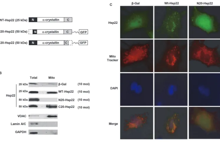
ofβ-galactosidase (β-Gal) was used as a negative control (Fig. 2 A). WT-Hsp22 and
Ad-N20-Hsp22 were infected at 10 moi in RNCMs. Overexpression of the corresponding protein
was comparable between WT-Hsp22 and N20-Hsp22-adenoviruses (Fig. 2 B). Mitochondria
were further separated by sub-cellular fractionation. Although cells over-expressing
Ad-WT-Fig 2. N-terminal deletion of the Hsp22 coding sequence prevents its mitochondrial localization.A. Hsp22 mutants were generated by deletion of 20 amino acids at either the N-terminus (Ad-N20-Hsp22) or C-terminus (Ad-C20-Hsp22), respectively, from the Hsp22 WT coding sequence (Ad-WT-Hsp22). B. Examples of the ratio of Hsp22 between the mitochondrial fraction and the total protein extract for the corresponding protein extracted from cardiac myocytes treated withβ-Gal, WT-Hsp22, N20-Hsp22 and C20-Hsp22 adenoviruses (10 moi). Markers include VDAC (mitochondria), lamin A/C (nucleus), and GAPDH (total extracts). C. Immunofluorescence on myocytes infected with 10 moiβ-Gals, Ad-WT-Hsp22 and Ad-N20-Hsp22. Staining is performed with Hsp22 or GFP antibodies (green), Mitotracker (red) and nuclear DAPI counterstaining (blue), followed by merging of the three signals.
Hsp22 exhibited a significant increase (2.5 fold) in mitochondrial Hsp22 protein as compared
to theβ-Gal control, such accumulation was not observed in myocytes treated with
Ad-N20-Hsp22 (Fig. 2 B). In comparison, myocytes treated with Ad-N20-Hsp22 actually showed a loss
of mitochondrial Hsp22 localization (Fig. 2 B). We also generated another mutant adenovirus
in which the last 20 amino acids at the C-terminus (Ad-C20-Hsp22) were deleted. A green fluorescent protein (GFP) coding sequence was fused at the C-terminus of mutant as the same as Ad-N20-Hsp22. Cells over-expressing Ad-C20-Hsp22 exhibited a significant increase in
mi-tochondrial Hsp22 protein as compared to theβ-Gal control which is similar to Ad-WT-Hsp2,
indicating that it is the deletion of the 20 amino acid putative N-terminal mitochondrial locali-zation sequence and not the GFP fusion protein that is responsible for the failure of
Ad-N20-Hsp22 mutant protein to be taken up into the mitochondria (Fig. 2A and B).
Immunofluorescence was performed on RNCMs infected with 10 moi of the respective ade-noviruses for 48 hours. Myocytes infected with Ad-WT-Hsp22 showed an increased Hsp22 ex-pression in a punctate pattern, which superimposed with the MitoTracker signal, indicating a
mitochondrial localization (Fig. 2 C). Myocytes infected with Ad-N20-Hsp22 showed an
in-crease in exogenous, GFP-tagged Hsp22, which did not superimpose with Mitotracker (red),
indicating a loss of the mitochondrial localization of the mutant Hsp22 (Fig. 2 C).
N-terminus deletion does not affect function of Hsp22 protein on gene
expression
We showed before that Hsp22 over-expression regulates gene expression through the
regula-tion of specific transcripregula-tion factors, including STAT3 [10]. Therefore, we investigated whether
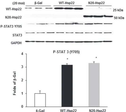
the N-terminal mutant Hsp22 protein preserved its biological function on transcriptional regu-lation. Ad-WT-Hsp22 and Ad-N20-Hsp22 were infected at 20 moi in RNCMs. Y705 phos-phorylation of the transcription factor STAT3, a target through which Hsp22 affects gene
expression [10], was used as a control to show the activation of the STAT3 protein. As shown
inFig. 3, upon a comparable amount of Hsp22 overexpression, myocytes infected with either Ad-WT-Hsp22 or Ad-N20-Hsp22 exhibited increased Y705 phosphorylation of STAT3 by
about 3.0 fold vsβ-Gal control (P<0.05). Therefore, the truncated Hsp22 protein retains its
na-tive function and activity on transcriptional regulation.
Mitochondrial localization of Hsp22 is necessary for cytoprotection
conferred by Hsp22
We have shown previously that overexpression of Hsp22 promotes cell survival by inhibiting cardiomyocyte apoptosis. We next tested whether the N-terminal mutant of Hsp22 influences its cytoprotection against apoptosis. Myocytes were infected with an adenovirus harboring the
Ad-WT-Hsp22 or Ad-N20-Hsp22 at 10 moi for 48 hours, and compared withβ-Gal.
Cheler-ythrine was subsequently added to induce cell apoptosis. Apoptosis was first measured by
TUNEL. As shown inFig. 4 A, over-expression of WT-Hsp22 reduced chelerythrine-induced
apoptosis by 60% (P<0.05) compared toβ-Gal. No protection was observed in myocytes
in-fected with the Ad-N20-Hsp22, which actually induced a 2.5-fold increase in apoptosis
com-pared with theβ-Gal control. A caspase-3 colorimetric assay confirmed the results found by
TUNEL. Again, while overexpression of Hsp22 markedly reduced chelerythrine-induced
cas-pase 3 activation compared to theβ-Gal group, overexpression of Ad-N20-Hsp22 significantly
N-terminus deletion abolishes Hsp22-stimulated oxidative
phosphorylation
Since our previous resultsin vivoshowed that manipulation of Hsp22 expression in the heart
stimulates mitochondrial respiration [10], we tested next whether preventing Hsp22
Fig 3. Regulation of gene expression of Hsp22 mutant proteins in cardiac myocytes.Immunoblotting of P-STAT3 (Y705) in myocytes treated with theβ-Gal, WT-Hsp22 or N20-Hsp22 adenoviruses (20 moi).
*P<0.05 vsβ-Gal, n = 6 per group.
doi:10.1371/journal.pone.0119537.g003
Fig 4. Mitochondrial localization of Hsp22 is necessary for cellular protection in cardiac myocytes.Apoptosis measured by TUNEL and caspase-3 activity in RNCMs infected with theβ-Gal control, Ad-WT-Hsp22 or Ad-N20-Hsp22 with chelerythrine.*, P<0.05 versusβ-Gal; #, P<0.05 vs Ad-WT-Hsp22. n = 5 per group.
mitochondrial localization with the N-terminal mutant blocks the effect of the protein on mito-chondria. A Clark-type oxygen electrode was used to measure the oxygen consumption rate (OCR) in intact RNCMs transfected with Ad-WT-Hsp22 or Ad-N20-Hsp22, and compared to
β-Gal (Fig. 5). The rate of mitochondrial respiration was measured under basal (pseudo State
4) conditions in the presence of oligomycin to block the F1FoATP synthase, followed by
addi-tion of the uncoupler FCCP. In intact cells, the difference between OCR in the presence of
oli-gomycin [18] and in the presence of rotenone, represents proton leak across the inner
mitochondrial membrane, while OCR in the presence of FCCP represents the maximal respira-tory capacity, and indicates the potential of the cell to maintain its energy balance in stress
con-ditions [18]. Therefore, the ratio of these two rates represents the "spare" respiratory capacity
of cells to respond to conditions of increased energy demand.
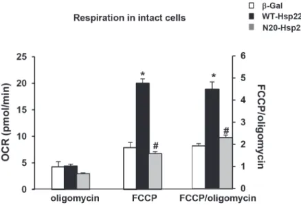
As shown inFig. 5, there was no significant difference in the leak rate in the three treatment
groups measured in the presence of oligomycin. The maximal OCR after addition of FCCP in-creased by about 2.5-fold in myocytes treated with Ad-WT-Hsp22 (10 moi) compared to the
β-Gal control (P<0.05), but this was not observed with the mutant Ad-N20-Hsp22.
Conse-quently, the FCCP/oligomycin OCR ratio increased significantly (P<0.05) in myocytes treated
with Ad-WT-Hsp22, but not in myocytes infected with Ad-N20-Hsp22 (Fig. 5). These data
show that Hsp22 overexpression does not uncouple the mitochondria, but rather it increases its maximally stimulated respiration, which was not observed upon overexpression of the Ad-N20-Hsp22 mutant. Thus, the effect of Hsp22 on mitochondrial respiration is dependent upon its translocation to the mitochondria via its N-terminus.
Hsp22 stimulates mitochondrial respiration through iNOS
Our previous studies showed that iNOS is central to the mechanism of cardioprotection by
Hsp22 [7], therefore we tested whether the effect of Hsp22 on mitochondrial respiration could
be mediated by iNOS. Myocytes were transfected with Ad-WT-Hsp22 for 24 hours, in the
ab-sence or preab-sence of the selective iNOS inhibitor 1400W [23]. Mitochondrial respiration was
Fig 5. Effect of N-terminus mutant Hsp22 on mitochondrial respiration in intact neonatal rat cardiac myocytes.Mitochondrial respiration measured by Clark electrode in intact myocytes transfected with Ad-WT-Hsp22 or Ad-N20-Hsp22 compared toβ-Gal. Mitochondrial respiration is presented on the left Y axis as OCR after addition of oligomycin and OCR after addition of FCCP, and, on the right Y axis, as the ratio of both rates (FCCP/oligomycin).*, P<0.05 versusβ-Gal; #, P<0.05 vs Ad-WT-Hsp22. n = 5 per group.
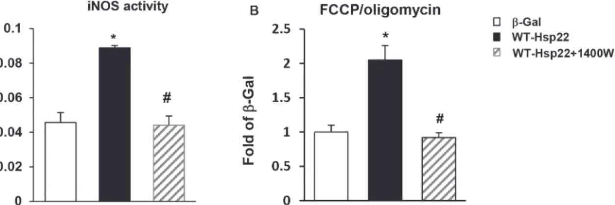
measured in these conditions by a Clark-type oxygen electrode (seeMethods). As shown in Fig. 6 A, Hsp22 overexpression increased iNOS activity by approximately 2-fold, which was
abolished upon addition of 1400W (Fig. 6 A). Consistent with the studies described above,
myocytes treated with Ad-WT-Hsp22 exhibited a 2-fold increase in OCR (P<0.05) compared
toβ-Gal, as represented by the FCCP/oligomycin ratio. This stimulation of mitochondrial
oxy-gen consumption by Ad-WT-Hsp22 was also abolished by 1400W (Fig. 6 B). Therefore, the
ef-fect of Hsp22 on mitochondrial respiration is shown to be dependent upon iNOS. This is
consistent with the data presented inFig. 5, showing that overexpression of Ad-WT-Hsp22 in
myocytes exhibited an increase in mitochondrial respiration compared toβ-Gal, which was
abolished upon the over-expression of Ad-N20-Hsp22.
The N-terminal mutant Hsp22 blocks the translocation of iNOS into
mitochondria
We showed above that the N-terminal deletion of Hsp22 prevented its translocation into
mito-chondria (Fig. 2), and abolished Hsp22-stimulated mitochondrial respiration (Fig. 5). The later
observation is consistent with the effect of the iNOS inhibitor on Hsp22-stimulated
mitochon-drial respiration as shown inFig. 6 B. Therefore, we tested whether the mitochondrial
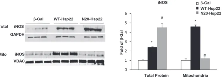
translo-cation of Hsp22 affects the expression and/or regulation of iNOS in the mitochondria. We first tested whether the Ad-N20-Hsp22 mutant affects iNOS expression at a global, cel-lular level. RNCMs were infected with 20 moi of Ad-WT-Hsp22 or Ad-N20-Hsp22, and
com-pared toβ-Gal control. Immunoblotting for iNOS was performed using total cell lysates.
Compared toβ-Gal control, myocytes infected with Ad-WT-Hsp22 exhibited a significant
2.5-fold increase in global iNOS expression. Myocytes treated with Ad-N20-Hsp22 exhibited a
sig-nificant global increase of iNOS expression by 4.6-fold in total cellular lysate (Fig. 7), indicating
that the mutant Hsp22 did not impair the regulation of Hsp22 on iNOS gene expression. This result is consistent with the observation that the mutant Hsp22 maintains the activation of
STAT3 (Fig. 3), a transcription factor known to up-regulate iNOS expression [6,10].
Next, we tested whether Hsp22 affects the mitochondrial translocation of iNOS. As shown inFig. 7, iNOS expression in the mitochondrial fraction was significantly increased by 4.5-fold
in myocytes infected with Ad-WT-Hsp22 (P<0.05 versusβ-Gal) but this increase was not
ob-served in mitochondria of myocytes treated with Ad-N20-Hsp22 (Fig. 7). These data indicate
Fig 6. Hsp22 stimulates mitochondrial respiration through iNOS.A. iNOS activity in myocytes transfected with Ad-WT-Hsp22 in presence or in absence of 1400W, compared toβ-Gal. Optical density represents the measured absorbance at 540 nm.*, P<0.05 versusβ-Gal; #, P<0.05 vs Ad-WT-Hsp22 without 1400W. n = 5 per group. B. Mitochondrial respiration measured by Clark electrode in intact myocytes transfected with Ad-WT-Hsp22 in presence or in absence of the iNOS inhibitor 1400W, and compared toβ-Gal control, as presented by the FCCP/oligomycin ratio.*, P<0.05 versusβ-Gal; #, P<0.05 vs Ad-WT-Hsp22 without 1400W. n = 4 per group.
that the mutant Hsp22 protein prevented mitochondrial localization of iNOS despite the
sub-stantial increase in total iNOS protein expression. Together with the data shown inFig. 2and
Fig. 5, these results demonstrate that the mitochondrial compartmentalization of Hsp22 is nec-essary for the migration of iNOS to mitochondria. In addition, these data show that the effect of Hsp22 on mitochondrial respiration requires an increase in mitochondrial localization of iNOS, but not necessarily a global increase in iNOS expression in the cell.
Discussion
In this study, we demonstrate that the mitochondrial localization of Hsp22 mediated by its N-terminal domain is required for the stimulation of mitochondrial respiration via a mitochon-drial iNOS-dependent mechanism.
The essential mitochondrial function of heat shock proteins has been demonstrated
previ-ously. For instance, it has been shown in optic lens that mitochondrial translocation ofα
B-crystallin preserves mitochondrial function during oxidative stress [24,25]. Mitochondrial and
cytosolic Hsp70 play a vital role in mediating the translocation of nuclear-encoded proteins across the mitochondrial membranes and into the matrix, and facilitating their folding and
as-sembly [26]. Mitochondrial Hsp10 and Hsp60 as well as Hsp70 are crucial for the folding and
assembly of nuclear- and mitochondrial DNA-encoded subunits of the oxidative
phosphoryla-tion complexes [26]. Hsp10 and Hsp60 are mitochondria-specific and protect against apoptosis
[27]. InDrosophila, mitochondrial Hsp22 increases longevity and promotes protection against
oxidative stress [9]. Our own previous study has shown that deletion of Hsp22 impairs
mito-chondrial respiration [10] and results in a deterioration of cardiac function under stress.
Although location of Hsp22 in mitochondrial has been studied in Drospohila Melanogaster
(DM) model[9], the sub mitochondrial distribution of Hsp22 in mammals has never been
re-vealed. In this study, we identified for the first time that mitochondrial Hsp22 is predominantly associated with the inner membrane (IM) in the mammalian heart. The significance of this unique location of Hsp22 can be extrapolated from the critical role of the IM where oxidative phosphorylation is taken place to reform ATP through the molecules in the IM, implying a po-tential role of Hsp22 in mitochondrial respiration. To support this concept, we found that Hsp22 is also acts as an important regulator of mitochondrial genes which includes mitochon-drial molecular carrier genes and heat shock proteins (Data not shown). One example from
Fig 7. Mitochondrial translocation of iNOS by Hsp22.iNOS expression in total cell lysates and mitochondrial fractions from myocytes treated with Ad-WT-Hsp22 or Ad-N20-Ad-WT-Hsp22 compared toβ-Gal.*, P<0.05 vsβ-Gal; #, P<0.05 vs Ad-WT-Hsp22. n = 6 per group.
our results showed that overexpression of Hsp22 not only upregulates several of SLC25 family members, that are responsible for the molecular transport along the IM of mitochondria but also that Hsp22 showed a physically interaction with one of its members SLC25A25 in an iso-lated mitochondrial fraction extracted from the mouse heart (Data not shown). These data in-dicates a necessity of the physical location of Hsp22 location along the mitochondrial IM area. Our data is partially different from the finding in DM model in which Hsp22 predominantly located in mitochondrial matrix. One of the explanations for this difference may be partially due to the species difference and tissue specificity between two models. Mammals have a more comprehensive and sensitive aerobic metabolism than DM, accordingly require a different reg-ulating mechanism. Also, the heart is one the most energy demanding tissues and totally de-pendent upon the oxidative phosphorylation chain to produce the large amounts of ATP required for the continuous contraction and relaxation.
In the present study, our first observation is that the mammalian Hsp22 translocates to the mitochondria of myocytes through its N-terminal domain. We show that N-terminal trunca-tion of Hsp22 blocked its mitochondrial translocatrunca-tion, and impaired the regulatrunca-tion of oxidative phosphorylation.
Our second observation is that the mitochondrial transfer of Hsp22 is necessary for the well-known cytoprotective effects of Hsp22. Previously, we showed that hearts from the Hsp22 TG mouse are characterized by a major reduction in infarct size upon reversible coronary ar-tery occlusion, and that such protection was lost after addition of the pan-NOS inhibitor
L-NNA[5,7]. We showed that such cardioprotective effects also apply to isolated cardiomyocytes
over-expressing Hsp22[12]. Accordingly, in the present study, when isolated myocytes were
treated with a pro-apoptotic stimulus, Ad-WT-Hsp22 conferred a protective effect. However, overexpression of Ad-N20-Hsp22 resulted in a loss of this cardioprotective effect and an actual increase in apoptosis. Importantly, although the truncated N-terminal mutant Hsp22 protein impairs its subcellular localization in mitochondria, its effect on gene regulation in the nucleus was not affected, as shown by the sustained activation of the transcription factor STAT3. It is therefore likely that the cardioprotective effect of Hsp22 results from a synergy between gene regulation and mitochondrial localization.
Our third observation is that the mitochondrial localization of iNOS depends on mitochon-drial translocation of Hsp22 itself, since overexpression of the Hsp22 N-terminal mutant, which fails to translocate to the mitochondria, increased iNOS expression in total protein but not in the mitochondrial compartment. These findings suggest the possibility of a co-transloca-tion mechanism of Hsp22 and iNOS. Several studies have shown before that NOS is involved in mitochondrial physiology. The first report of the presence of a NOS isoform in the mito-chondria was demonstrated by immunocytochemical localization of nitric oxide synthase in rat brain and liver mitochondria, which became known as mtNOS (mitochondrial nitric oxide
synthase) [28–30]. In addition, NO stimulates mitochondrial biogenesis in cardiac muscle [31]
through activation of several transcription factors, including PGC-1α, NRF-1 and the
mito-chondrial transcription factor A (TFAM).
Our fourth observation is that the mitochondrial transfer of iNOS and Hsp22 is necessary for the stimulatory effect of Hsp22 on mitochondrial respiration. Previously, we showed that increased Hsp22 expression in hearts from Hsp22 TG mice stimulates mitochondrial oxidative
phosphorylationin vivo, whereas its deletion in a knockout model has the opposite effect [10].
Accordingly, in the present study, cardiac myocytes overexpressing Ad-WT-Hsp22 exhibited a
significant increase in maximally stimulated mitochondrial respiration compared toβ-Gal
on respiration was prevented upon iNOS inhibition, and the N-terminal Hsp22 mutant, which does not stimulate mitochondrial respiration, increased total iNOS expression in myocytes but failed to promote INOS translocation to the mitochondria. Taken together, these data demon-strate that the effect of Hsp22 on oxidative phosphorylation requires the mitochondrial trans-location of both Hsp22 and iNOS. Importantly, although the truncated N-terminal mutant Hsp22 protein demonstrated an impaired mitochondria subcellular localization, its effect on gene regulation was not affected, as shown by the sustained activation of the transcription
fac-tor STAT3. Since STAT3 is known to promote iNOS gene expression [6,10], this may explain
why, in the presence of Ad-N20-Hsp22, global iNOS abundance was still significantly in-creased although its mitochondrial translocation was impaired. It is therefore likely that the ef-fect of Hsp22 on mitochondrial respiration results from a synergy between gene regulation and mitochondrial localization. However, the precise molecular mechanism controlling the interac-tion and importainterac-tion of Hsp22 and iNOS in the mitochondria will require more studies at the subcellular level, since the main purpose of the present study was to elucidate the physiological
consequence of this mechanism on mitochondrial respiration observed in ourin vivomodels.
The effects of NO on mitochondrial respiration are controversial. Some studies suggest that
NO from either the endothelial or the inducible NOS causes decreased oxygen consumptionin
vivo[32]. For example, stimulation of the perfused rat heart with bradykinin or carbachol,
which activate endothelial NOS, causes a decrease in oxygen consumption in cardiomyocytes
[32,33], probably by inhibiting cytochrome oxidase. Increased iNOS levels have also been
pre-viously associated with reduction of mitochondrial respiration in pathological conditions, such
as during hypoxia [34]. By contrast, other studies have shown that a moderate increase in
iNOS expression in the heart, such as found during the second window of ischemic
precondi-tioning or in our Hsp22 TG mouse model [5], prevents cell damage by promoting
mitochon-drial respiration [35]. There might be several reasons for such divergent results. First, most of
the detrimental effects of iNOS on mitochondrial respiration were observed under stress
condi-tions, especially when oxygen supply was low or interrupted [35]. In addition, iNOS exhibits
variable tissue and cellular localizations. For example, under permanent coronary occlusion,
iNOS increases predominantly in inflammatory cells but not in cardiac myocytes[35], while
is-chemic preconditioning increases iNOS in cardiac myocytes [35]. Therefore, depending on the
model used, there can be a differential pattern of iNOS expression, as well as variable levels of NO production, which might explain the conflicting effects of iNOS on mitochondrial tion. In our study, we clearly show that iNOS is necessary to promote mitochondrial respira-tion in a model of isolated myocytes, and, even more importantly, we determine that it is not the total cellular iNOS levels but rather the presence of iNOS in the mitochondria that is crucial for such stimulation. Increased mitochondrial translocation of iNOS by Hsp22, and the result-ing enhanced mitochondrial respiration, may prove critical durresult-ing cardiac stress. However, we
also showed before [5] that Hsp22 blocks the mitochondrial pathway of apoptosis via
caspase-9, Bad and Bcl2. It is therefore possible that the overall survival effect mediated by Hsp22
in-volves several mechanisms interacting inside the mitochondria.
Although other sources of NO production, such as eNOS, for example, may also be involved in the biological function of Hsp22, our investigation most likely associates specifically with the effects of iNOS, first because there is an excellent correlation between the mitochondrial trans-location of iNOS and the observed physiological effects, and also because the inhibitor used in
our study is acknowledged to be highly selective for iNOS [23]. Since NO production by iNOS
is about 100-fold higher than that by eNOS [23], we are also confident that the nitrate assay
In conclusion, our study demonstrates a synergistic effect of Hsp22 and iNOS located inside the mitochondria in accelerating oxidative phosphorylation in cardiac stress, a condition
dur-ing which the expression of both proteins is increasedin vivo.
Author Contributions
Conceived and designed the experiments: ER PL AT CKS CD HQ. Performed the experiments: ER HD. Analyzed the data: ER PL AT CKS CD HQ. Contributed reagents/materials/analysis tools: ER PL HD AT CKS CD HQ. Wrote the paper: ER PL AT CKS CD HQ.
References
1. Depre C, Vanoverschelde JL, Taegtmeyer H. Glucose for the heart. Circulation. 1999; 99: 578–588. PMID:9927407
2. Lesnefsky EJ, Moghaddas S, Tandler B, Kerner J, Hoppel CL. Mitochondrial dysfunction in cardiac dis-ease: ischemia—reperfusion, aging, and heart failure. J Mol Cell Cardiol. 2001; 33: 1065–1089. PMID: 11444914
3. Depre C, Kim SJ, John AS, Huang Y, Rimoldi OE, Pepper JR, et al. Program of cell survival underlying human and experimental hibernating myocardium. Circ Res. 2004; 95: 433–440. PMID:15242971 4. Depre C, Tomlinson JE, Kudej RK, Gaussin V, Thompson E, Kim SJ, et al. Gene program for cardiac
cell survival induced by transient ischemia in conscious pigs. Proc Natl Acad Sci USA. 2001; 98: 9336– 9341. PMID:11481491
5. Depre C, Wang L, Sui X, Qiu H, Hong C, Hedhli N, et al. H11 kinase prevents myocardial infarction by preemptive conditioning of the heart. Circ Res. 2006; 98: 280–288. PMID:16373598
6. Bolli R. Cardioprotective function of inducible nitric oxide synthase and role of nitric oxide in myocardial ischemia and preconditioning: an overview of a decade of research. J Mol Cell Cardiol 2001; 33: 1897– 1918. PMID:11708836
7. Chen L, Lizano P, Zhao X, Sui X, Dhar SK, Shen YT, et al. Pre-emptive Conditioning of the Swine Heart by H11 Kinase/Hsp22 Provides Cardiac Protection Through Inducible Nitric Oxide Synthase. Am J Phy-siol Heart Circ PhyPhy-siol. 2011; 300: H1303–1310. doi:10.1152/ajpheart.00979.2010PMID:21317305 8. Lizano P, Rashed E, Kang H, Dai H, Sui X, Yan L, et al. The valosin-containing protein promotes
cardi-ac survival through the inducible isoform of nitric oxide synthase. Cardiovasc Res. 2013; 99: 685–693. doi:10.1093/cvr/cvt136PMID:23737493
9. Morrow G, Inaguma Y, Kato K, Tanguay RM. The small heat shock protein Hsp22 of Drosophila mela-nogaster is a mitochondrial protein displaying oligomeric organization. J Biol Chem. 2000; 275: 31204– 31210. PMID:10896659
10. Qiu H, Lizano P, Laure L, Sui X, Rashed E, Park JY, et al. H11 kinase/heat shock protein 22 deletion im-pairs both nuclear and mitochondrial functions of STAT3 and accelerates the transition into heart failure on cardiac overload. Circulation. 2011; 124: 406–415. doi:10.1161/CIRCULATIONAHA.110.013847 PMID:21747053
11. Depre C, Hase M, Gaussin V, Zajac A, Wang L, Hittinger L, et al. H11 kinase is a novel mediator of myo-cardial hypertrophy in vivo. Circ Res. 2002; 91: 1007–1014. PMID:12456486
12. Sui X, Li D, Qiu H, Gaussin V, Depre C. Activation of the bone morphogenetic protein receptor by H11kinase/Hsp22 promotes cardiac cell growth and survival. Circ Res. 2009; 104: 887–895. doi:10. 1161/CIRCRESAHA.108.192328PMID:19246680
13. Depre C, Wang L, Tomlinson JE, Gaussin V, Abdellatif M, Topper JN, et al. Characterization of pDJA1, a cardiac-specific chaperone found by genomic profiling of the post-ischemic swine heart. Cardiovasc Res. 2003; 58: 126–135. PMID:12667953
14. Hase M, Depre C, Vatner SF, Sadoshima J. H11 has dose-dependent and dual hypertrophic and proa-poptotic functions in cardiac myocytes. Biochem J. 2005; 388: 475–483. PMID:15656793
15. Hedhli N, Wang L, Wang Q, Rashed E, Tian Y, Sui X, et al. Proteasome activation during cardiac hyper-trophy by the chaperone H11 Kinase/Hsp22. Cardiovasc Res. 2008; 77: 497–505. PMID:18006445 16. Schnaitman C, Erwin VG, Greenawalt JW. The submitochondrial localization of monoamine oxidase.
An enzymatic marker for the outer membrane of rat liver mitochondria. J Cell Biol. 1967; 32: 719–735. PMID:4291912
18. Turner JD, Gaspers LD, Wang G, Thomas AP. Uncoupling protein-2 modulates myocardial excitation-contraction coupling. Circ Res. 2010; 106: 730–738. doi:10.1161/CIRCRESAHA.109.206631PMID: 20056920
19. Nicholls DG, Darley-Usmar VM, Wu M, Jensen PB, Rogers GW, Ferrick DA. Bioenergetic profile exper-iment using C2C12 myoblast cells. J Vis Exp. 2010; 6;(46). pii: 2511. doi:10.3791/2511
20. Minghetti L, Nicolini A, Polazzi E, Creminon C, Maclouf J, Levi G. Inducible nitric oxide synthase ex-pression in activated rat microglial cultures is downregulated by exogenous prostaglandin E2 and by cyclooxygenase inhibitors. Glia. 1997; 19: 152–160. PMID:9034831
21. Lin MW, Tsao LT, Chang LC, Chen YL, Huang LJ, Kuo S, et al. Inhibition of lipopolysaccharide-stimu-lated NO production by a novel synthetic compound CYL-4d in RAW 264.7 macrophages involving the blockade of MEK4/JNK/AP-1 pathway. Biochem Pharmacol. 2007; 73: 1796–1806. PMID:17379190 22. Wang MJ, Huang HM, Chen HL, Kuo JS, Jeng KC. Dehydroepiandrosterone inhibits
lipopolysaccha-ride-induced nitric oxide production in BV-2 microglia. J Neurochem. 2001; 77: 830–838. PMID: 11331412
23. Garvey EP, Oplinger JA, Furfine ES, Kiff RJ, Laszlo F, Whittle BJ, et al. 1400W is a slow, tight binding, and highly selective inhibitor of inducible nitric-oxide synthase in vitro and in vivo. J Biol Chem. 1997; 272: 4959–4963. PMID:9030556
24. McGreal RS, Kantorow WL, Chauss DC, Wei J, Brennan LA, Kantorow M. alphaB-crystallin/sHSP pro-tects cytochrome c and mitochondrial function against oxidative stress in lens and retinal cells. Biochim Biophys Acta. 2012; 1820: 921–930. doi:10.1016/j.bbagen.2012.04.004PMID:22521365
25. McGreal RS, Brennan LA, Kantorow WL, Wilcox JD, Wei J, Chauss D, et al. Chaperone-independent mitochondrial translocation and protection by alphaB-crystallin in RPE cells. Exp Eye Res. 2013; 110:10–17. doi:10.1016/j.exer.2013.02.016PMID:23466869
26. Herrmann JM, Stuart RA, Craig EA, Neupert W. Mitochondrial heat shock protein 70, a molecular chap-erone for proteins encoded by mitochondrial DNA. J Cell Biol. 1994; 127: 893–902. PMID:7962074 27. Danan IJ, Rashed ER, Depre C. Therapeutic potential of H11 kinase for the ischemic heart. Cardiovasc
Drug Rev. 2007; 25: 14–29. PMID:17445085
28. Bates TE, Loesch A, Burnstock G, Clark JB. Immunocytochemical evidence for a mitochondrially locat-ed nitric oxide synthase in brain and liver. Biochem Biophys Res Commun. 1995; 213: 896–900. PMID: 7544582
29. Bates TE, Loesch A, Burnstock G, Clark JB. Mitochondrial nitric oxide synthase: a ubiquitous regulator of oxidative phosphorylation? Biochem Biophys Res Commun. 1996; 218: 40–44. PMID:8573169 30. Lacza Z, Snipes JA, Zhang J, Horvath EM, Figueroa JP, Szabo C, et al. Mitochondrial nitric oxide
synthase is not eNOS, nNOS or iNOS. Free Radic Biol Med. 2003; 35: 1217–1228. PMID:14607521 31. Nisoli E, Carruba MO. Nitric oxide and mitochondrial biogenesis. J Cell Sci. 2006; 119: 2855–2862.
PMID:16825426
32. Poderoso JJ, Peralta JG, Lisdero CL, Carreras MC, Radisic M, Schopfer F, et al. Nitric oxide regulates oxygen uptake and hydrogen peroxide release by the isolated beating rat heart. Am J Physiol. 1998; 274: C112–119. PMID:9458719
33. Li W, Jue T, Edwards J, Wang X, Hintze TH. Changes in NO bioavailability regulate cardiac O2 con-sumption: control by intramitochondrial SOD2 and intracellular myoglobin. Am J Physiol Heart Circ Phy-siol. 2004; 286: H47–54. PMID:12919935
34. Borutaite V, Moncada S, Brown GC. Nitric oxide from inducible nitric oxide synthase sensitizes the in-flamed aorta to hypoxic damage via respiratory inhibition. Shock. 2005; 23: 319–323. PMID:15803054 35. Wang Y, Guo Y, Zhang SX, Wu WJ, Wang J, Bao W, et al. Ischemic preconditioning upregulates