Green Synthesis of AgNPs Stabilized with biowaste and their antimicrobial activities
Nakuleshwar Dut Jasuja
1, Deepak Kumar Gupta
2, Mohtashim Reza
3, Suresh C. Joshi
4 1School of Science, Suresh Gyan Vihar University, Mahal, Jagatpura, Jaipur, India.
2
Centre for Converging Technologies, University of Rajasthan, Jaipur, India.
3
University Science Instrumentation Centre (USIC), University of Rajasthan, Jaipur, India.
4
Department of Zoology, University of Rajasthan, Jaipur, India.
Submitted: December 21, 2013; Approved: April 17, 2014.
Abstract
In the present study, rapid reduction and stabilization of Ag+ ions with different NaOH molar con-centration (0.5 mM, 1.0 mM and 1.5 mM) has been carried out in the aqueous solution of silver nitrate by the bio waste peel extract ofP.granatum. Generally, chemical methods used for the synthesis of AgNPs are quite toxic, flammable and have adverse effect in medical application but green synthesis is a better option due to eco-friendliness, non-toxicity and safe for human. Stable AgNPs were syn-thesized by treating 90 mL aqueous solution of 2 mM AgNO3with the 5 mL plant peels extract (0.4%
w/v) at different NaOH concentration (5 mL). The synthesized AgNPs were characterized by UV-Vis spectroscopy, TEM and SEM. Further, antimicrobial activities of AgNPs were performed on Gram positivei.e.Staphylococcus aureus,Bacillus subtiliusand Gram negativei.e. E. coli,Pseudomonas aeruginosabacteria. The AgNPs synthesized at 1.5 mM NaOH concentration had shown maximum zone of inhibition (ZOI)i.e.49±0.64 inE. coli, whereasPseudomonas aeruginosa,Staphylococcus aureusandBacillus subtiliushad shown 40±0.29 mm, 28±0.13 and 42±0.49 mm ZOI respectively. The MIC value of 30mg/mL observed forE. coliWhereas,Staphylococcus aureus,Bacillus subtilius andPseudomonas aeruginosahad shown 45mg/mL, 38mg/mL, 35mg/mL respectively. The study re-vealed that AgNPs had shown significant antimicrobial activity as compared to Streptomycin.
Key words:Silver nanoparticles, biowaste, antibacterial activity, MIC, SEM, TEM.
Introduction
Recently, nanoparticles are used in multidisciplinary areas such as biomedicine, biocatalysis, electronics, chem-istry and energy due to their extensive applicability. These particles have small size (1-100 nm) and elevated surface area which resulted in increase reactivity, spectacular alter-ation in optical, electronic and chemical properties which are significantly different from bulk materials (Catauroet al., 2004; Stevanovic et al., 2012; Vijayakumar et al., 2013). Silver nanoparticles (AgNPs) have more concerned as compare to other metallic nanoparticles (MNPs) due to their unique properties like magnetic and optical polari-zability, electrical conductivity and antimicrobial activities (Evanoff and Chumanov, 2005). As Inorganic agents (i.e. Ag) have already been used in various medical and indus-trial processes for an inhibitory effect towards many
bacte-rial strains and microorganisms (Saxenaet al., 2010; Jainet al., 2009; Latha and Kannabiran, 2006; Krishnamurthyet al., 2012). AgNPs can be used to destroy microorganisms on textile fabrics (Viveket al., 2011; Whiteet al., 2012) or they can be employed for water treatment (Binupriyaet al., 2010). The capability of pathogenic bacteria to get resis-tance against antibacterial agents is a tremendous problem in medical practice which limits the efficacy of these drugs (Quelemeset al., 2013). These drawbacks give researchers tremendous opportunities to develop new substances like AgNPs to combat them. Green synthesis of nanoparticles using plants or plant derived extracts is good option over chemical and physical methods because it is rapid, non toxic, eco-friendly, cost effective, don’t require high pres-sure, temperature, toxic chemicals and compatible for phar-maceutical and biomedical applications (Vivek et al., 2011). Plant-based nanoparticles synthesis has advantages
Send correspondence to N.D. Jasuja. School of Science, Suresh Gyan Vihar University, Mahal, Jagatpura, Jaipur, India. E-mail: [email protected].
over other biological methods because of their rapid reac-tion rate for the synthesis of nanoparticles (Whiteet al., 2012).
In the present study, rapid reduction and stabilization of Ag+ ions with different NaOH molar concentration in the aqueous solution of silver nitrate by the bio waste peel extract ofP.granatumreported. Further, the anti-bacterial activity of these biologically synthesized nanoparticles per-formed against Gram positive (G+) and Gram negative (G-) bacteria.
Experimental
Punica granatum were collected from the National Institute of Ayurveda, Jaipur. Further, plant was identified and registered (Reg. No. RUBL21110) by Herbarium, De-partment of Botany, University of Rajasthan, Jaipur, India. Punica granatum peels were removed and dried under shade at room temperature for about 10 days. The dried peels were powdered by mechanical grinder and sieved to give particle size 50-150 mm. Powder (34 g) was filled in the thimble and extracted successively with 70% ethanol in soxhlet extractor at 40 °C for 48 h. The extracts were con-centrated to dryness using rotary evaporator and used as re-ducing and capping agent. The stable AgNPs were synthesized by treating 90 mL aqueous solution of AgNO3 (2 mM) with 5 mL filtered (0.45 mm) peels extract (0.4% w/v) and 5 mL NaOH of different molar concentra-tion (0.5 mM, 1.0 mM and 1.5 mM) at room temperature (25 °C) for 20 min (Vasireddyet al., 2012). The obtained solutions were centrifuged at 15,000 rpm for 20 min (Vijayakumaret al., 2013; Ganet al., 2012) subjected to purification and dried for the analysis of the prepared AgNPs. UV-Vis spectral analysis was done between a range of 300-600 nm using a double-beam spectrophoto-meter (Hitachi, U-3010) with all the samples dispersed in distilled water and kept in a quartz cuvette with a path length of 10 mm (Vasireddyet al., 2012). Scanning elec-tron microscopy (Carl Zeiss EVO® 18 elecelec-tron micro-scope) and Transmission electron microscopy (FEI Tecnai T20 TEM System) for the morphological analysis of the prepared AgNPs samples was performed (Vasireddyet al., 2012).
Screening of nanoparticles using disc diffusion method
The antibacterial activities of the synthesized AgNPs were studied against four bacteria, viz. Staphylococcus aureus(G+),Bacillus subtilis(G+),Escherichia coli(G-), and Pseudomonas aeruginosa (G-) by discs diffusion method (Gould, 1952; Rioset al., 1988; Kimet al., 2007). Standard size Whatman No. 1 filter paper discs, 6.0 mm in diameter, sterilized by moist heat at 121 lb in an autoclave for 15 min were used to determine antimicrobial activity of AgNPs (Bhadauria and Kumar, 2012). Muller Hinton Agar
(MHA) medium was poured into autoclaved petriplates and allowed to solidify. The homogeneous suspension (100mL) of test inoculums 1-5 x 10⁶cfu/mL was used for inoculation over the respective agar medium plates. Sterilized filter pa-per discs were impregnated with 50 mL of AgNPs (100mg/mL) and placed over the surface of agar plates con-taining bacterial culture. Negative controls were prepared in the same way but using 50 mL of pure solvent (auto-claved distilled water) on sterile discs. Similarly, The disc of control antibioticsi.e.Streptomycin sulphate (100 g/mL) for antibacterial activity were also aseptically placed over the seeded agar plates for comparison of antibacterial activ-ity of AgNPs. The plates were incubated at 37 °C for 24 h after which the average diameter of the inhibition zone sur-rounding the disk was measured with a ruler with up to 1 mm resolution. The mean and standard deviation (SD) re-ported for each type of nanoparticles (0.5 mM, 1.0 mM, 1.05 mM) and with each microbial strain were based on six replicates (Qiet al., 2004). The activity index was calcu-lated on the basis of the size of the inhibition zone by the following formula:
Activity index Inhibition zone of sample (mm) Inhib
=
ition zone of standart (mm).
Determination of the minimum inhibitory concentration (MIC)
with that of the nanoparticles-free control in order to deter-mine inhibition after 24 and 48 hours of incubation.
Statistical analysis
Statistical analysis was carried out by SPSS version 16.0 software. The result express as arithmetic mean±SD.
Results and Discussion
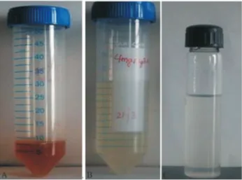
In the present study, the AgNO3solution immediately
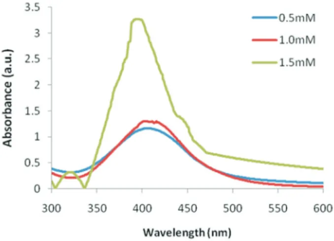
turned dark brown after the addition of P. garnatum peel ex-tract as a reducing and stabilizing agent in all of the samples of different NaOH molar concentrations, which shows the formation of AgNPs (Figure 1). The oxidation reaction of phenol groups (Figure 2 a-d) in peel extract was responsible for the reduction of silver ions (Wanget al., 2007; Soun-darrajanet al., 2012). It is observed that addition of 0.5 mM (I) NaOH showed broadening of the surface plasmon reso-nance (SPR) peak at 406 nm. Whereas, addition of 1.0 mM (II) NaOH shifted the absorption peak at 401 nm and 1.5 mM (III) NaOH resulted in a blue shift of lmax to 395 nm (Figure 3). Study revealed that increase in NaOH concentration may accelerate the nucleation process which increases the absorption intensity and the shifting of ab-sorption peaks may be due to decrease in the particle size of Ag-NPs (Vasireddy et al., 2012). Further, the phenolic groups of flavonoids and glycosides (Figure 2 a-d) of P. granatumpeels(Van Elswijket al., 2004; Jasujaet al., 2012) act as a reducing agent may be ionized at higher mo-lar NaOH concentration which leads rapid reduction reac-tion and synthesized spherical particles of AgNPs. The mechanistic reaction of the formation of AgNPs is ex-pressed in Figure 4.
The electrons moves freely in conduction band and valence band which lie very close to each other in Metal NPs i.e. AgNPs. The collective oscillations of electrons
(Plasmon) generate surface plasmon resonance (SPR) ab-sorption band (Taleb et al., 1998; Noginov et al., 2007; Link and El-Sayed, 2003; Kreibig and Vollmer, 1995) oc-curring due to the resonance with the incident light wave (Nathet al., 2007). The electric field of an incident wave in-duces a polarization of these electrons with respect to much heavier ionic core of AgNPs (Daset al., 2009). UV-Visible wave induces a polarization of the loosely bound surface electron due to low penetration depth (approximate 50 nm). As a result the net charge difference take place which acts as a restoring force. This creates a dipolar oscillation of all the electrons with the same phase (Inbakandanet al., 2010). A strong absorption takes place when the frequency of the electromagnetic field becomes resonant with the coherent electron motion, which may be the origin of dark brown colour. Due to the localized SPR, Metal NPs Shows strong absorption peak while bulk metal particles shows propagat-ing SPR. This absorption strongly depends on the particle size, dielectric medium and chemical surroundings (Nogi-novet al., 2007; Link and El-Sayed, 2003; Umashankariet al., 2012). The UV/Vis absorption spectra of the silver nano particles dispersed in water is shown in the Figure 1. When the size of particles is smaller than the average free path of the electrons (52 nm for silver metal (Abdullinet al., 1998; Henglein, 1998), silver dielectric function modifies which leads to an increased Plasmon bandwidth with decreasing size of particle (Basetet al., 2011).
SEM and TEM analysis
Figures 5 (A) and (B) showed the SEM and typical bright-field TEM micrographs of the synthesized AgNPs. The micrographs of AgNPs found polydisperse and mostly spherical in shape. In some places, Agglomeration of AgNPs may be due to possible sedimentation at a later time. The average size estimated was 15 nm for AgNPs. It is reported earlier that proteins can bind to nanoparticles ei-ther through free amine groups and ei-therefore, stabilization of the AgNPs by protein is a possibility (Daniel and Astruc, 2004; Kawsaret al., 2009; Shahverdiet al., 2007; Chienet al., 2007; Ahmad and Sharma, 2012).
Antibacterial activity
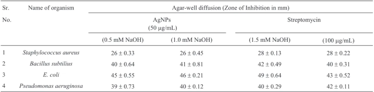
The study demonstrated the synergistic activity of AgNPs against gram-positive and gram-negative bacteria. The maximum inhibitory effects of AgNPs observed when prepared with higher NaOH (1.5 mM) molar concentration. The study revealed that AgNPs (50mg/mL) had shown sig-nificant inhibitory effect against E.coli and Bacilus subtiliusi.e. 49 ±0.64 and 42 ±0.49 mm when compared with Streptomycin (100mg/mL) i.e.43 ±0.52 and 40±0.31 mm respectively Figure 6 (a-d) and Table 1.
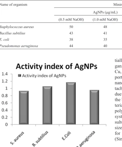
The MIC value of 30 mg Ag/mL was observed in E. coli. Whereas,Staphylococcus aureus,Bacillus subtilius and Pseudomonas aeruginosa had shown 45 mg/mL,
38 g/mL, 35mg/mL MIC respectively (Table 2). AgNPs had shown more than 1 active index for E. coli and Bacillus subtiliuswhen compared with standard drug (Fig-ure 7).
The screening results indicated that AgNPs disc (50mg/mL) were more active against gram-negative bacte-riai.e. Escherichia coliwith a mean zone of inhibition 49 ±0.64 mm (Table 1). This may be due to the differences in
the cell wall of gram-positive and gram negative bacteria. The cell wall of gram positive bacteria is wider than the gram-negative bacteria (Thielet al., 2007; Martinez-Casta-nonet al., 2008; Kimet al., 2007). Gram negative bacteria have a layer of lipopolysaccharide (LPS) which surrounded by a thin layer of peptidoglycan (7-8 nm) (Kreibig and Vollmer, 1995). The overall charge of bacterial cells at bio-logical pH values is negative because of excess number of carboxylic groups, which upon dissociation make the cell surface negative (Raffiet al., 2008). Weak positive charges present on silver nanoparticles (Schultzet al., 2000) are at-tracted towards negative charges on the LPS. Moreover, Excess formation of free radicals may attack LPS which lead to a breakdown of membrane function. Increased per-meability of the cell membrane or leakage of cell contents could be caused by Reactive Oxygen Species (ROS) (Men-diset al., 2005). This also leads morphological changes of bacterial cells and growth inhibition (Amroet al., 2000; Danilczuket al., 2006; Sondi and Salopek-Sondi, 2004). It is logical to state that binding of the nanoparticles to the bacteria depends on the surface area available for interac-tion. Nanoparticles have larger surface area available for interaction which enhances bactericidal effect than the large sized particles (Raffiet al., 2008) eg. Inorganic sub-stances or antibiotics; hence AgNPs exhibits more toxicity to the microorganism (Bakeret al., 2005).
Figure 2- (a-d) Flavonoids and their glycosides fromP. granatumpeels (Van Elswijket al., 2004; Jasujaet al., 2012).
Figure 3- UV-Visible spectra of AgNPs prepared at different NaOH (0.5 mM, 1.0 mM, and 1.5 mM) molar concentrations.
Figure 5- (a) Scanning electron micrograph of AgNPs synthesized by green methods (b) Transmission Electron Microscopy (TEM) image of AgNPs (scale bar 100 nm).
Figure 6- (a-d) Antibacterial Activities of Streptomycin sulphate disc (1 mg/10 mL) on (a) Staphylococcus aureus (b) Bacillus subtilius
(c)E.coli(d)Pseudomonas aeruginosa. (e-h) Antibacterial Activities of AgNPs (60mg/10 mL) on (e)Staphylococcus aureus(f)Bacillus subtilius
(g)E. coli(h)Pseudomonas aeruginosa.
Table 1- Antibacterial activity of Streptomycin (100mg/mL) and AgNPs (100mg/mL) against bacterial species tested by disc diffusion assay.
Sr. Name of organism Agar-well diffusion (Zone of Inhibition in mm)
No. AgNPs
(50mg/mL)
Streptomycin
(0.5 mM NaOH) (1.0 mM NaOH) (1.5 mM NaOH) (100mg/mL)
1 Staphylococcus aureus 26±0.33 26±0.45 28±0.13 28±0.22
2 Bacillus subtilius 40±0.64 41±0.81 42±0.49 40±0.31
3 E. coli 45±0.55 46±0.21 49±0.64 43±0.52
Conversely, the cell wall in gram-positive bacteria is composed of a thick layer (about 20-80 nm) of peptido-glycan, consisting of linear polysaccharide chains cross-linked by short peptides to form a three dimensional rigid structure (Wileyet al., 2006). The rigidity and extended cross-linking not only provide the cell walls with fewer an-choring sites for the silver nanoparticles but also make them difficult to penetrate. Earlier studies also revealed that silver species release Ag+ ions which interact with the thiol groups of bacterial proteins, may retard or change the repli-cation of DNA (Mariniet al., 2007; Martinez-Castanonet al., 2008). Somehow, it may be the reason that the G+ Ba-cillus subtiliusalso inhibited by AgNPs significantly when compared with control antibiotics.
Conclusions
It is concluded that the extract ofP. granatumare ca-pable of producing stable AgNPs by reduction of aqueous Ag+ ions in to Ag0. This green chemistry approach toward the synthesis of AgNPs has various advantages i.e rapid re-duction, economic viability etc. Applications of such eco-friendly nanoparticles in bactericidal, wound healing, med-ical and electronic applications, makes this method
poten-tially exciting for the large-scale synthesis of other inor-ganic nanomaterials (Ankannaet al., 2010)e.g.Au, Fe, Zn, Cu, Graphenes etc. The increase in zone of inhibition re-ported in this study was dependent on the concentration of nanoparticles due to higher NaOH molar concentration. At-tachment of nanoparticles by cell wall of bacteria would be due to negative charges and specific functional groups on the bacterial surface. AgNPs after penetration into the bac-terial cell may disturb the rigidity of cell wall or lipo-polysaccharides membrane, inactivate their transport system, enzymes functioning, generate H2O2 which
re-sulted in bacterial death. The silver nanoparticles synthe-sized via green route are highly toxic to G-ve and somehow for G+ve bacteria can be used in medical applications (Singhet al., 2010).
Acknowledgments
The authors are sincerely thankful to Mr. Sunil Sharma, Chancellor and Dr. Sudhanshu Sharma, Chief Mentorof Suresh Gyan Vihar University for providing a platform for this research. The authors also appreciation vows to USIC, University of Rajasthan, Jaipur, India for providing SEM and TEM facilities.
References
Abdullin SN, Stepanov AL, Osin YU, Khaibullin IB (1998) Ki-netics of silver nanoparticle formation in a viscous-flow polymer. Surface science 395:242-245.
Ahmad N, Sharma S (2012) Biosynthesis of silver nanoparticles from biowaste pomegranate peels. International Journal of Nanoparticles 5:185-195.
Amro NA, Kotra LP, Mesthrige KW, Bulychev A, Mobashery S, Liu G (2000) High-resolution atomic force microscopy stud-ies of theEscherichia coliouter membrane: Structural basis for permeability. Langmuir 16:2789-2796.
Ankanna S, Prasad TNVKV, Elumalai EK, Savithramma N (2010) Production of biogenic silver nanoparticles using boswellia ovalifoliolata stem bark. Digest Journal of Nano-materials & Biostructures 5:369-372.
Baker C, Pradhan A, Pakstis L, Pochan DJ, Shah SI (2005) Syn-thesis and antibacterial properties of silver nanoparticles. J Nanosci Nanotechnol 5:244-249.
Baset S, Akbari H, Zeynali H, Morteza S (2011) Size measure-ment of metal and semiconductor nanoparticles via uv-vis
Figure 7- Activity index of AgNPs compared with Streptomycin.
Table 2- Minimum inhibition concentrations (MIC) of AgNPs at different NaOH molar concentration.
Name of organism Minimum inhibition concentration
AgNPs (mg/mL) AgNO3(mg/mL) Peel extract (mg/mL) (0.5 mM NaOH) (1.0 mM NaOH) (1.5 mM NaOH)
Staphylococcus aureus 50 48 45 120 0.40
Bacillus subtilius 43 41 38 108 0.50
E. coli 38 35 30 102 0.85
absorption spectra. Digest J Nanomater Biostructures 6:709-716.
Bhadauria S, Kumar P (2012) Broad spectrum antidermatophytic drug for the control of tinea infection in human beings. Mycoses 55:339-343.
Binupriya AR, Muthuswamy S, Soon-In Y (2010) Myco-crystallization of silver ions to nano-sized particles by live and dead cell filtrates ofAspergillus oryzaevar viridis and its bactericidal activity towards Staphylococcus aureus KCCM 12256. Ind Eng Chem Res 49:852-858.
Catauro M, Raucci MG, De Gaaetano F, Marotta A (2004) Anti-bacterial and bioactive silver-containingNa2O•CaO•2SiO2
glass prepared by sol-gel method. J Mater Sci 15:831-837. Chien SW, Wang MC, Huang CC, Seshaiah K (2007)
Character-ization of humic substances derived from swine manure-based compost and correlation of their characteristics with reactivities with heavy metals. J Agric Food Chem 55:4820-4827.
Daniel MC, Astruc D (2004) Gold Nanoparticles: Assembly, Supramolecular Chemistry, Quantum-Size-Related Pro-perties and Applications toward Biology, Catalysis and Nanotechnology. Chem Rev 104:293-346.
Danilczuk M, Lund A, Sadlo J, Yamada H, Michalik J (2006) Conduction electron spin resonance of small silver particles. Spectrochimica Acta A 63:189-191.
Das R, Nath SS, Chakdar D, Gope G, Bhattacharjee R (2009) Preparation of silver nanoparticles and their characteriza-tion. J Nanotechnol 5:1-6.
Evanoff Jr DD, Chumanov G (2005) Synthesis and optical proper-ties of silver nanoparticles and arrays. Chem Phys Chem 6:1221-1231.
Gan PP, Ng SH, Huang Y, Li SF (2012) Green synthesis of gold nanoparticles using palm oil mill effluent (POME): A low-cost and eco-friendly viable approach. Bioresour Technol 113:132-137.
Gould JC (1952) The determination of bacterial sensitivity of anti-biotics. Edinburgh Med J 59 :178-199.
Henglein A (1998) Colloidal silver nanoparticles: Photochemical preparation and interaction with O2, CCl4, and some metal
ions. Chem Mater 10:444-450.
Inbakandan D, Venkatesan R, Khan SA (2010) Biosynthesis of gold nanoparticles utilizing marine sponge Acanthella elongata (Dendy, 1905). Colloids Surf B Biointerfaces 81:634-639.
Jain D, Daima HK, Kachnwaha S, Kothari SL (2009) Synthesis of plant-mediated silver nanoparticles using papaya fruit ex-tract and evaluation of their anti microbial activities. Digest Journal of Nanomaterials and Biostructures 4:557-563. Jasuja ND, Saxena R, Chandra S, Sharma R (2012)
Pharmacolog-ical characterization and beneficial uses of Punica granatum. Asian J Plant Sci 11:251-267.
Kawsar SMA, Mostafa G, Huq E, Nahar N, Ozeki Y (2009) Chemical Constituents and Hemolytic Activity of Macro-tyloma uniflorum L. International Journal of Biological Chemistry 3:42-48.
Kim JS, Kuk E, Yu KN, Kim JH, Park SJ, Lee HJ, Kim SH, Park YK, Park YH, Hwang CY, Kim YK, Lee YS, Jeong DH, Cho MH (2007) Antimicrobial effects of silver nano-particles. Nanomedicine 3:95-101.
Kreibig U, Vollmer M (1995) Optical Properties of Metal Clusters Springer Berlin, 535.
Krishnamurthy NB, Nagaraj B, Barasa M, Liny P, Dinesh R (2012) Green synthesis of gold nanoparticles using Tagetes erectal (mari gold) flower extract and evaluation of their antimicrobial activities. Int J Pharm Bio Sci 3:212-221. Latha PS, Kannabiran K (2006) Antimicrobial activity and
phyto-chemicals of Solanum trilobatumLinn. Afr J Biotechnol 5:2402-2404.
Link S, El-Sayed MA (2003) Optical Properties and ultrafast dy-namics of metallic nanocrystals. Annu Rev Phys Chem 54:331-366.
Marini M, De Niederhausern N, Iseppi R, Bondi M, Sabia C, Toselli M, Pilati F (2007) Antibacterial activity of plastics coated with silver-doped organic-inorganic hybrid coatings prepared by sol-gel processes. Biomacromolecules 8:1246-1254.
Martinez-Castanon GA, Nino-Martinez N, Martinez-Gutierrez F, Martinez-Mendoza JR, Ruiz F (2008) Synthesis and anti-bacterial activity of silver nanoparticles with different sizes. Journal of Nanoparticle Research 10:1343-1348.
Mendis E, Rajapakse N, Byun HG, Kim SK (2005) Investigation of jumbo squid (Dosidicus gigas) skin gelatin peptides for their in vitro antioxidant effects. Life Sci 77:2166-2178. Nath SS, Chakdar D, Gope G (2007) Synthesis of CdS and ZnS
quantum dots and their applications in electronics. Nano-trends 2:1-5.
Noginov MA, Zhu G, Bahoura M, Adegoke J, Small C (2007) The effect of gain and absorption on surface plasmos in metal nanoparticles. Applied Physics B-Lasers and Optics 86:455-460.
Qi L, Xu Z, Jiang X, Hu C, Zou X (2004) Preparation and antibac-terial activity of chitosan nanoparticles. Carbohydr Res 339:2693-2700.
Quelemes PV, Araruna FB, de Faria BE, Kuckelhaus SA, da Silva DA, Mendonça RZ, Eiras C, Dos S Soares MJ, Leite JR (2013) Development and antibacterial activity of cashew gum-based silver nanoparticles. Int J Mol Sci14:4969-4981. Raffi M, Hussain F, Bhatti TM, Akhter JI, Hameed A, Hasan MM
(2008) Antibacterial characterization of silver nanoparticles against E coli ATCC-15224. J Mater Sci Technol 24:192-196.
Rios JL, Recio MC, Villar A (1988) Screening methods for natu-ral products with antimicrobial activity: a review of the liter-ature. J Ethnopharmacol 23:127-149.
Ruparelia JP, Chatterjee AK, Duttagupta SP, Mukherji S (2008) Strain specificity in antimicrobial activity of silver and cop-per nanoparticles. Acta Biomaterialia 4:707-716.
Saxena A, Tripathi RM, Singh RP (2010) Biological synthesis of silver nanoparticles by using onion (Allium cepa) extract and their antibacterial activity. Digest J Nanomater Bio-structures 5:427-432.
Schultz S, Smith DR, Mock JJ, Schultz DA, (2000) Single-target molecule detection with no bleaching multicolor optical immunolabels. Proc Natl Acad Sci 97:996-1001.
Shahverdi AR, Minaeian S, Shahverdi HR, Jamalifar H, Nohi AA (2007) Rapid synthesis of silver nanoparticles using culture supernatants of Enterobacteria: A novel biological ap-proach. Process Biochemistry 42:919-923.
anti-microbial activities. Digest Journal of Nanomaterials & Biostructures (DJNB) 5:483-489.
Sondi I, Salopek-Sondi B (2004) Silver nanoparticles as anti-microbial agent: a case study on E coli as a model for Gram-negative bacteria. J Colloid Interface Sci 275:177-182.
Soundarrajan C, Sankari A, Dhandapani P, Maruthamuthu S, Ravichandran S, Sozhan G, Palaniswamy N (2012) Rapid biological synthesis of platinum nanoparticles using Ocimum sanctum for water electrolysis applications. Bio-process Biosyst Eng 35:827-833.
Stevanovic M, Savanovic I, Uskokovic V, Skapin SD, Bracko I, Jovanovic U, Uskokovic D (2012) A new, simple, green and one-pot four-component synthesis of bare and poly-(a,g,l-glutamic acid)-capped silver nanoparticles. Colloid Polym Sci 290:221-231.
Taleb A, Petit C, Pileni MP (1998) Optical Properties of self as-sembled 2D and 3D superlattices of silver nanoparticles. J Phys Chem B 102:2214-2220.
Thiel J, Pakstis L, Buzby S, Raffi M, Ni C, Pochan DJ, Shah SI (2007) Antibacterial properties of silver-doped titania. Small 3:799-803.
Umashankari J, Inbakandan D, Ajithkumar TT, Balasubramanian T (2012) Mangrove plant, Rhizophora mucronata (Lamk, 1804) mediated one pot green synthesis of silver nano-particles and its antibacterial activity against aquatic patho-gens. Aquat Biosyst 8:11.
Van Elswijk DA, Schobel UP, Lansky EP, Irth H, van de Greef J (2004) Rapid dereplication of estrogenic compounds in po-megranate (Punica granatum) using on-line biochemical de-tection coupled to mass spectrometry. Phytochemistry 65:233-241.
Vasireddy R, Paul R, Mitra AK (2012) Green synthesis of silver nanoparticles and the study of optical properties. Nanomater Nanotechnol 2:1-6.
Vijayakumar M, Priya K, Nancy FT, Noorlidaha A, Ahmed ABA (2013) Biosynthesis, characterisation and anti-bacterial ef-fect of plant-mediated silver nanoparticles using Artemisia nilagirica. Ind Crops Prod 41:235-240.
Vivek M, Kumar PS, Steffi S, Sudha S (2011) Biogenic silver nanoparticles by Gelidiella acerosa extract and their anti-fungal effects. Avicenna J Med Biotechnol 3:143-148. Wang W, Chen Q, Jiang C, Yang D, Liu X, Xu S (2007) One step
synthesis of biocompatible gold nanoparticles using gallic acid in the presence of poly-(N-vinyl-2-pyrrolidone). Colloids Surf A 301:73-79.
White GV, Kerscher P, Brown RM, Morella JD, McAllister W, Dean D, Kitchens CL (2012) Green synthesis of robust, biocompatible silver nanoparticles using garlic extract. J Nanomater 730746:1-12.
Wiley BJ, Im SH, Li ZY, McLellan J, Siekkkinen A, Xia Y (2006) Maneuvering the Surface Plasmon Resonance of Silver Nanoparticles through Shape- Controlled Synthesis. J Phys Chem B 110:15666-15675.