Oxidative Stress and Subclinical Inflammation in
Nonobese Women (Menopausal Status)
Jean Kyung Paik1,2., Ji Young Kim1,2., Oh Yoen Kim1,2
, Yonghee Lee2, Tae-Sook Jeong3, Gary Sweeney4, Yangsoo Jang1,5,6,7, Jong Ho Lee1,2*
1Yonsei University Research Institute of Science for Aging, Yonsei University, Seoul, Korea,2National Research Laboratory of Clinical Nutrigenetics/Nutrigenomics, Department of Food and Nutrition, College of Human Ecology, Yonsei University, Seoul, Korea,3National Research Laboratory of Lipid Metabolism and Atherosclerosis, Korea Research Institute of Bioscience and Biotechnology, Daejeon, Korea,4Institut Pasteur Korea, Seoul, Korea & Department of Biology, York University, Toronto, Canada,5Cardiology Division, Yonsei University College of Medicine, Seoul, Korea,6Cardiovascular Genome Center, Yonsei University College of Medicine, Seoul, Korea, 7Severance Medical Research Institute, Yonsei University College of Medicine, Seoul, Korea
Abstract
Background:This study aimed to determine the association of lipoprotein-associated phospholipase A2(Lp-PLA2) activity in
circulation and peripheral blood mononuclear cells (PBMCs) with inflammatory and oxidative stress markers in nonobese women and according to menopausal status. Lp-PLA2 activity, a marker for cardiovascular risk is associated with
inflammation and oxidative stress.
Methodology/Principal Findings: Eighty postmenopausal women (53.064.05 yr) and 96 premenopausal women (39.769.25 yr) participated in this study. Lp-PLA2 activities, interleukin (IL)-6, tumor necrosis factor (TNF)-a, and IL-1b in
plasma as well as in PBMCs were measured. Plasma ox-LDL was also measured. Postmenopausal women demonstrated higher circulating levels of ox-LDL and IL-6, as well as IL-6, TNF-a, and IL-1bin PBMCs, than premenopausal women. In both groups, plasma Lp-PLA2activity positively correlated with Lp-PLA2activity in PBMCs and plasma ox-LDL. In premenopausal
women, Lp-PLA2 activities in plasma and PBMCs positively correlated with IL-6, TNF-a, and IL-1b in PBMCs. In
postmenopausal women, plasma ox-LDL positively correlated with PBMC cytokine production. In subgroup analysis of postmenopausal women according to plasma ox-LDL level (median level: 48.715 U/L), a significant increase in Lp-PLA2
activity in the plasma but not the PBMCs was found in the high ox-LDL subgroup. Plasma Lp-PLA2 activity positively
correlated with unstimulated PBMC Lp-PLA2activity in the low ox-LDL subgroup (r = 0.627, P,0.001), whereas in the high
ox-LDL circulating Lp-PLA2 activity positively correlated with plasma ox-LDL (r = 0.390, P = 0.014) but not with Lp-PLA2
activity in PBMCs.
Conclusions/Significance:The lack of relation between circulating Lp-PLA2 activity and Lp-PLA2 activity in PBMCs was
found in postmenopausal women with high ox-LDL. This may indicate other sources of circulating Lp-PLA2activity except
PBMC in postmenopausal women with high ox-LDL. We also demonstrated that circulating Lp-PLA2and PBMC secreted
Lp-PLA2associate differently with markers of oxidative stress and sub clinical inflammation in nonobese women, particularly
according to the menopausal states.
Citation:Paik JK, Kim JY, Kim OY, Lee Y, Jeong T-S, et al. (2012) Circulating and PBMC Lp-PLA2Associate Differently with Oxidative Stress and Subclinical Inflammation in Nonobese Women (Menopausal Status). PLoS ONE 7(2): e29675. doi:10.1371/journal.pone.0029675
Editor:Philippe Rouet, I2MC INSERM UMR U1048, France
ReceivedApril 18, 2011;AcceptedDecember 2, 2011;PublishedFebruary 16, 2012
Copyright:ß2012 Paik et al. This is an open-access article distributed under the terms of the Creative Commons Attribution License, which permits unrestricted use, distribution, and reproduction in any medium, provided the original author and source are credited.
Funding:This work was supported by the National Research Foundation, Ministry of Education, Science and Technology (2010-0000317, 2010-0015017, and M10642120002-06N4212-00210), Seoul, Korea. The funders had no role in study design, data collection and analysis, decision to publish, or preparation of the manuscript.
Competing Interests:The authors have declared that no competing interests exist. * E-mail: [email protected]
.These authors contributed equally to this work.
Introduction
Lipoprotein-associated phospholipase A2(Lp-PLA2), also known
as plasma platelet activating factor acetylhydrolase (PAF-AH), is unique among members of the phospholipase A2superfamily due to
its origin, its association with circulating lipoproteins, and its substrate preference for polar phospholipids, including those generated during the oxidation of low-density lipoprotein (LDL) [1]. Lp-PLA2is secreted by monocytes, macrophages, T
lympho-cytes, and mast cells [2], and catalyzes the hydrolysis of oxidized LDL (ox-LDL) [3], which produces the proinflammatory mediators lysophosphatidylcholine and oxidized fatty acid [3]. There has been a growing interest in Lp-PLA2 because of its key role in lipid
metabolism and in initiating inflammation [4]. Epidemiological and clinical studies have indicated that Lp-PLA2 is a marker for
cardiovascular risk, with higher plasma Lp-PLA2mass or activity
Many studies found correlations between Lp-PLA2 and
triglycerides, LDL-cholesterol, high-density lipoprotein (HDL)-cholesterol, body mass index (BMI), age, sex, use of postmeno-pausal hormones, and smoking [6,9,12–19]. Lp-PLA2 has been
associated with an increased incidence of ischemic stroke among nonusers of hormone therapy in postmenopausal women indepen-dent of traditional cardiovascular risk factors. Furthermore, Keyzer et al. [20] found a positive association between Lp-PLA2activity
and inflammation and oxidative stress in a hypercholesterolemic swine model for atherosclerosis. Wang et al. [21] reported the stimulatory effect of ox-LDL on the expression of Lp-PLA2 in
monocytes, which are a primary source of this enzyme. These recent findings in animal and in vitro studies may provide insight into the interaction between Lp-PLA2activity and oxidative stress
in the context of atherosclerosis. Therefore, our aim was to study the relationship of Lp-PLA2 activity in plasma and the enzyme
activity in supernatants from nonstimulated peripheral blood mononuclear cell (PBMC) cultures. Plasma ox-LDL and cytokine production from PBMCs in healthy nonobese women and also according to the menopausal status were evaluated.
Methods
Study participants
A total of 176 healthy, nonobese women aged 20–68 years were recruited during routine check-ups at a health promotion center at Yonsei University Hospital. Postmenopausal status (n = 80) was defined as an absence of menstruation for at least 12 months and the presence of estrogen deficiency symptoms, including hot flushes, increased sweating, nervousness, irritability, depression, palpitations, insomnia, headaches, dyspareunia, and joint pains. Premenopausal status (n = 96) was defined as the presence of regular menses. At the time of subject enrollment, subjects were interviewed about smoking status (non-/ex-smoker and current smoker), and frequency of alcohol intake. Alcohol consumption was calculated as the grams of ethanol ingested per day and cigarettes smoking data were reported as the number of cigarettes smoked per day. All participants were clinically healthy and were not taking any medications known to affect the immune system, such as oral contraceptives, lipid-lowering agents, anti-hyperten-sive drugs, functional foods, or vitamin and/or mineral supple-ments. The purpose of the study was carefully explained to all participants and their written consent was obtained prior to their participation. The study design was approved by the Institutional Review Board of Yonsei University.
Anthropometric parameters, blood pressure, and blood collection
Body weight and height were measured in the morning, lightly clothed without shoes and the BMI was calculated as body weight in kilograms divided by height in meters squared. Waist circumference was measured at the umbilical level with the subjects standing after normal expiration and the hip girth was measured at the widest part of the hip and, the waist and hip ratio (WHR) was calculated.
Blood pressure (BP) was measured in the left arm of seated patients with an automatic blood pressure monitor (TM-2654, A&D, Tokyo, Japan) after a 20-min rest. After a 12-hour fast, venous blood specimens were collected in EDTA-treated or untreated tubes. The blood specimens collected in the EDTA-treated tubes were used for the isolation of PBMCs or separated into plasma and stored at270uC until further analysis. The blood samples collected in non-treated tubes were separated into serum and stored until further analysis.
Serum lipid profile, fasting glucose, free fatty acid, and white blood cell count
Fasting total-cholesterol and triglyceride levels were measured using commercially available kits on a Hitachi 7150 Autoanalyzer (Hitachi Ltd., Tokyo, Japan). After precipitation of serum chylomicrons with dextran sulfate magnesium, the concentrations of LDL- and HDL-cholesterol in the supernatants were enzymat-ically measured. Fasting glucose levels were measured using a glucose oxidase method with a Beckman Glucose Analyzer (Beckman Instruments, Irvine, CA, USA). Free fatty acids were analyzed with a Hitachi 7150 autoanalyzer (Hitachi Ltd, Tokyo, Japan).White blood cell (WBC) count was determined using the HORIBA ABX diagnostic (HORIBA ABX SAS, Parc Euro-medicine, France).
Cytokine secretion in non-stimulated PBMCs
Whole blood was mixed with the same volume of RPMI 1640 (Gibco, Invitrogen, Carlsbad, CA, USA) and gently laid on a histopaque-1077 (Sigma-Aldrich, St. Louis, MO, USA). The sample was then centrifuged at 2000 rpm for 20 min at 10uC. After the separation, a thin layer of PBMCs was isolated and washed twice with RPMI 1640. The pellet was resuspended in RPMI 1640 with streptomycin. Isolated PBMCs were cultured in RPMI 1640 supplemented with 10% fetal bovine serum, seeded in 12-well plates (16106cells/mL; Nunc, Roskilde, Denmark), and incubated at 37uC with 5% CO2for 24 hours. After a 24-hour
incubation, supernatants were collected and stored at280uC until interleukin (IL)-1b, IL-6, tumor necrosis factor (TNF)-a, and Lp-PLA2activity levels were assayed [22,23].
Cytokine assay for IL-1b, IL-6, and TNF-alevels in serum and PBMC supernatants
Levels of IL-1b, IL-6, and TNF-a in serum and PBMC supernatants were measured using the Bio-PlexTMReagent Kit on the Bio-PlexTM (Bio-Rad Laboratories, Hercules, CA, USA), according to the manufacturer’s instructions.
Lp-PLA2activity in plasma and PBMC supernatants
Lp-PLA2 activity in plasma and PBMC supernatants was
measured by using a modification of a previously described high-throughput radiometric activity assay [11].
Plasma-oxidized LDL and serum high sensitivity-C-reactive protein (hs-CRP)
Plasma ox-LDL was measured using an enzyme immunoassay (Mercodia, Uppsala, Sweden). The resulting color reaction was read at 450 nm with a Wallac Victor2multilabel counter (Perkin Elmer Life Sciences, Turku, Finland). Serum hs-c-reactive protein (CRP) levels were measured with an Express PlusTMauto-analyzer (Chiron Diagnostics Co., Walpole, MA, USA) using a commer-cially available, high-sensitivity CRP-Latex(II) 62 kit (Seiken Laboratories Ltd., Tokyo, Japan).
Urinary 8-epi-prostaglandin F2a(8-epi-PGF2a) levels
Serum follicle stimulating hormone (FSH) and 17ß-estradiol levels
Serum levels of FSH and 17ß-estradiol were measured using commercially-available kits (ADIVIA Centaur FSH and ADIVIA Centaur Estradiol, respectively, Siemens, USA) on the ADIVIA Centaur (ADIVIA Centur, Siemens).
Data analysis
Statistical analyses were performed using SPSS version 12.0 for Windows (SPSS Inc., Chicago, IL, USA). The independent t-test was used to compare parameters between the two groups. One-way analysis of variance (ANOVA) with Bonferroni correction was used to test whether there were effects from menopausal state and plasma ox-LDL levels (below or above the median level) in postmenopausal women. General linear model (GLM) analysis was also performed with adjustment for age or BMI and alcohol consumption. Frequency was tested with the chi-square test. Pearson and partial correlation coefficients were used to examine relationships between variables. For descriptive purposes, mean values are presented using untransformed values. Results are expressed as the mean6standard deviation (SD). A two-tailed value of P,0.05 was considered statistically significant.
Results
Clinical characteristics of study participants
In this study, postmenopausal women had a significantly higher BMI and included a lower percentage of alcohol drinkers (Table 1). Before and after adjusting for age, BMI and percentage of alcohol drinkers, postmenopausal women demonstrated significantly higher WHRs in addition to higher serum levels of total-cholesterol, LDL-total-cholesterol, and glucose, and lower serum levels of hs-CRP, than premenopausal women. Postmenopausal women also had significantly lower serum levels of 17b-estradiol and higher FSH levels than premenopausal women. These differences confirmed that postmenopausal women had estrogen deficiency. Premenopausal and postmenopausal women did not differ in terms of diastolic BP, or serum levels of triglycerides, HDL-cholesterol, and free fatty acid. Premenopausal and postmeno-pausal women also did not differ in terms of the number of circulating leukocytes (Table 1).
Oxidative stress markers, cytokines, and Lp-PLA2activity
according to menopausal status
Table 2 provides data for oxidative stress markers, circulating levels of cytokines, and Lp-PLA2 activity in the plasma and in
supernatants from non-stimulated PBMC cultures from premen-opausal and postmenpremen-opausal women. Significant differences between the two groups are indicated in the last column before and after adjustment for age, BMI and percentage of alcohol drinkers. Plasma ox-LDL and serum IL-6 levels in the postmenopausal group were significantly higher than those in premenopausal women. Urinary levels of 8-epi-PGF2a, serum levels of TNF-aand IL-1b, and Lp-PLA2activities in plasma and
cultured nonstimulated PBMC supernatants did not significantly differ between the two groups. Additionally, plasma Lp-PLA2
activity was not significantly difference between the two groups after further adjustment for LDL-cholesterol (p = 0.746). Non-stimulated PBMCs from postmenopausal women secreted significantly higher amounts of IL-6, TNF-a, and IL-1b into the culture media than those from premenopausal women (Table 2).
Correlations among oxidative stress markers, cytokines, and Lp-PLA2activity in the circulation and in PBMCs
according to menopausal status
In both premenopausal and postmenopausal women, plasma Lp-PLA2activity positively correlated with plasma ox-LDL and
supernatant Lp-PLA2 activity from nonstimulated PBMC
cultures (Table 3). In premenopausal women, Lp-PLA2activity
in plasma and PBMC supernatants positively correlated with IL-6, TNF-a, and IL-1blevels in nonstimulated PBMCs. After the adjustment for age, BMI, alcohol consumption, the relationships were still retained (IL-6 and IL-1b). In postmenopausal women, plasma ox-LDL positively correlated with IL-6, TNF-a, and IL-1blevels in cultured nonstimulated PBMC supernatants. After the adjustment, the relationships were still retained (TNF-aand IL-1b) (Table 3). IL-6, TNF-a, and IL-1b levels in nonstimu-lated PBMCs were linearly interrenonstimu-lated for both premenopausal and postmenopausal women (data not shown). In the postmen-opausal women, serum IL-6 levels positively correlated with serum IL-1b(r = 0.396, P,0.001) and serum TNF-a(r = 0.296, P = 0.012). Serum IL-6 levels negatively correlated with PBMC levels of IL-6 (r =20.299, P = 0.005), TNF-a (r =20.359, P = 0.001), and IL-1b(r =20.302, P = 0.005) in premenopausal women, whereas serum IL-6 levels positively correlated with PBMC levels of IL-6 (r = 0.317, P = 0.006), TNF-a (r = 0.313, P = 0.007), and IL-1b (r = 0.316, P = 0.006) in postmenopausal women.
Lp-PLA2activity, oxidative stress markers, and
LDL-cholesterol according to menopausal status and plasma ox-LDL levels
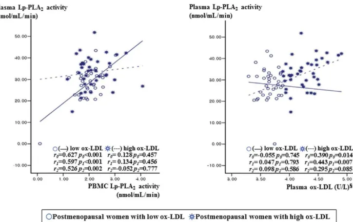
Since Lp-PLA2 is known to hydrolyze the sn2 ester bond of
oxidized phospholipids including ox-LDL [12], the postmeno-pausal women were subdivided into two groups according to plasma ox-LDL level: high ox-LDL ($48.715 U/L, n = 40) and low ox-LDL (,48.715 U/L, n = 40) according to the median level of LDL. In the postmenopausal women with low ox-LDL, plasma Lp-PLA2 activity positively correlated with
unstimulated PBMC Lp-PLA2 activity (r0= 0.627, P0,0.001;
r1= 0.597, P1,0.001) before and after the adjustment for age,
BMI, and alcohol consumption (Fig. 1). In postmenopausal women with high ox-LDL, however, circulating Lp-PLA2activity
positively correlated with plasma ox-LDL (r0= 0.390, P0= 0.014;
r1= 0.443, P1= 0.007) but not with Lp-PLA2activity in PBMCs.
Additionally partial correlation coefficient with adjust for age, BMI, alcohol consumption, and LDL-cholesterol level were also used to examine the relationships between variables. In the postmenopausal women with low ox-LDL, plasma Lp-PLA2
activity positively correlated with non-stimulated PBMC Lp-PLA2 activity (r2= 0.526, P2= 0.002) but not correlated with
plasma ox-LDL (r2= 0.098, P2= 0.586). In postmenopausal
women with high ox-LDL, the positive correlation between circulating Lp-PLA2 activity and plasma ox-LDL (r2= 0.295,
P2= 0.085) was a little bit attenuated, but is in a positive
direction. On the other hand, circulating Lp-PLA2 did not
correlation with Lp-PLA2 activity in PBMCs (r2=20.052,
P2= 0.777). The postmenopausal group with high ox-LDL had
Discussion
The major finding of this study is the lack of relation between circulating Lp-PLA2activity and Lp-PLA2activity in PBMCs in
postmenopausal women with high ox-LDL ($48.715 U/L, above median). A significant increase in Lp-PLA2activity in the plasma
but not the PBMCs of postmenopausal women with high ox-LDL may indicate other sources of Lp-PLA2 production except
PBMC. The extent of the increase in plasma Lp-PLA2 may
depend not only on the levels of lipoproteins carrying Lp-PLA2in
circulation but also on the cellular synthesis of this enzyme [24]. Monocytes, macrophages, T-lymphocytes, mast cells, and liver
cells are known as the main sources of Lp-PLA2 [25,26].
Recently, Keyzer et al. [20] found increased circulating Lp-PLA2activity with increased ox-LDL levels in
hypercholesterol-emic pigs and the main source of increased circulating Lp-PLA2
activity were plaque macrophages [20,27,28]. Therefore, the Lp-PLA2production in plaque macrophages could partly explain the
positive correlation of circulating Lp-PLA2 activity with plasma
ox-LDL but not with Lp-PLA2 activity in PBMCs from
postmenopausal women with high ox-LDL in this study. However, we could not measure Lp-PLA2 activity in plaque
macrophages, or the plaque or intima itself, where it may be of most biological relevance.
Table 1.Clinical characteristics of the study participants according to menopausal status.
Premenopausal women (n = 96) Postmenopausal women (n = 80) P0 P1
Age (yr) 39.7 6 9.25 53.0 6 4.05 ,0.001
-Years since menopause - 3.49 6 3.87 -
-Body Mass Index (kg/m2) 21.9
6 2.84 22.8 6 2.27 0.024
-Cigarette smoker, n (%) 1 (1.0) 2 (2.5) 0.592
-Alcohol drinker, n (%) 62 (64.6) 37 (46.3) 0.022
-Waist hip ratio 0.84 6 0.05 0.88 6 0.05 ,0.001 0.042
Systolic BP (mmHg) 109.3 6 14.2 118.6 6 12.0 ,0.001 0.614
Diastolic BP (mmHg) 74.2 6 10.6 76.0 6 8.75 0.238 0.592
Triglyceride (mg/dL)$ 90.7 6 41.6 98.2 6 45.4 0.489 0.340
Total-cholesterol (mg/dL) 182.6 6 26.4 210.3 6 32.7 ,0.001 0.004
LDL-cholesterol (mg/dL) 109.1 6 23.4 132.5 6 28.6 ,0.001 0.006
HDL-cholesterol (mg/dL) 55.4 6 12.8 58.1 6 14.1 0.185 0.145
Glucose (mg/dL)$ 85.5 6 7.98 92.1 6 12.3 ,0.001 0.016
Free fatty acid (uEq/L)$ 407.1 6 191.8 409.5 6 158.2 0.488 0.548
hs-CRP (mg/dL)$ 0.51 6 0.75 0.36 6 0.92 ,0.001 ,0.001
Serum FSH (IU/L) 10.6 6 17.9 75.2 6 28.9 ,0.001 ,0.001
Serum 17ß-estradiol (pg/mL)$ 134.6 6 126.9 18.2 6 24.8 ,0.001 ,0.001
White blood cells (6109/L)$ 4.94 6 1.01 4.98 6 1.15 0.785 0.948
Means6SD. Tested by independent t-test or general linear model with the adjustment.
$tested by log-transformed P
0: unadjusted, P1: adjusted for age, BMI, and alcohol consumption. doi:10.1371/journal.pone.0029675.t001
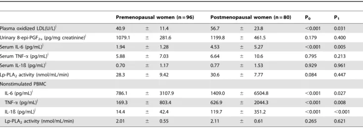
Table 2.Oxidative stress markers, cytokines, and Lp-PLA2activity according to menopausal status.
Premenopausal women (n = 96) Postmenopausal women (n = 80) P0 P1
Plasma oxidized LDL(U/L)$ 40.9 6 11.4 56.7 6 23.8 ,0.001 0.031
Urinary 8-epi-PGF2a(pg/mg creatinine)$ 1079.1 6 281.6 1199.8 6 461.5 0.179 0.400
Serum IL-6 (pg/mL)$ 1.94 6 1.28 4.53 6 5.27 ,0.001 0.005
Serum TNF-a(pg/mL)$ 5.88 6 7.03 6.64 6 10.6 0.795 0.213
Serum IL-1ß (pg/mL)$ 0.70 6 1.17 0.77 6 1.53 0.929 0.961
Lp-PLA2activity (nmol/mL/min) 28.3 6 9.42 30.6 6 7.77 0.084 0.447
Nonstimulated PBMC
IL-6 (pg/mL)$ 786.1 6 3107.9 1409.0 6 6504.8 ,0.001 0.027
TNF-a(pg/mL)$ 169.3 6 803.4 626.9 6 2044.3 ,0.001 0.008
IL-1ß (pg/mL)$ 14.4 6 42.4 119.7 6 351.2 ,0.001 ,0.001
Lp-PLA2activity (nmol/mL/min) 2.01 6 0.55 2.11 6 0.61 0.265 0.621
Mean6SD. Tested by independent t-test or general linear model with the adjustment.
$
Table 3.Correlations among oxidative stress markers, cytokines, and Lp-PLA2activity in the circulation and in PBMCs according to
menopausal status.
Premenopausal women (n = 96) Postmenopausal women (n = 80)
Plasma ox-LDL
Plasma Lp-PLA2 activity
Nonstimulated
PBMC Lp-PLA2 Plasma ox-LDL
Plasma Lp-PLA2 activity
Nonstimulated PBMC Lp-PLA2
r0 r1 r0 r1 r0 r1 r0 r1 r0 r1 r0 r1
Plasma ox- LDL(U/L)$ - - 0.244* 0.247* 0.022 0.006 - - 0.390*** 0.394** 0.215 0.154 Serum IL-6 (pg/mL)$ 0.042 0.051 20.150 20.154 20.020 20.032 0.171 0.143 0.022 0.016 0.019 20.017 Serum TNF-a(pg/mL)$ 20.037 20.016 20.178 20.202 0.101 0.068 0.090 0.031 20.022 20.018 0.030 20.008 Serum IL-1ß (pg/mL)$ 20.018 20.020 0.042 0.033 0.073 0.042 20.061 20.062 20.050 20.046 20.022 20.048 Lp-PLA2activity (nmol/
mL/min)
0.244* 0.247* - - 0.427*** 0.422*** 0.390*** 0.394** - - 0.380** 0.374**
Nonstimulated PBMCs
IL-6 (pg/mL)$ 0.058 0.042 0.326** 0.318** 0.363*** 0.353** 0.230* 0.222 0.051 0.073 0.127 0.121 TNF-a(pg/mL)$ 0.106 0.072 0.209* 0.202 0.205* 0.198 0.308** 0.307** 0.171 0.208 0.032 0.022 IL-1ß (pg/mL)$ 0.056 0.035 0.277** 0.269** 0.272** 0.255* 0.304** 0.301** 0.220 0.251* 0.080 0.067 Lp-PLA2activity (nmol/
mL/min)
0.022 0.006 0.427*** 0.422*** - - 0.215 0.154 0.380** 0.374** -
-Pearsonandpartialcorrelation analysis, r0: unadjusted, r1: adjusted for age, BMI, and alcohol consumption, *P,0.05,
**P,0.01, ***P,0.001,
$tested by log-transformed.
doi:10.1371/journal.pone.0029675.t003
Figure 1. Relationship between Lp-PLA2activity from PBMCs or plasma ox-LDL and plasma Lp-PLA2activity according to plasma
ox-LDL (below or above the median level of 48.715 U/L) in postmenopausal women.1tested by log-transformed. Tested by Pearson correlation (r0) or partial correlation analysis (r1, r2).r0: correlation coefficient, unadjusted.r1: correlation coefficient after adjusted for age, BMI, and
alcohol consumption.r2: correlation coefficient after adjusted for age, BMI, alcohol consumption, and LDL-cholesterol.
Lp-PLA2is thought to play an atherogenic role by hydrolyzing
oxidized phospholipids in ox-LDL, resulting in the generation of two bioactive lipid mediators, lysophosphatidyl choline, and oxidized free fatty acids [3,24,29]. The biological role of Lp-PLA2is also controversial; initial reports indicated an
antiathero-genic effect, whereas growing evidence has demonstrated a role for Lp-PLA2as a proinflammatory molecule and an independent risk
factor for CVD [5,11,14,30,31]. Lp-PLA2 belongs to the
expanding superfamily of structurally diverse phospholipase A2
enzymes, also known as PAF-AH [32]. It travels mainly with LDL in the blood, and less than 20% is associated with HDL [1]. In mice, the majority of plasma PAF-AH is bound to HDL, this was considered as possible antiatherogenic effect [33]. In human atherosclerotic lesions, two main sources of Lp-PLA2 were
identified in human atherosclerotic lesions, including that which is brought into the intima bound to LDL from the circulation, and that which is synthesized de novo by plaque inflammatory cells (i.e. macrophages, T cells, mast cells) [1,32,34]. Lp-PLA2hydrolyzes
the sn-2 ester bond of oxidized phospholipids, generating bioactive oxidized free fatty acids and lysophosphatidyl choline, which are potent proinflammatory and proatherogenic products [1]. Indeed, Stafforini et al. [35] showed that the secreted form of Lp-PLA2
released F2-isoprostanes, the end-products of lipid oxidation, from
the sn-2 position of phosphatidylcholine with high affinity. Kono et al. [36] reported that intracellular type II Lp-PLA2, which
shares homology with the plasma enzyme Lp-PLA2, is involved in
the metabolism of esterified 8-iso-PGF2a. We also showed that mean levels of plasma Lp-PLA2activity and urinary 8-epi-PGF2a were higher among postmenopausal women with high ox-LDL than those with low ox-LDL or in premenopausal women.
Ox-LDL stimulates Lp-PLA2expression in monocytes through
the pathway of phosphatidylinositol 3-kinase and p38 mitogen-activating protein kinase [21], which mediates the expression of
many genes involved in stress-induced responses (e.g., IL-1b) [37]. Shi et al. [24] found that the activation of leukocytes in the experimental model of diabetes and hypercholesterolemia is associated with a rapid increase in circulating Lp-PLA2 and an
upregulation in a range of inflammatory mediators, including IL-6, TNF-a, and IL-1b. In the postmenopausal women in this study, plasma ox-LDL also positively correlated with IL-6, TNF-a, and IL-1blevels in cultured, nonstimulated PBMC supernatants. This observation supports the previous finding that the presence of ox-LDL significantly induced the expression of IL-1b, IL-6, and TNF-a, whereas unmodified LDL had no effect on the expression of inflammatory mediators [24]. This correlation, however, was not found in premenopausal women. Additionally, the postmen-opausal women showed higher levels of plasma ox-LDL and serum IL-6, and more cytokine production from PBMCs than the premenopausal women. This result may be partly related to the exaggerated production of cytokines caused by estrogen depriva-tion after menopause. Rachon et al. [22] showed that estrogen deprivation after menopause enhanced IL-6 production by PBMCs and increased serum IL-6 levels in postmenopausal women. It has been also suggested that the increased production of cytokines in healthy, older individuals results from the loss of sex steroids [38]. Estradiol acts to inhibit pro-inflammatory cytokine gene expression, NF-kB binding, and production of proinflamma-tory cytokines [39,40]. In addition, the levels of IL-6, TNF-a, and IL-1b in supernatants from nonstimulated PBMC cultures of the postmenopausal women positively correlated with serum IL-6 levels [41]. This finding supports the previous suggestion that locally-produced TNF-a and IL-1b do not escape into the circulation, although they induce a strong systemic IL-6 response [41]. Therefore, enhanced cytokine production by PBMCs in postmenopausal women, may not only result from the activation of leukocytes in the presence of high ox-LDL after menopause but Figure 2. Lp-PLA2activity in plasma and supernatants from nonstimulated PBMC cultures and oxidative stress markers according
to menopausal status and plasma ox-LDL levels (below or above the median level of 48.715 U/L).Data are means6SD.1
tested by log-transformed. P0: unadjusted, tested by one-way ANOVA with Bonferroni method P1: adjusted for BMI and alcohol consumption, tested by general linear model (GLM) analysis. P2: adjusted for age, BMI, and alcohol consumption, tested by GLM analysis.
also may be the consequence of the decrease in estrogen levels or other factors that contribute to the aging process [12,22,39]. On the other hand, cytokine production particularly IL-6 and IL-1b
from PBMCs in premenopausal women, but not in postmeno-pausal women positively correlated with Lp-PLA2 activity in
plasma and PBMC supernatants. The biological role of Lp-PLA2
or the activity levels according to the menopausal states are still controversial, but our results may be partly explained by the recent studies demonstrating that Lp-PLA2works as an proinflammatory
molecules or initiate inflammatory response, and is an indepen-dent risk factor for CVD [5,11,14,30,31]. It is known that circulating Lp-PLA2activity is derived from atherosclerotic plaque
cells, however our study subjects had not carried cardiovascular disease including atherosclerosis or other chronic diseases (i.e. diabetes, or dyslipidemia). Therefore, we could observe the clear positive correlation in premenopausal women rather than postmenopausal women whose metabolisms are interfered by the stressful condition (i.e. oxidative stress, estrogen deprivation).
In addition, the prospective observation needs to be performed in the future to investigate if Lp-PLA2is a physiological responder
or an inducer of vascular inflammation. Actually, the Lp-PLA2
activity may be different according to the ethnicities, for example,
rare homozygous mutation of Lp-PLA2V279F polymorphism, the
F/F genotype indicating the loss of function of Lp-PLA2activity
and the less atherogenic properties is found in Korean but not in Western people [42]. Our study was designed for the cross-sectional observation, not for prospective observation, thus it is not easy to determine the casual relationship between Lp-PLA2 and
inflammatory response. In summary, the lack of relationship between circulating Lp-PLA2 activity and Lp-PLA2 activity in
PBMCs was found in postmenopausal women with high ox-LDL. This may indicate other sources of circulating Lp-PLA2activity
except PBMC in postmenopausal women with high ox-LDL. We also demonstrated that circulating Lp-PLA2and PBMC secreted
Lp-PLA2associate differently with markers of oxidative stress and
sub clinical inflammation in nonobese women, particularly according to the menopausal states.
Author Contributions
Conceived and designed the experiments: JKP JYK OYK YJ JHL GS. Performed the experiments: JKP JYK YL. Analyzed the data: JKP OYK. Contributed reagents/materials/analysis tools: T-SJ. Wrote the paper: JHL.
References
1. Zalewski A, Macphee C (2005) Role of lipoprotein-associated phospholipase A2 in atherosclerosis: biology, epidemiology, and possible therapeutic target. Arterioscler Thromb Vasc Biol 25: 923–931.
2. Caslake MJ, Packard CJ (2003) Lipoprotein-associated phospholipase A2 (platelet-activating factor acetylhydrolase) and cardiovascular disease. Curr Opin Lipidol 14: 347–352.
3. MacPhee CH, Moores KE, Boyd HF, Dhanak D, Ife RJ, et al. (1999) Lipoprotein-associated phospholipase A2, platelet-activating factor acetylhydro-lase, generates two bioactive products during the oxidation of low-density lipoprotein: use of a novel inhibitor. Biochem J 338: 479–487.
4. Mohler ER, 3rd, Ballantyne CM, Davidson MH, Hanefeld M, Ruilope LM, et al. (2008) The effect of darapladib on plasma lipoprotein-associated phospholipase A2 activity and cardiovascular biomarkers in patients with stable coronary heart disease or coronary heart disease risk equivalent: the results of a multicenter, randomized, double-blind, placebo-controlled study. J Am Coll Cardiol 51: 1632–1641.
5. Packard CJ, O’Reilly DS, Caslake MJ, McMahon AD, Ford I, et al. (2000) Lipoprotein-associated phospholipase A2 as an independent predictor of coronary heart disease. West of Scotland Coronary Prevention Study Group. N Engl J Med 343: 1148–1155.
6. Oei HH, van der Meer IM, Hofman A, Koudstaal PJ, Stijnen T, et al. (2005) Lipoprotein-associated phospholipase A2 activity is associated with risk of coronary heart disease and ischemic stroke: the Rotterdam Study. Circulation 111: 570–575.
7. Winkler K, Winkelmann BR, Scharnagl H, Hoffmann MM, Grawitz AB, et al. (2005) Platelet-activating factor acetylhydrolase activity indicates angiographic coronary artery disease independently of systemic inflammation and other risk factors: the Ludwigshafen Risk and Cardiovascular Health Study. Circulation 111: 980–987.
8. Gerber Y, McConnell JP, Jaffe AS, Weston SA, Killian JM, et al. (2006) Lipoprotein-associated phospholipase A2 and prognosis after myocardial infarction in the community. Arterioscler Thromb Vasc Biol 26: 2517–2522. 9. Brilakis ES, McConnell JP, Lennon RJ, Elesber AA, Meyer JG, et al. (2005)
Association of lipoprotein-associated phospholipase A2 levels with coronary artery disease risk factors, angiographic coronary artery disease, and major adverse events at follow-up. Eur Heart J 26: 137–144.
10. Winkler K, Hoffmann MM, Winkelmann BR, Friedrich I, Scha¨fer G, et al. (2007) Lipoprotein-associated phospholipase A2 predicts 5-year cardiac mortality independently of established risk factors and adds prognostic information in patients with low and medium high-sensitivity C-reactive protein (the Ludwigshafen risk and cardiovascular health study). Clin Chem 53: 1440–1447.
11. Kim JY, Hyun YJ, Jang Y, Lee BK, Chae JS, et al. (2008) Lipoprotein-associated phospholipase A2 activity is associated with coronary artery disease and markers of oxidative stress: a case-control study. Am J Clin Nutr 88: 630–637. 12. Hatoum IJ, Nelson JJ, Cook NR, Hu FB, Rimm EB (2010) Dietary, lifestyle, and
clinical predictors of lipoprotein-associated phospholipase A2 activity in individuals without coronary artery disease. Am J Clin Nutr 91: 786–793. 13. Detopoulou P, Nomikos T, Fragopoulou E, Panagiotakos DB, Pitsavos C, et al.
(2009) Lipoprotein-associated phospholipase A2 (Lp-PLA2) activity, platelet-activating factor acetylhydrolase (PAF-AH) in leukocytes and body composition in healthy adults. Lipids Health Dis 8: 19.
14. Koenig W, Khuseyinova N, Lo¨wel H, Trischler G, Meisinger C (2004) Lipoprotein-associated phospholipase A2 adds to risk prediction of incident coronary events by C-reactive protein in apparently healthy middle-aged men from the general population: results from the 14-year follow-up of a large cohort from southern Germany. Circulation 110: 1903–1908.
15. Persson M, Nilsson JA, Nelson JJ, Hedblad B, Berglund G (2007) The epidemiology of Lp-PLA(2): distribution and correlation with cardiovascular risk factors in a population-based cohort. Atherosclerosis 190: 388–396. 16. Iribarren C, Gross MD, Darbinian JA, Jacobs DR, Jr., Sidney S, et al. (2005)
Association of lipoprotein-associated phospholipase A2 mass and activity with calcified coronary plaque in young adults: the CARDIA study. Arterioscler Thromb Vasc Biol 25: 216–221.
17. Ballantyne CM, Hoogeveen RC, Bang H, Coresh J, Folsom AR, et al. (2005) Lipoprotein-associated phospholipase A2, high-sensitivity C-reactive protein, and risk for incident ischemic stroke in middle-aged men and women in the Atherosclerosis Risk in Communities (ARIC) study. Arch Intern Med 165: 2479–2484.
18. Yang EH, McConnell JP, Lennon RJ, Barsness GW, Pumper G, et al. (2006) Lipoprotein-associated phospholipase A2 is an independent marker for coronary endothelial dysfunction in humans. Arterioscler Thromb Vasc Biol 26: 106–111. 19. Khuseyinova N, Imhof A, Rothenbacher D, Trischler G, Kuelb S, et al. (2005) Association between Lp-PLA2 and coronary artery disease: focus on its relationship with lipoproteins and markers of inflammation and hemostasis. Atherosclerosis 182: 181–188.
20. De Keyzer D, Karabina SA, Wei W, Geeraert B, Stengel D, et al. (2009) Increased PAFAH and oxidized lipids are associated with inflammation and atherosclerosis in hypercholesterolemic pigs. Arterioscler Thromb Vasc Biol 29: 2041–2046.
21. Wang WY, Li J, Yang D, Xu W, Zha RP, et al. (2010) OxLDL stimulates lipoprotein-associated phospholipase A2 expression in THP-1 monocytes via PI3K and p38 MAPK pathways. Cardiovasc Res 85: 845–852.
22. Rachon´ D, Mys´liwska J, Suchecka-Rachon´ K, Wieckiewicz J, Mys´liwski A (2002) Effects of oestrogen deprivation on interleukin-6 production by peripheral blood mononuclear cells of postmenopausal women. J Endocrinol 172: 387–395. 23. von Haehling S, Genth-Zotz S, Sharma R, Bolger AP, Doehner W, et al. (2003) The relationship between age and production of tumour necrosis factor-alpha in healthy volunteers and patients with chronic heart failure. Int J Cardiol 90: 197–204.
24. Shi Y, Zhang P, Zhang L, Osman H, Mohler ER, 3rd, et al. (2007) Role of lipoprotein-associated phospholipase A2 in leukocyte activation and inflamma-tory responses. Atherosclerosis 191: 54–62.
25. Asano K, Okamoto S, Fukunaga K, Shiomi T, Mori T, et al. (1999) Cellular source(s) of platelet-activating-factor acetylhydrolase activity in plasma. Biochem Biophys Res Commun 261: 511–514.
26. Tarbet EB, Stafforini DM, Elstad MR, Zimmerman GA, McIntyre TM, et al. (1991) Liver cells secrete the plasma form of platelet-activating factor acetylhydrolase. J Biol Chem 266: 16667–16673.
27. Karasawa K, Harada A, Satoh N, Inoue K, Setaka M (2003) Plasma platelet activating factor-acetylhydrolase (PAF-AH). Progress in Lipid Research 42: 93–114.
29. Carpenter KL, Dennis IF, Challis IR, Osborn DP, Macphee CH, et al. (2001) Inhibition of lipoprotein-associated phospholipase A2 diminishes the death-inducing effects of oxidised LDL on human monocyte-macrophages. FEBS Lett 505: 357–363.
30. Karabina SA, Ninio E (2006) Plasma PAF-acetylhydrolase: an unfulfilled promise?. Biochim Biophys Acta 1761: 1351–1358.
31. Silva IT, Mello AP (2011) Damasceno NR. Antioxidant and inflammatory aspects of lipoprotein-associated phospholipase A2 (Lp-PLA2): a review. Lipids Health Dis [Epub ahead of print].
32. Six DA, Dennis EA (2000) The expanding superfamily of phospholipase A(2) enzymes: classification and characterization. Biochim Biophys Acta 1488: 1–19. 33. Tsaoussis V, Vakirtzi-Lemonias C (1994) The mouse plasma PAF acetylhy-drolase: II. It consists of two enzymes both associated with the HDL. J Lipid Mediat Cell Signal 9: 317–331.
34. Liapikos TA, Antonopoulou S, Karabina SP, Tsoukatos DC, Demopoulos CA, Tselepis AD (1994) Platelet-activating factor formation during oxidative modification of low-density lipoprotein when PAF-acetylhydrolase has been inactivated. Biochim Biophys Acta 1212: 353–360.
35. Stafforini DM, Sheller JR, Blackwell TS, Sapirstein A, Yull FE, et al. (2006) Release of free F2-isoprostanes from esterified phospholipids is catalyzed by intracellular and plasma platelet-activating factor acetylhydrolases. J Biol Chem 281: 4616–4623.
36. Kono N, Inoue T, Yoshida Y, Sato H, Matsusue T, et al. (2008) Protection against oxidative stress-induced hepatic injury by intracellular type II PAF-acetylhydrolase by metabolism of oxidized phospholipids in vivo. J Biol Chem 283: 1628–1636.
37. Caivano M, Cohen P (2000) Role of mitogen-activated protein kinase cascades in mediating lipopolysaccharide-stimulated induction of cyclooxygenase-2 and IL-1 beta in RAW264 macrophages. J Immunol 164: 3018–3025.
38. Ershler WB, Keller ET (2000) Age-associated increased interleukin-6 gene expression, late-life diseases, and frailty. Annu Rev Med 51: 245–270. 39. Liu H, Liu K, Bodenner DL (2005) Estrogen receptor inhibits interleukin-6 gene
expression by disruption of nuclear factor-kB transactivation. Cytokine 31: 251–257.
40. Ray P, Ghosh SK, Zhang DH, Ray A (1997) Repression of interleukin-6 gene expression by 17 b-estradiol: inhibition of the DNA-binding activity of the transcription factors NF-IL6 and NF-kB by the estrogen receptor. FEBS Lett 409: 79–85.
41. Papanicolaou DA, Vgontzas AN (2000) Interleukin-6: the endocrine cytokine. J Clin Endocrinol Metab 85: 1331–1333.
42. Paik JK, Chae JS, Jang Y, Kim JY, Kim OY, Jeong TS, et al. (2010) Effects of V279F in the Lp-PLA2gene on markers of oxidative stress and inflammation in