Arch Bone Jt Surg. 2014;2(3):185-191. http://abjs.mums.ac.ir
the online version of this article abjs.mums.ac.ir
Mark van Suchtelen, BSc; Stéphanie J.E. Becker, MD; Jillian S. Gruber, BA; David Ring, MD, PhD
Research performed at Harvard Medical School, Massachusetts General Hospital, Boston, United States
Introduction
C
arpal tunnel syndrome (CTS) is the most common peripheral mononeuropathy (1, 2). Some evidence suggests that CTS is genetically mediated, structural, and progressive (3, 4). Reported improvement in some patients tends to focus on symptoms rather than pathophysiology (5).There is evidence suggesting that nonoperative treatments for CTS are palliative at best and do not consistently alter the course of the pathophysiology: (1) corticosteroid injections; (2) immobilization of the hand by splinting in the neutral position; (3) non-steroidal
anti-inflammatory drugs (NSAIDs); and (4) exercises
(6-8). Division of the transverse carpal ligament (carpal
tunnel release; CTR) may be the only treatment that can alter the natural history of CTS (9). There is substantial variation in symptom frequency and intensity in patients with CTS that does not correlate well with objective pathophysiology (10).
The purpose of this study was to look for progression of measurable pathophysiology of the median nerve in a convenience sample of patients with CTS that had more than one electrodiagnostic test. Our primary null hypothesis was that patients with two or more electrodiagnostic tests who had not been treated operatively would not have an increase in median nerve distal motor latency (DML) over time. Secondary research questions addressed changes in
Corresponding Author: David Ring, Orthopaedic Hand and
Upper Extremity Service, Harvard Medical School, Massachusetts
General Hospital, Boston, MA, USA. Email: [email protected]
Received: 30 March 2014 Accepted: 28 May 2014
Progression of Carpal Tunnel Syndrome
According to Electrodiagnostic Testing in
Nonoperatively Treated Patients
Abstract
Background: This study tested the null hypothesis that nonoperatively treated patients would not show disease progression of carpal tunnel syndrome (CTS) over time according to median nerve distal motor latency (DML) on two electrodiagnostic tests.
Methods: This retrospective study analyzed sixty-two adult nonoperatively treated patients who were diagnosed with
CTS conirmed by a minimum of two electrodiagnostic tests at our institution between December 2006 and October 2012. A Wilcoxon signed-rank test was conducted to test the difference between electrodiagnostic measurements between the irst and last test.
Results: The mean time between the irst and last electrodiagnostic test was 26±12 months (range, 12 to 55 months).
The only electrodiagnostic measurement that increased signiicantly was the difference between median and ulnar DML on the same side (r=0.19, P=0.038). The time between the electrodiagnostic tests was signiicantly longer for
patients with at least 10% worsening of the DML at the second test compared to cases of which the DML did not worsen or improve a minimum of 10% (P=0.015).
Conclusions: There is evidence that—on average—idiopathic median neuropathy at the carpal tunnel slowly
progresses over time, and this can be measured with electrodiagnostics, but studies with a much longer interval between electrodiagnostic tests may be needed to determine if it always progresses.
pathophysiology as measured by median distal sensory
latency (DSL), median/ulnar mixed palmar sensory
latency, median motor amplitude, and the difference between median DML and ulnar DML. We also evaluated
the influence of documented nonopererative treatments
(e.g., corticosteroid injections, wrist splints, medication, or a combination of these nonoperative treatments). Finally, predictors of change in DSL and DML according to multiple electrodiagnostic tests over time were sought
Materials and Methods Study design
Patients who underwent electrodiagnostic testing between December 2006 and October 2012 were
identified based on their visits. Three thousand
two hundred and eight patients underwent 3407 electrodiagnostic tests at our institution and were diagnosed with CTS according to the test results.
The TECA Synergy N2 EMG, by Oxford Instruments
Medical was used in an outpatient setting to carry out all electrodiagnostic tests. The protocol including hand temperature was standardized. Patients were included based on the following criteria: 1) age 18 years or older; 2) patients who were diagnosed with CTS and had more than one electrodiagnostic test performed between December 2006 and October 2012. Patients were
excluded based on the following criteria: 1) time between the first and last electrodiagnostic test was less than 12
months; 2) prior surgery for CTS; 3) pregnancy during one of the electrodiagnostic tests (mandated by our
Human Research Committee); 4) systemic inflammatory illness that could involve the upper extremity (e.g., SLE,
rheumatoid arthritis, psoriatic arthritis, etc.); and 5) recent trauma of the wrist (within two months before
the first electrodiagnostic test). The medical records
were reviewed by a research assistant not involved in patient care. The Human Research Committee at our institution approved this study.
Chart review and definitions
Given that the more severely affected hand was likely to have operative treatment shortly after electrodiagnostic testing, we chose to evaluate the less severe hand of patients who had bilateral CTS.
The following electrodiagnostic criteria for CTS were
used in our institution: 1) median/ulnar mixed palmar
sensory latencies (palm to wrist stimulation over 8 cm
distance) ≥ 0.4 milisecond (ms); 2) difference of median nerve distal motor latencies (DML) between sides ≥
1.0 ms; and 3) difference between median DML and
ulnar DML of same limb ≥ 1.8 ms. Electrodiagnostic test results were recorded for the first and last test:
electrodiagnosis, median distal sensory latency (DSL),
median sensory amplitude, median/ulnar mixed palmar
sensory nerve studies, median distal motor latency (DML), median motor amplitude, ulnar DML, difference between median and ulnar DML on the same side. The presence of the following concomitant electrodiagnoses was recorded if present at the last electrodiagnostic test: cubital tunnel syndrome, nonlocalizing ulnar
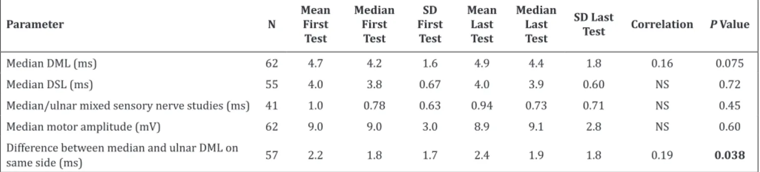
Table 1. Measurements of First Electrodiagnostic Test Versus Last Electrodiagnostic Test
Parameter N
Mean First
Test
Median First Test
SD First Test
Mean Last Test
Median Last Test
SD Last
Test Correlation P Value
Median DML (ms) 62 4.7 4.2 1.6 4.9 4.4 1.8 0.16 0.075
Median DSL (ms) 55 4.0 3.8 0.67 4.0 3.9 0.60 NS 0.72
Median/ulnar mixed sensory nerve studies (ms) 41 1.0 0.78 0.63 0.94 0.73 0.71 NS 0.45
Median motor amplitude (mV) 62 9.0 9.0 3.0 8.9 9.1 2.8 NS 0.60
Difference between median and ulnar DML on
same side (ms) 57 2.2 1.8 1.7 2.4 1.9 1.8 0.19 0.038
N=Number; SD=Standard Deviation; DML=Distal Motor Latency; DSL=Distal Sensory Latency; NS=Not Significant; ms=milisecond; mV=milivolt. The number in bold indicates a significant P value (P<0.05).
Table 2. Difference Between First and Last DML (n = 62)
Parameter N Months Between Tests (SD) P Value
Difference in DML between electrodiagnostic tests 0.048
>10% deterioration* 16 33 (13)
>10% improvement 9 28 (13)
Neither 10% improvement or detoration* 37 23 (9.3)
DML = Distal Motor Latency; SD = Standard Deviation.
neuropathy, cervical radiculopathy, and polyneuropathy. Polyneuropathy was only considered an electrodiagnosis if more than two nerves were involved (e.g., the presence of CTS and cubital tunnel syndrome did not qualify as a polyneuropathy).
Data recorded from the patient’s medical records as described at time of the last electrodiagnostic test were:
sex, age, symptomatic side, paresthesias/numbness,
previous CTR, and any concomitant electrodiagnosis.
The number of months between the first and last
electrodiagnostic test, and treatment for CTS between the
first and last electrodiagnostic test were also collected.
Treatment was divided into the following categories: 1) no treatment; 2) splint; 3) medication (e.g., non-steroidal
anti-inflammatory drugs [NSAIDs]); and 4) splint and
corticosteroid injection(s) and/or medication. The following data was collected from the medical records before or at the time of the last electrodiagnostic test: myelopathy, diabetes mellitus, stroke, hypothyroidism,
depression and wrist trauma. None of the patients that
were included had insulin-dependent diabetes mellitus.
Outcome measures
Given that the DSL is often unrecordable in patients with carpal tunnel syndrome, our primary outcome variable was the quantitative difference in median nerve DML
between the first and last performed electrodiagnostic
test. Secondary outcome measurements were median
DSL, median/ulnar mixed palmar sensory latency,
median motor amplitude, and difference between median DML and ulnar DML on the same side over time.
All other variables were considered explanatory.
Statistical analysis
An a-priori sample size analysis using a two-tailed paired t-test showed that a sample size of 44 patients
would achieve 90% power at a 0.05 significance level to
detect a 10% difference with a standard deviation of 1.0 ms in the median DML between two electrodiagnostic tests.
The data was assessed for normality with the Kolmogorov-Smirnov test. The data was not normally distributed and therefore nonparametric tests were
used. A Wilcoxon signed-rank test was conducted to
test the hypothesis that patients who had two or more electrodiagnostic tests and have not been treated surgically for CTS will not show disease progress
according to the median DML over time. A Wilcoxon
signed-rank test was also used to test the hypothesis that patients with two or more electrodiagnostic tests who have not been treated surgically will not show disease progress according to their median distal
sensory latency (DSL), median/ulnar mixed palmar
sensory latency, median motor amplitude, and difference between median DML and ulnar DML on the same side over time.
Mann-Whitney U tests were conducted to test the association between dichotomous variables and continuous variables. Kruskal-Wallis tests were performed to determine the relationship between categorical variables with more than two categories and continuous variables. The Tukey test was used to judge
the statistical significance of significant Kruskal-Wallis
test results.
All explanatory variables with P<0.10 in bivariable analysis would be entered into a backwards stepwise multivariable linear regression analysis to assess predictors. Cases with missing data for either one or both
of the variables in a specific analysis were excluded. A P value of < 0.05 was considered statistically significant.
Results
From the total of 199 patients that had two or more electrodiagnostic tests over time, 62 patients were eligible for analysis according to the inclusion and
exclusion criteria. There were 21 men (34%) and 41
women (66%) in the study population. The average age was 55±13 years (range, 30 to 87 years). The mean time
between the first and last electrodiagnostic test was
26±12 months (range, 12 to 55 months). One patient was
only tested on one side during the first test and another
14 patients were only tested on one side during the last test. Eight patients had unilateral electrodiagnostically
confirmed CTS at the first test and one other patient Table 3. Overview of Nonoperative Treatments (n = 62)
Nonoperative Treatment Number %
No treatment 22 35.5
Splint 31 50.0
Medication 4 6.5
Splint & injection and/or medication 5 8.1
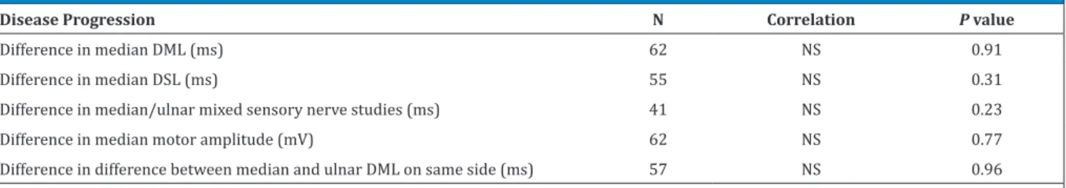
Table 4. Relationship between Different Nonoperative Treatments and Disease Progression of CTS over Time
Disease Progression N Correlation P value
Difference in median DML (ms) 62 NS 0.91
Difference in median DSL (ms) 55 NS 0.31
Difference in median/ulnar mixed sensory nerve studies (ms) 41 NS 0.23
Difference in median motor amplitude (mV) 62 NS 0.77
Difference in difference between median and ulnar DML on same side (ms) 57 NS 0.96
(seven in total) had unilateral electrodiagnostically
confirmed CTS at the last test. Fifty-three patients had bilateral electrodiagnostically confirmed CTS at the first test and another two patients (41 in total) had bilateral electrodiagnostically confirmed CTS at the
last test. Five patients had a nonrecordable DSL at the
first test and another two patients (seven in total) had a
nonrecordable DSL at the last test.
Differences in measurements between the first and last electrodiagnostic test
For 21 patients no median/ulnar mixed palmar
sensory latencies were recorded, and the difference between median and ulnar DML on the same side
was not recorded for five patients. There was no significant difference in DML between the first and last electrodiagnostic test (r=0.16, P=0.075). Only the
difference between median and ulnar DML on the same
side (r=0.19, P=0.038) reached significance with the
numbers available (Table 1).
The last DML improved >10% compared to the first
DML in 9 patients, worsened >10% in 16 patients, and neither improved nor deteriorated >10% in 37 patients (Table 2). The time between the electrodiagnostic tests
was significantly longer for patients in whom the DML
worsened >10% compared to cases of which the DML
neither improved nor deteriorated (P=0.015), but was not significantly different from patients that improved
with the numbers available (P=0.52).
Difference between nonoperative treatments
Half of all patients received a splint and another 36% did not receive any treatment (Table 3). There was no correlation between the different nonoperative treatments and disease progression over time (all
P≥0.23) (Table 4).
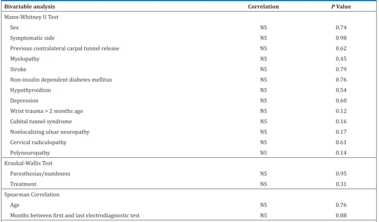
Predictors of difference in median DSL between first and last electrodiagnostic test
In bivariable analysis, none of the explanatory
variables correlated with the difference in DSL between
the first and last electrodiagnostic test (P≥0.12). No
multivariable analysis was conducted for the difference in DSL since there were no variables that met the criterion to be entered into a regression analysis (Table 5).
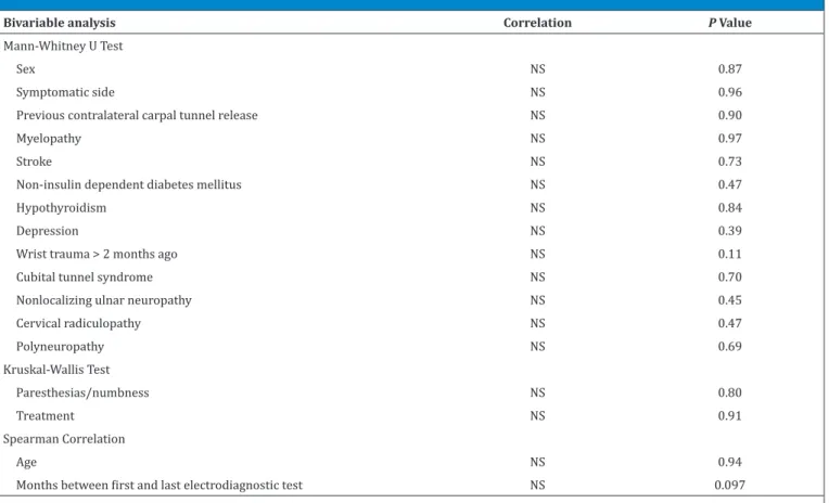
Predictors of difference in median DML between first and last electrodiagnostic test
In bivariable analysis, only the correlation between difference in DML and months between the first and last
electrodiagnostic test met the criterion for entry into
Table 5. Bivariable and Multivariable Analysis - Difference in Median DSL (n = 57)
Bivariable analysis Correlation P Value
Mann-Whitney U Test
Sex NS 0.74
Symptomatic side NS 0.98
Previous contralateral carpal tunnel release NS 0.62
Myelopathy NS 0.45
Stroke NS 0.79
Non-insulin dependent diabetes mellitus NS 0.76
Hypothyroidism NS 0.54
Depression NS 0.60
Wrist trauma > 2 months ago NS 0.12
Cubital tunnel syndrome NS 0.16
Nonlocalizing ulnar neuropathy NS 0.17
Cervical radiculopathy NS 0.61
Polyneuropathy NS 0.14
Kruskal-Wallis Test
Paresthesias/numbness NS 0.95
Treatment NS 0.31
Spearman Correlation
Age NS 0.76
Months between first and last electrodiagnostic test NS 0.88
a multivariable model (r=0.21, P=0.097) so none was
constructed (Table 6).
Discussion
Currently, the average patient and hand surgeon seem to evaluate treatment options based on symptoms more than measurable pathophysiology. Evidence that median nerve pathology slowly but inevitably progresses in CTS might lead patients and surgeons to select operative treatment for moderate CTS before weakness, atrophy, or static numbness develop, even if the moderate CTS is producing few symptoms.
The primary null hypothesis of this was not rejected:
there was no significant difference in the DML of the median nerve between the first and last electrodiagnostic
tests on average, although one might interpret the
findings as a trend towards significance given the limited number of patients in our series. In secondary
analyses the difference between median and ulnar DML
on the same side showed significant progression over time. In addition, time between tests was significantly
longer for cases in which the DML deteriorated >10% compared to cases in which the DML neither improved nor deteriorated >10%. Lastly, there might be a trend
towards a significant correlation between deterioration in DML over time and months between the first and last
electrodiagnostic test. These observations suggest that
an average of 26 months between tests is insufficient.
The measurable pathophysiology of CTS may indeed
wax and wane as it eventually progresses, but over a
much longer time period on average.
Consistent with the theory that CTS is progressive
when measured over a sufficiently long time interval,
Seror performed serial electrodiagnostic tests in 36 untreated wrists and noted that the mean sensory
nerve conduction velocity (NCV) was not significantly
different with an average of eight months between
electrodiagnostic tests but was significantly different
when there was an average interval of 33 months between tests (11). Among 56 patients treated with a corticosteroid injection, there was evidence of a slight transient improvement in electrophysiology in some patients, but at about a year injections did not alter the progressive course of the disease (11).
Among the studies that support the concept that CTS is not inevitably progressive and can improve without surgery all have short study intervals, many used arbitrary categorical ratings, and some include patients with normal electrophysiology (5, 12, 13). Padua et al. repeated electrodiagnostic testing after 10 to 15 months of nonoperative treatment in 274 hands of 196 patients with a clinical diagnosis of CTS (4.5% with
Table 6. Bivariable and Multivariable Analysis - Difference in Median DML (n = 62)
Bivariable analysis Correlation P Value
Mann-Whitney U Test
Sex NS 0.87
Symptomatic side NS 0.96
Previous contralateral carpal tunnel release NS 0.90
Myelopathy NS 0.97
Stroke NS 0.73
Non-insulin dependent diabetes mellitus NS 0.47
Hypothyroidism NS 0.84
Depression NS 0.39
Wrist trauma > 2 months ago NS 0.11
Cubital tunnel syndrome NS 0.70
Nonlocalizing ulnar neuropathy NS 0.45
Cervical radiculopathy NS 0.47
Polyneuropathy NS 0.69
Kruskal-Wallis Test
Paresthesias/numbness NS 0.80
Treatment NS 0.91
Spearman Correlation
Age NS 0.94
Months between first and last electrodiagnostic test NS 0.097
References
Spector TD. The genetic contribution to carpal tunnel syndrome in women: a twin study. Arthritis Rheum. 2002; 47: 275-9.
4. Kulick RG. Carpal tunnel syndrome. Orthop Clin
North Am. 1996; 27:345-54.
5. Ortiz-Corredor F, Enrí�quez F, Dí�az-Ruí�z J, Calambas
N. Natural evolution of carpal tunnel syndrome
1. Diagnosis of the carpal tunnel syndrome. Lancet. 1985; 1:854-5.2.
2. Papanicolaou GD, McCabe SJ, Firrell J. The prevalence and characteristics of nerve compression symptoms in the general population. J Hand Surg Am. 2001; 26:460-6.
3. Hakim AJ, Cherkas L, El Zayat S, MacGregor AJ, normal electrodiagnostic testing) and found that 27% improved (12). Factors associated with neurophysiologic improvement were short duration of symptoms, young age, unilateral symptoms, and negative Phalen test all of which suggest that a substantial percentage of these patients may have had a diagnosis other than median neuropathy or they may have had very mild abnormalities
in the context of atypical symptoms (e.g. pain with
typing) that might have been a variation of normal (12). Ortiz-Corridor et al. followed 132 untreated patients for an average of two years (5). Eight percent deteriorated to a worse electrophysiological severity level (divided into
normal; mild; moderate A; moderate B; severe; extreme),
67% remained at the same level, and 25% improved to a lower level. Another study compared electrophysiological tests over a mean interval of 15 months (range 5 to 30 months) in 52 nonoperatively treated and 99 operatively
treated wrists with CTS and found a significant decrease
in distal motor latency in both groups (13). Goodman and Gilliatt evaluated serial electrodiagnostic tests with an interval between tests of 9 to 126 weeks in 25 splinted and 23 operated hands (14). Using 4 categories (severe, moderate, mild and normal) follow-up motor latency outcomes improved in 19, deteriorated in 1, and remained the same in 5 in the splinted patients and improved consistently after operative treatment (14). A study by Hardoim et al. analyzed the difference in electrodiagnostic measurements between two tests more than one year apart in 210 nonoperatively treated hands (15). The DML worsened in 52.9%, did not change in 7.6% and improved in 39.5%. They found similar results for the difference in sensory conduction velocities.
A strength of this study is that all electrodiagnostic tests
were conducted in one office. Other strengths are that 1.
The electrodiagnostic tests were all reported in the same format and 2. That all medical records were reviewed by one research assistant who was not involved in patient care.
Limitations include our use of a sample of convenience resulting in a small sample and a time between tests that is probably too short for a disease that may progress over decades. We do not have information regarding why patients underwent repeat electrodiagnostic testing nor on the number of nonoperatively treated patients that did not have repeat testing. A better study would prospectively evaluate electrodiagnostic tests over a minimum of 10 years in patients treated nonoperatively. Another limitation is our focus on the median DML for the primary null hypothesis. Median DML not only evaluates the nerve but also neuromuscular transmission and
Mark van Suchtelen BSc Stéphanie J.E. Becker MD Jillian S. Gruber BA David Ring MD, PhD
Orthopaedic Hand and Upper Extremity Service, Harvard
Medical School, Massachusetts General Hospital, Boston, MA, USA
muscular fiber conduction velocity (16). This is why
sensory nerve conduction studies are more sensitive than motor studies in detecting early abnormalities in affected patients (17). Patients with a normal DML can have an abnormal DSL. With DML as our primary outcome measure it is possible that we missed patients who had very mild or mild DSL changes that were not yet detectable with DML. We decided on DML as our primary outcome measure because DSL is more often nonrecordable than DML, and this way we would minimize loss of patients due to no available quantitative
data. We did not detect a significant progress in DSL between the first and last electrodiagnostic test, but we
had less power for that determination because of the number of patients with nonrecordable DSL. Another limitation is the fact that there was a wide variability of intervals between electrodiagnostic tests. The
time between the first and last electrodiagnostic test
ranged from 12 to 55 months and the time between the electrodiagnostic tests showed to be a predictor of the difference in DML.
At short intervals between electrodiagnostic tests it may
be difficult to separate true changes in pathophysiology (signal) from normal fluctuations in electrodiagnostic
measurements (noise). A study repeating electrodiagnostic tests in untreated patients at least 5 or 10 years after
abnormal median nerve function at the carpal tunnel is first
documented might better address whether this disease is
always progressive. This is difficult to study in symptomatic
patients as a high percentage request surgery within a few years of diagnosis (18). Such a study might be most feasible in patients with unilateral symptoms diagnosed with asymptomatic contralateral disease.
Acknowledgements
SJEB is supported by Dutch research grants from
Anna Foundation|NOREF, Genootschap Noorthey, and Stichting Vreedefonds, the Netherlands, for Scientific
in untreated patients.Clin Neurophysiol. 2008;
119:1373-8.
6. Graham RG, Hudson DA, Solomons M, Singer M. A prospective study to assess the outcome of steroid injections and wrist splinting for the treatment of carpal tunnel syndrome. Plast Reconstr Surg. 2004; 113:550-6.
7. Graham B. Nonsurgical treatment of carpal tunnel syndrome. J Hand Surg Am. 2009; 34:531-4.
8. Sevim S, Dogu O, Camdeviren H, Kaleagasi H, Aral M, Arslan E, et al. Long-term effectiveness of steroid injections and splinting in mild and moderate carpal
tunnel syndrome. Neurol Sci. 2004; 25:48-52.
9. Brooks JJ, Schiller JR, Allen SD, Akelman E. Biomechanical and anatomical consequences of carpal tunnel release. Clin Biomech. 2003; 18:685-93.
10. Nunez F, Vranceanu AM, Ring D. Determinants of pain in patients with carpal tunnel syndrome. Clin Orthop Relat Res. 2010; 468:3328-32.
11. Seror P. Nerve conduction studies after treatment for carpal tunnel syndrome. J Hand Surg Br. 1992; 17:641-5.
12. Padua L, Padua R, Aprile I, Pasqualetti P, Tonali
P, Italian CTS Study Group, et al. Multiperspective
follow-up of untreated carpal tunnel syndrome: a
multicenter study. Neurology. 2001; 56:1459-66.
13. Todnem K, Lundemo G. Median nerve recovery
in carpal tunnel syndrome. Muscle Nerve. 2000;
23:1555-60.
14. Goodman HV, Gilliatt RW. The effect of treatment on median nerve conduction in patients with the carpal tunnel syndrome. Rheumatology. 1961; 6:137-55. 15. Hardoim DG, de Oliveira GB, Kouyoumdjian JA. Carpal
tunnel syndrome: long-term nerve conduction
studies in 261 hands. Arq Neuropsiquiatr. 2009; 67:
69-73.
16. Seror P. Sensitivity of the various tests for the diagnosis of carpal tunnel syndrome. J Hand Surg Br. 1994; 19:725-8.
17. Cioni R, Passero S, Paradiso C, Giannini F, Battistini
N, Rushworth G. Diagnostic specificity of sensory and
motor nerve conduction variables in early detection
of carpal tunnel syndrome. J Neurol. 1989;
236:208-13.