https://doi.org/10.1590/0004-282X20170152
REVIEW
Conservative therapeutic management of
carpal tunnel syndrome
Manejo terapêutico conservador da síndrome do túnel do carpo
Roberto Sérgio Martins1, Mário Gilberto Siqueira1
In the wrist, the carpal bones are arranged to form the
loor and sides of an arch-shaped channel. he lexor reti-naculum, or transverse carpal ligament, is a ibrous band that
attaches the medial and lateral eminences of this bone arch, forming the palmar roof of a narrow opening called the car -pal tunnel1. his tunnel encloses the median nerve and nine
tendons of the lexor inger muscles. Any pathophysiological condition or anatomic abnormality that causes enlargement of the carpal tunnel components, reduces its cross-section or raises the pressure inside it, can lead to symptomatic com-pression of the median nerve – carpal tunnel syndrome (CTS). Carpal tunnel syndrome is the most common compressive neuropathy, with a prevalence of 5.8% in adult women and 0.6%
for men2. Generally, CTS is characterized by an insidious onset and
the diagnosis is completely clinical. Usually, the patient complains of tingling and numbness in the hand, especially in the irst three ingers and radial half of the fourth inger (the cutaneous distribu-tion of the median nerve), commonly associated with worsening at night. Physical examination frequently reveals positive provoca-tive test results, such as Tinel’s sign, Phalen’s test and the carpal compression test. Sensory loss and, less frequently, motor deicit and thenar atrophy, can be observed in neurologic examination1.
In most cases, conservative treatment is the irst thera-peutic alternative, especially in patients without signiicant sensory or motor deicits. However, despite its prevalence and the impact of CTS on health systems, there is still much controversy regarding optimal therapy. his can be explained by the fact that most of our knowledge about the treatment of CTS has been based on uncontrolled trials and retrospective studies leading to conlicting conclusions. Accordingly, this review aims to provide a comprehensive and critical analy-sis of conservative treatment in the management of the CTS.
Corticosteroid injection
Corticosteroid injection has been used in the conservative treatment of CTS to reduce symptoms. he exact mechanism of this therapy remains unclear but the anti-inlammatory efect is probably the most signiicant factor in relieving symptoms3.
Diferent types of corticoids have been used, such as hydro-cortisone, dexamethasone, methylprednisolone or triamcino-lone, usually in association with a local anesthetic, but there is no objective standard for deining the ideal dose or speciic
drug3,4. A unique study showed no diference when comparing
shorter- versus longer-acting corticosteroids as well as low and
1Universidade de São Paulo, Faculdade de Medicina, Unidade de Cirurgia do Nervo Periférico, Divisão de Neurocirurgia Funcional, Instituto de Psiquiatria, São Paulo SP, Brasil.
Correspondence: Roberto Sergio Martins; Rua Oscar Freire, 2250; 5409-011 São Paulo SP, Brasil; E-mail: [email protected]
Conflict of interest: There is no conlict of interest to declare. Received 16 August 2017; Accepted 22 August 2017. ABSTRACT
Carpal tunnel syndrome is the most prevalent nerve compression and can be clinically or surgically treated. In most cases, the irst therapeutic alternative is conservative treatment but there is still much controversy regarding the most effective modality of this treatment. In this study, we critically evaluated the options of conservative treatment for carpal tunnel syndrome, aiming to guide the reader through the conventional options used in this therapy.
Keywords: carpal tunnel syndrome; median nerve; conservative treatment.
RESUMO
A síndrome do túnel do carpo é a compressão de nervo mais prevalente e seu tratamento pode ser clínico ou cirúrgico. Na maioria dos casos o tratamento conservador é a primeira alternativa terapêutica mas ainda há muitas controvérsias a respeito do tratamento mais eicaz. Neste estudo avaliamos de forma crítica as opções de tratamento conservador da síndrome do túnel do carpo, objetivando guiar o leitor no uso racional deste tipo de terapêutica.
high doses5 and, therefore, the authors recommended the for
-mer due to the potentially-less collateral efects. Usually, we use 1 ampoule with 4 mg dexamethasone with 5 ml of xylocaine.
Corticosteroid injections are usually performed according to speciic parameters. he injection can be targeted at the ante-rior lexor crest of the wrist on the ulnar side of the palmar ten-don, an easily-palpable structure. Due to the high risk of nerve injury, injection between the tendons of the radial long carpal and the long palmar muscle should be avoided. he angle of insertion of the needle is 45° and it is introduced about 1 cm in depth. he most common risks of corticosteroid injection are nerve or tendon injuries. Inadvertent injection of the median nerve causes immediate shock pain, with the risk of sensory and motor deicits and persistent neuropathic pain. Use in dia-betic patients should be avoided and some patients may experi-ence temporary worsening of pain for two to three days after the injection. Manual efort should be avoided for one to two days
after the procedure3. he application can be repeated in one to
three months but more than two or three applications are not
recommended owing to the potential adverse efects3.
In seven studies of adequate methodology6,7,8,9,10,11,12,13,
Piazzini et al.11 concluded that corticosteroid injection in the
carpal tunnel can be considered as an efective treatment. However, no signiicant clinical beneit was found for corti-costeroid injections compared with other treatments, includ-ing splint immobilization14,15. In addition, adverse efects such
as infection, allergic reaction, osteonecrosis, tendon rupture,
and nerve or tendon injury should be considered3. herefore,
this treatment has been used as a temporary solution, as the efects are short term (months) and, as such, it is a good option in transient conditions that lead to CTS, such as pregnancy or in patients who present with local or systemic risks for surgery.
Oral supplements and medications
Vitamin B6 (pyridoxine) acts as a coenzyme in numerous enzymatic reactions of lipid, amino acids and glucose
metab-olism that are part of the neural function16,17. Conclusions
about the efects of vitamin B6 in CTS patients were
estab-lished based on case reports or researches with small num -bers of participants. he improvement observed in some
cases was thought to be due to resolution of some previ -ously-undiagnosed neuropathy or by analgesic action on the painful pathway17. Despite this fact, some authors still
rec-ommend this therapy, based on clinical response, and the dose usually used is 200 mg/day. he most common adverse efects are numbness, paraesthesia, and other symptoms related to sensory neuropathy, which usually disappear upon
discontinuation of the supplement17.
Oral diuretics have been used in the treatment of
CTS to reduce edema and hence the tunnel’s content. Trichlormethiazide, a diuretic with properties similar to those of hydrochlorothiazide, was preferentially used in some compara-tive studies but the results were inefeccompara-tive in the management of the CTS4,11. Oral steroids, even at low doses, are more efective
than nonsteroidal anti-inlammatory drugs (NSAIDs) and diuret-ics in the conservative treatment of CTS11. However, the risk of
side efects limits their long-term use18. Although NSAIDs are
not efective, they may be useful in patients with CTS-associated tendinitis or tenosynovitis to relieve symptoms4,18.
Exercise therapy and mobilization techniques
he rationale for using dynamic exercises as a treatment of CTS is derived from cadaver and in vivo ultrasound stud
-ies showing median nerve and tendon excursions through the carpal tunnel during the wrist and/or inger
move-ment19,20,21,22. According to several authors, gliding exercises
improve symptoms by preventing, or stretching, the
adhe-sions among the tendons and median nerve, decreasing teno
-synovial edema, improving venous return and, thus, reducing
pressure inside the carpal tunnel19,20,21,22,23,24,25,26,27.
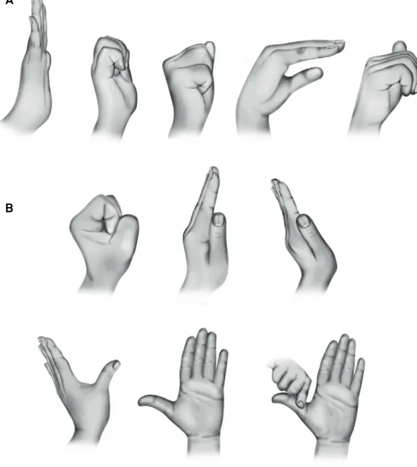
Basically, the exercises involve a sequence of inger move-ments (for tendon gliding) and wrist and ingers movemove-ments (for median nerve gliding) (Figures 1 A and B). In general, both exercises had been applied concurrently but in some studies, one of the therapies was adopted as the only task23. Patients were
instructed to practice each exercise, with ten repetitions, three to ive times daily. Each position was sustained for ive seconds23.
Conlicting results concerning this therapy have been published. According to Huisstede et al.3, this fact is explained
because some studies had poor methodological quality and evaluated this therapy in association with other
manage-ments, such as the combination of a splint, ultrasound ther -apy, and tendon and nerve gliding exercises3,25. Some studies
did not show improvement23 and one even showed worsening
of the functional status28. According to Ashworth, nerve and
tendon gliding exercises were less efective than splint immo-bilization in relieving symptoms and improving hand
func-tion4. In the 2012 Cochrane Review, Page et al.29 concluded
that there was limited evidence that justiied the use of exer-cise and mobilization interventions for CTS. However, the authors suggested that consideration of this therapy should be based on clinician’s experience and patient’s preference.
Wrist immobilization
Wrist immobilization is the most frequently-adopted con-servative therapy for CTS30. he rationale for splinting was
established based on the following: avoiding the extremes of
wrist position reduces the pressure within the carpal tunnel
and the neutral wrist position improves hemodynamic param-eters, reducing the edema and minimizing nerve friction and
compression31,32. Two major types of orthoses used as a
in-gerless glove are described in the treatment of carpal tunnel
syndrome: the hand brace and wrist splint. he brace, less fre-quently used, is made of soft materials without rigid compo-nents, unlike the splint33. he purpose of both devices is the
Wrists splints can be prefabricated or produced by mod-eling a heated thermoplastic component on the patient’s wrist (Figure 2). Discontinuation of treatment due to
cutane-ous intolerance to the splint has been reported in less than
1% of patients33. Usually, prefabricated splints contain a volar
rod that gives irmness, but may cause discomfort in some patients. However, in general, both models are well tolerated. Typically, the splints are manufactured for use in the neutral
position but this recommendation is based on a single ran -domized controlled trial considered to be of poor quality3,30,34.
Wrists splints usually are used for at least a three-month
period30,33 at night-time. According to Huisstede et al.,29 there
is no evidence supporting the full-time use of wrist splint compared to a night-only period.
Despite the prevalence of CTS, the eicacy of diferent thera-peutic modalities, including wrist immobilization, lacks prospec-tive and well-controlled studies. Although widely used in clinical practice, the full efectiveness of the splint has not yet been consis-tently demonstrated in systematic reviews. Page et al.35, in a 2012
Cochrane review, found limited evidence for splinting over a short period versus no treatment or other conservative treatments. However, previous reviews have shown moderate evidence supporting the use of wrist splints in the treatment of CTS11,30.
Furthermore, as noted by Roll and Hardison, a randomized and controlled trial was not included in any of these reviews36,37. A trial
by Hall et al. showed that the use of a wrist splint improved symp-toms in CTS patients37. here was no support for the long-term
use of wrist splints in a review by Page et al.38.
Figure 1. A) Example of the sequence of inger movements during tendon gliding exercises; B) Example of the sequence of inger movements during median nerve gliding exercises. In the last sequence, the thumb is pulled back gently. Adapted from: Akalin E et al.23
A
Other conservative treatments
Low-level laser therapy
In general, laser therapy acts by transferring energy, induc-ing local efects includinduc-ing increased production of endorphins, serotonin and several mediators reducing the inlammatory
reaction and increasing analgesia39. Low power lasers have been
used in the treatment of CTS, suitable for biostimulating action. An important detail in the analysis of the treatment is the power density of the dose, which should be suicient to cross through the diferent tissues to the target organ. he dose generally used is 8–10 J/cm2 with a wavelength ranging from 830–904 nm39.
his low-level laser therapy is applied through a speciic probe at diferent points (usually three) in the wrist where the median nerve travels supericially40. he duration of
applica-tion at each point is 90 seconds40. he purpose of the
treat-ment is to reduce the inlammatory process and edema, but
some authors have used the laser as a tool to stimulate points used in acupuncture with the aim of improving associated pain41. Positive efects of laser on axonal regeneration have
been demonstrated in experimental studies42,43, however it is
not yet clear whether the laser energy is suicient to reach
the median nerve in humans39.
he results with this type of therapy are conlicting and may be explained by the heterogeneity of factors such as laser font, dose intensity, duration of application, and diferent
method-ologies used to evaluate the results39. here is still uncertainty
whether the structures that are the target of treatment in
humans are, in fact, reached by the transferred energy39. hus,
evidence of treatment eicacy still needs to be demonstrated and cost-efectiveness needs to be assessed11,40,41,42,43.
Ultrasound
herapeutic ultrasound is a physical therapy that uses sound waves administered by a speciic transducer and absorbed by surrounding tissues. he pathophysiologi-cal premise that justiies its use as a therapeutic option in patients with CTS is still controversial38. Some authors argue
that the efect of ultrasound is secondary to increased local temperature resulting in increased blood low rates,
metabo-lism and neural regeneration, while other researchers claim
that the action is due to an anti-inlammatory efect44,45.
Because of the limited number of studies, there are still a number of doubts about the therapeutic use of ultrasound
in CTS, especially with regard to the number and duration of sessions required. According to the systematic review carried out by Page et al.38, there is poor quality evidence to
recom-mend therapeutic ultrasound over other conservative treat
-ments used in CTS, a conclusion also observed in another
recent trial44. No adverse efects have been reported in the
reviews by Page et al.38, but only three reports were evaluated.
In conclusion, due to the scarcity of quality evidence in the current literature, doubts still exist about which conservative treatment is most appropriate for CTS, especially to treat CTS in the long term. he recommendation should be based on the intensity of symptoms, severity of the clinical presentation and the patient’s preference. Despite the low level of evidence, cor-ticosteroid injection and wrist immobilization are the tools to be used preferentially in the conservative treatment of CTS.
References
1. Middleton SD, Anakwe RE. Carpal tunnel syndrome. BMJ. 2014;6;349:g6437. https://doi.org/10.1136/bmj.g6437.
2. Krom MC, Knipschild PG, Kester AD, Thijs CT, Boekkooi PF, Spaans F. Carpal tunnel syndrome: prevalence in the general population. J Clin Epidemiol. 1992;45(4):373-6. https://doi.org/10.1016/0895-4356(92)90038-O
3. Huisstede BM, Fridén J, Coert JH, Hoogvliet P, European HANDGUIDE Group. Carpal tunnel syndrome: hand surgeons, hand therapists, and physical medicine and rehabilitation physicians agree on a multidisciplinary treatment guideline-results from the European Handguide Study. Arch Phys Med Rehabil. 2014;95(12):2253-63. https://doi.org/10.1016/j.apmr.2014.06.022
4. Ashworth NL. Carpal tunnel syndrome. Am Fam Physician. 2016;94(10):830-1.
5. O’Gradaigh D, Merry P. Corticosteroid injection for the treatment of carpal tunnel syndrome. Ann Rheum Dis. 2000;59(11):918-9. https://doi.org/10.1136/ard.59.11.918
6. Armstrong T, Devor W, Borschel L, Contreras R. Intracarpal steroid injection is safe and effective for short-term management of carpal tunnel syndrome. Muscle Nerve. 2004;29(1):82-8. https://doi.org/10.1002/mus.10512
7. Breuer B, Sperber K, Wallenstein S, Kiprovski K, Calapa A, Snow B et al. Clinically signiicant placebo analgesic response in a pilot trial of botulinum B in patients with hand pain and carpal tunnel syndrome. Pain Med. 2006;7(1):16-24. https://doi.org/10.1111/j.1526-4637.2006.00084.x
8. Dammers JW, Veering MM, Vermeulen M. Injection with methylprednisolone proximal to the carpal tunnel: randomized double blind trial. BMJ. 1999;319(7214):884-6. https://doi.org/10.1136/bmj.319.7214.884
9. Gökoğlu F, Fındıkoğlu G, Yorgancoğlu ZR, Okumuş M, Ceceli E, Kocaoğlu S. Evaluation of iontophoresis and local corticosteroid injection in the treatment of carpal tunnel syndrome. Am J Phys Med Rehabil. 2005;84(2):92-6. https://doi.org/10.1097/01.PHM.0000151942.49031.DD
10. Nalamachu S, Crockett RS, Mathur D. Lidocaine patch 5% for carpal tunnel syndrome: how it compares with injections: a pilot study. J Fam Pract. 2006;55(3):209-14.
11. Piazzini DB, Aprile I, Ferrara PE, Bertolini C, Tonali P, Maggi L et al. A systematic review of conservative treatment of carpal tunnel syndrome. Clin Rehabil. 2007;21(4):299-314. https://doi.org/10.1177/0269215507077294
12. Wong SM, Hui AC, Tang A, Ho PC, Hung LK, Wong KS et al. Local vs. systemic corticosteroids in the treatment of carpal tunnel syndrome. Neurology. 2001;56(11):1565-7. https://doi.org/10.1212/WNL.56.11.1565
13. Wong SM, Hui ACF, Lo SK, Chiu JH, Poon WF, Wong L. Single vs. two steroid injections for carpal tunnel syndrome: a randomised clinical trial. Int J Clin Pract. 2005;59(12):1417-21. https://doi.org/10.1111/j.1368-5031.2005.00696.x
14. Marshall S, Tardif G, Ashworth N. Local corticosteroid injection or carpal tunnel syndrome. Cochrane Database Syst Rev. 2007(2):CD001554.
15. Sevim S, Dogu O, Camdeviren H, Kaleagasi H, Aral M, Arslan E et al. Long-term effectiveness of steroid injections and splinting in mild and moderate carpal tunnel syndrome. Neurol Sci. 2004;25(2):48-52. https://doi.org/10.1007/s10072-004-0229-0
16. Amadio PC. Pyridoxine as an adjunct in the treatment of carpal tunnel syndrome. J Hand Surg Am. 1985;10(2):237-41. https://doi.org/10.1016/S0363-5023(85)80112-X
17. Ryan-Harshman M, Aldoori W. Carpal tunnel syndrome and vitamin B6. Can Fam Physician. 2007;53(7):1161-2.
18. O’Connor D, Marshall S, Massy-Westropp N. Non-surgical treatment (other than steroid injection) for carpal tunnel syndrome. Cochrane Database Syst Rev 2003(1):CD003219. https://doi.org/10.1002/14651858.CD003219
19. Horng YS, Hsieh SF, Lin MC, Chang YW, Lee KC, Liang HW.
Ultrasonographic median nerve changes under tendon gliding exercise in patients with carpal tunnel syndrome and healthy controls. J Hand Ther. 2014;27(4):317-23. https://doi.org/10.1016/j.jht.2014.07.007
20. Szabo RM, Bay BK, Sharkey NA, Gaut C. Median nerve displacement through the carpal canal. J Hand Surg Am. 1994;19(6):901-6. https://doi.org/10.1016/0363-5023(94)90087-6
21. Wright WT, Glowczewskie F, Wheeler D, Miller G, Cowin D. Excursion and strain of the median nerve. J Bone Joint Surg Am. 1996;78(12):1897-903. https://doi.org/10.2106/00004623-199612000-00013
22. Yoshii Y, Villarraga HR, Henderson J, Zhao C, An KN, Amadio PC. Ultrasound assessment of the displacement and deformation of the median nerve in the human carpal tunnel with active inger motion. J Bone Joint Surg (Am). 2009;91(12):2922-30. https://doi.org/10.2106/JBJS.H.01653
23. Akalin E, El O, Peker O, Senocak O, Tamci S, Gülbahar S et al. Treatment of carpal tunnel syndrome with nerve and tendon gliding exercises. Am J Phys Med Rehabil. 2002;81(2):108-13. https://doi.org/10.1097/00002060-200202000-00006
24. Heebner ML, Roddey TS. The effects of neural mobilization in addition to standard care in persons with carpal tunnel syndrome from a community hospital. J Hand Ther. 2008;21(3):229-40. https://doi.org/10.1197/j.jht.2007.12.001
25. Pinar L, Enhos A, Ada S, Güngör N. Can we use nerve gliding exercises in women with carpal tunnel syndrome? Adv Ther. 2005;22(5):467-75. https://doi.org/10.1007/BF02849867
26. Rozmaryn LM, Dovelle S, Rothman ER, Gorman K, Olvey KM, Bartko JJ. Nerve and tendon gliding exercises and the conservative management of carpal tunnel syndrome. J Hand Ther.
1998;11(3):171-9. https://doi.org/10.1016/S0894-1130(98)80035-5
27. Wehbé MA, Hunter JM. Flexor tendon gliding in the hand. Part II. Differential gliding. J Hand Surg Am. 1985;10(4):575-9. https://doi.org/10.1016/S0363-5023(85)80086-1
28. Horng YS, Hsieh SF, Tu YK, Lin MC, Horng YS, Wang JD. The comparative effectiveness of tendon and nerve gliding exercises in patients with carpal tunnel syndrome: a randomized trial. Am J Phys Med Rehabil. 2011;90(6):435-42. https://doi.org/10.1097/PHM.0b013e318214eaaf
29. Page MJ, O’Connor D, Pitt V, Massy-Westropp N. Exercise and mobilisation interventions for carpal tunnel syndrome. Cochrane Database Syst Rev. 2012;13(6):CD009899. https://doi.org/10.1002/14651858.CD009899
30. Huisstede BM, Hoogvliet P, Randsdorp MS, Glerum S, van Middelkoop M, Koes BW. Carpal tunnel syndrome. Part I: effectiveness of nonsurgical treatments: a systematic review. Arch Phys Med Rehabil. 2010;91(7):981-1004. https://doi.org/10.1016/j.apmr.2010.03.022
31. Manente G, Torrieri F, Di Blasio F, Staniscia T, Romano F, Uncini A. An innovative hand brace for carpal tunnel syndrome: a randomized controlled trial. Muscle Nerve. 2001;24(8):1020-5. https://doi.org/10.1002/mus.1105
32. Schmid AB, Elliott JM, Strudwick MW, Little M, Coppieters MW. Effect of splinting and exercise on intraneural edema of the median nerve in carpal tunnel syndrome: an MRI study to reveal therapeutic mechanisms. J Orthop Res. 2012;30(8):1343-50. https://doi.org/10.1002/jor.22064
33. De Angelis MV, Pierfelice F, Di Giovanni P,
Staniscia T, Uncini A. Eficacy of a soft hand brace and a wrist splint for carpal tunnel syndrome: a randomized controlled study. Acta Neurol Scand. 2009;119(1):68-74. https://doi.org/10.1111/j.1600-0404.2008.01072.x
34. Burke DT, Burke MM, Stewart GW, Cambré A. Splinting for carpal tunnel syndrome: in search of the optimal angle. Arch Phys Med Rehabil. 1994;75(11):1241-4. https://doi.org/10.1016/0003-9993(94)90012-4
35. Page MJ, Massy-Westropp N, O’Connor D, Pitt V. Splinting for carpal tunnel syndrome. Cochrane Database Syst Rev. 2012;11(7):CD010003. https://doi.org/10.1002/14651858.CD010003
36. Roll SC, Hardison ME. Effectiveness of occupational therapy interventions for adults with musculoskeletal conditions of the forearm, wrist, and hand: a systematic review. Am J Occup Ther. 2017;71(1):7101180010p1-12. https://doi.org/10.5014/ajot.2017.023234
37. Hall B, Lee HC, Fitzgerald H, Byrne B, Barton A, Lee AH. Investigating the effectiveness of full-time wrist splinting and education in the treatment of carpal tunnel syndrome: a randomized controlled trial. Am J Occup Ther. 2013;67(4):448-59. https://doi.org/10.5014/ajot.2013.006031
38. Page MJ, O’Connor D, Pitt V, Massy-Westropp N. Therapeutic ultrasound for carpal tunnel syndrome. Cochrane Database Syst Rev. 2013;3(3):CD009601. https://doi.org/10.1002/14651858.CD009601
39. Chang WD, Wu JH, Jiang JA, Yeh CY, Tsai CT. Carpal tunnel syndrome treated with a diode laser: a controlled treatment of the transverse carpal ligament. Photomed Laser Surg. 2008;26(6):551-7. https://doi.org/10.1089/pho.2007.2234
40. Yagci I, Elmas O, Akcan E, Ustun I, Gunduz OH, Guven Z. Comparison of splinting and splinting plus low-level laser therapy in idiopathic carpal tunnel syndrome. Clin Rheumatol 2009;28(9):1059-65. https://doi.org/10.1007/s10067-009-1213-0
42. Gigo-Benato D, Geuna S, de Castro Rodrigues A, Tos P, Fornaro M, Boux E et al. Low-power laser biostimulation enhances nerve repair after end-to-side neurorrhaphy: a double-blind randomized study in the rat median nerve model. Lasers Med Sci. 2004;19(1):57-65. https://doi.org/10.1007/s10103-004-0300-3
43. Oliveira FB, Pereira VM, Trindade AP, Shimano AC, Gabriel RE, Borges AP. Action of therapeutic laser and ultrasound in peripheral nerve regeneration. Acta Ortop Bras. 2012;20(2):98-103. https://doi.org/10.1590/S1413-78522012000200008
44. Armagan O, Bakilan F, Ozgen M, Mehmetoglu O, Oner S. Effects of placebo-controlled continuous and pulsed ultrasound treatments on carpal tunnel syndrome: a randomized trial. Clinics (São Paulo). 2014;69(8):524-8. https://doi.org/10.6061/clinics/2014(08)04