Submitted 10 September 2015 Accepted 3 November 2015 Published7 December 2015
Corresponding author Jasper M. de Goeij, [email protected]
Academic editor Mar´ıa ´Angeles Esteban
Additional Information and Declarations can be found on page 12
DOI10.7717/peerj.1430
Copyright 2015 Alexander et al.
Distributed under
Creative Commons CC-BY 4.0
OPEN ACCESS
Biofouling of inlet pipes a
ff
ects water
quality in running seawater aquaria and
compromises sponge cell proliferation
Brittany E. Alexander1,∗, Benjamin Mueller2, Mark J.A. Vermeij2,3, Harm H.G. van der Geest1and Jasper M. de Goeij1,2,∗
1Department of Aquatic Environmental Ecology, Institute for Biodiversity and Ecosystem
Dynamics, University of Amsterdam, Amsterdam, Netherlands
2CARMABI Foundation, Willemstad, Curac¸ao
3Department of Aquatic Microbiology, Institute for Biodiversity and Ecosystem Dynamics,
University of Amsterdam, Amsterdam, Netherlands ∗
These authors contributed equally to this work.
ABSTRACT
Marine organism are often kept, cultured, and experimented on in running seawater aquaria. However, surprisingly little attention is given to the nutrient composition of the water flowing through these systems, which is generally assumed to equal
in situconditions, but may change due to the presence of biofouling organisms.
Significantly lower bacterial abundances and higher inorganic nitrogen species (nitrate, nitrite, and ammonium) were measured in aquarium water when biofouling organisms were present within a 7-year old inlet pipe feeding a tropical reef running seawater aquaria system, compared with aquarium water fed by a new, biofouling-free inlet pipe. These water quality changes are indicative of the feeding activity and waste production of the suspension- and filter-feeding communities found in the old pipe, which included sponges, bivalves, barnacles, and ascidians. To illustrate the physiological consequences of these water quality changes on a model organism kept in the aquaria system, we investigated the influence of the presence and absence of the biofouling community on the functioning of the filter-feeding spongeHalisarca caerulea, by determining its choanocyte (filter cell) proliferation rates. We found a 34% increase in choanocyte proliferation rates following the replacement of the inlet pipe (i.e., removal of the biofouling community). This indicates that the physiological functioning of the sponge was compromised due to suboptimal food conditions within the aquarium resulting from the presence of the biofouling organisms in the inlet pipe. This study has implications for the husbandry and performance of experiments with marine organisms in running seawater aquaria systems. Inlet pipes should be checked regularly, and replaced if necessary, in order to avoid excessive biofouling and to approachin situwater quality.
Subjects Aquaculture, Fisheries and Fish Science, Cell Biology, Ecology, Environmental Sciences, Marine Biology
INTRODUCTION
Running seawater aquaria are frequently used to study the physiology of marine organisms under controlled,ex situcondition (e.g.,Wilkerson & Muscatine, 1984;Enr´ıquez, M´endez & Prieto, 2005;Anthony et al., 2008;Duckworth & Peterson, 2013). In the experimental design and set-up of such studies, ambient physical abiotic factors, such as light, temperature, and water flow are given the most attention since these are well known to deviate fromin situ
conditions. However, surprisingly little attention is given to biotic and chemical abiotic factors in running seawater aquaria systems, which are usually only monitored in specific feeding or nutrient-enrichment experiments (e.g.,Tacon et al., 2002;Jim´enez & Ribes, 2007;
Bracken, 2004). It is generally assumed that the chemical and biological composition of the seawater flowing through aquaria matchesin situambient water. The extent to which changes in water quality occur within running seawater aquaria and the potential effect of this on the physiology of experimental marine organisms remains largely unknown.
The motivation for the present study was a large discrepancy in the number of prolif-erative cells measured in the spongeHalisarca caerulea(Porifera: Demospongiae) during two distinct fieldwork periods of several months, using the same running seawater aquaria system and identical methodology. In the first series of experiments, the proliferation rate ofH. caeruleafilter cells (choanocytes), i.e., the percentage of proliferative choanocytes after 6 h, was estimated to be 46.6±2.6% (mean±95%-CI) under steady-state (negligible growth) conditions (De Goeij et al., 2009). During the second series of experiments, which were performed five to seven years later, we measured a significantly lower choanocyte pro-liferation rate of 17.6±3.3% for the same species (Alexander et al., 2014;Alexander et al.,
2015). We discussed and hypothesized the possible cause of this altered cell proliferation to be a suboptimal food supply to the aquaria during the latter fieldwork period (Alexander et al., 2014;Alexander et al., 2015). Preliminary tests also showed that during that second fieldwork period the bacterial abundances in the aquaria water were approximately three times lower (3.0×10⁵per mL) than in water samples taken at the reef entrance of the
inlet pipe (8.8×10⁵per mL). Bacterial numbers are a good proxy indicating the food
availability to sponges. The natural diet of these filter-feeding organisms mainly consists of bacterio- and phyto-plankton (e.g.,Pile et al., 1997;Ribes, Coma & Gili, 1999), and dissolved organic matter (Yahel et al., 2003;De Goeij et al., 2008;Mueller et al., 2014). The average bacterial retention efficiency is high, ranging between 68 and 95% for a wide range of tropical-(Mueller et al., 2014), temperate- (Pile, Patterson & Witman, 1996), and cold-water (Yahel et al., 2007) sponge species. The low bacterial abundances observed in our running seawater aquaria could therefore indeed point toward suboptimal nutritional conditions and may explain the compromised physiology of our experimental organisms.
months, whereas the inlet pipe in the latter study (Alexander et al., 2014) had been in place for seven years, allowing much more time for the establishment of biofouling com-munities. In addition, water flowing through running seawater aquaria may experience an increase in inorganic nutrients due to excreted waste products from these biofouling communities (e.g.,Smaal & Prins, 1993;Southwell et al., 2008). However, limited data is currently available on the effects of biofouling on the water quality of running seawater aquaria used to conduct physiological and ecological experiments on marine organisms.
To gain a better insight into the effect of biofouling on water quality and the outcome of experiments held in running seawater aquaria we asked the following research questions: Is water quality within our running seawater aquaria system affected by biofouling communities? If so, do bacterial abundances increase and nutrient concentrations decrease after the removal of such communities, i.e., by replacing the old inlet pipe? Does the presence of biofouling communities significantly hamper the physiology of experimental organisms kept in open seawater aquaria? In order to answer these questions, the bacterial abundances and inorganic nutrient concentrations (nitrate, nitrite, ammonium, and phosphate) were assessed in the reef water flowing along the length of the inlet pipe and in the flow-through aquarium fed by the inlet pipe. Subsequently, the presence and distribution of biofouling communities inside the 7-year old pipe was investigated. After replacing the old inlet pipe, the aforementioned water quality assessments were repeated, and the choanocyte proliferation rates for our model organismH. caeruleawere determined before and after the installation of the new inlet pipe.
MATERIALS AND METHODS
Fieldwork was performed under the research permit (#2012/48584) issued by the Curac¸aoan Ministry of Health, Environment and Nature (GMN) to the CARMABI foundation.
Running seawater aquaria system
The running seawater aquaria system is located on the Southern Caribbean island of Curac¸ao at the CARMABI research station (12◦
12′ N, 68◦
56′
W). The land-based facility consists of 18 glass flow-through aquaria ranging in volume from 50 to 160 L. Seawater is pumped (Hayward Super Pump SP0150Z1CM; capacity∼400 L min−1) from 10 m water depth at the reef slope through a 100-m long polypropylene pipe (5 cm inner Ø). The first 60 m of the pipe lies underwater, whereas the last 40 m, including the pump, is located above-water, partially underground. Water flow is regulated separately for each flow-through aquarium. The last time the old inlet pipe of the running seawater aquaria system had been replaced was in 2006. The new inlet pipe (only the first 60 m of the underwater section) was replaced on April 6th, 2013.
Sponge collection
All specimens of the encrusting spongeH. caeruleawere collected from the fringing coral reefs on the leeward coast of Curac¸ao (Southern Caribbean, 12◦
12′ N, 68◦
56′
replacement of the old pipe. Pieces of sponge were chiseled from the reef framework and the attached substrate was cleared of other organisms. All sponges were trimmed to a size of approximately 25 cm2and subsequently kept in 100-L running seawater aquaria, with a flow rate of 3 L min−1. Aquarium water was at ambient seawater temperature (26–27◦C) and kept under natural light cycles (the semi-enclosed aquarium building receives natural daylight). Semi-transparent black plastic sheets were used to imitatein situcryptic (i.e., the collected sponges inhabited coral cavity walls) light conditions that sponges experienced (photosynthetically active radiation (PAR) level 5–15µmol photons m−2 s−1during
daylight hours). Prior to cell proliferation experiments, sponges acclimatized to aquarium conditions for a minimum of 1 week before and after the old pipe was replaced by the new pipe to ensure they fully recovered from the collection and transportation to the aquaria (De Goeij et al., 2009;Alexander et al., 2014;Alexander et al., 2015).
Water sample collection
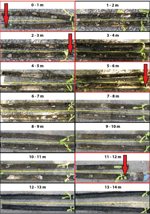
Water samples were taken inside the old pipe on March 22, 2013 and inside the new pipe on April 9, 2013, 3 d after its installation. To sample water from the center of the inlet pipe, Ø1.5 mm holes were drilled underwater using a hand drill through which samples were collected using 20 mL syringes with needle. The holes were sealed afterwards with marine PC-11 two-component epoxy (Protective Coating Company, Allentown, PA, USA). Triplicate 20 mL water samples were taken to determine bacterial abundances and inorganic nutrient concentrations along the length of the old and new inlet pipes at 0 (a few cm inside the inlet pipe), 3, 6, 12, 24, and 60 m from the inlet pipe entrance (Fig. 1and Fig. S1) as well as in one of the flow-through aquaria at 100 m from the entrance of the inlet pipe.
Analysis of bacterial abundance
Ten mL of the water sample was fixed immediately with 0.57 mL 35% formaldehyde solution (final concentration∼2%) for 1 h at 4◦C in the dark. Fixed samples were then filtered on 0.2µm polycarbonate filters (25 mm, Nuclepore Track-Etch; Whatman, Kent,
UK) with 0.45µm cellulose nitrate support filters (25 mm; Sartorius Stedim Biotech
GmbH, Goettingen, Germany). The polycarbonate filters were air-dried and stored at −20◦C until further processing. Filters were mounted on microscope slides in DAPI-mix (final concentration 1µg L−1) and bacteria were counted using an epifluorescence
microscope (×1,250). At least ten fields (each 0.0025 mm2) of a counting grid were counted per slide, or up to a minimum of 200 bacteria, when ten fields were not sufficient.
Analysis of inorganic nutrients
Five mL of the water sample was filtered using a 0.2µm syringe filter (25 mm Puradisc,
BrdU-labeling and sponge tissue sampling
Sponges (n=3 before andn=3 after installation of the new pipe; all different individuals) were enclosed in incubation chambers (3 L) with magnetic stirring devices (De Goeij et al., 2009;Alexander et al., 2014;Alexander et al., 2015), which were kept in the aquaria during the experiments to maintain ambient seawater temperature. In order to measure cell proliferation, 5-bromo-2′-deoxyuridine (BrdU; Sigma-Aldrich, Waltham, MA, USA) was added to incubation chambers containing the sponges. Sponges were incubated in seawater containing 50µmol L−1BrdU for 6 h (continuous labeling) in order to estimate
choanocyte proliferation rates (Nowakowski, Lewin & Miller, 1989;De Goeij et al., 2009;
Alexander et al., 2014). Immediately after the incubations, one tissue sample (∼0.5 cm2) was taken from each sponge and fixed in 4% paraformaldehyde in phosphate-buffered saline (PFA/PBS; 4 h at 4◦
C), rinsed in PBS, dehydrated through a graded series of ethanol and stored in 70% ethanol at 4◦C until further processing.
Sponge cell proliferation
Histological sections (3µm) of BMM-embedded sponge tissue were cut on a pyramitome
(LKB 11800, UK) using glass knives and collected on glass slides (StarFrost; Knittelglass, Braunschweig, Germany). BrdU immunohistochemistry was performed according to Alexander and colleagues (2014;2015) using a mouse anti-BrdU monoclonal antibody (MUB0200S, Nordic-MUbio, Susteren, The Netherlands), which was detected with an avidin-biotin enzyme complex (Vectastain Elite ABC Kit; Vector Laboratories, Burlingame, CA, USA). BrdU-positive cells were visualized with DAB (DAKO, Glostrup, Denmark) on haematoxylin-counterstained sections, and mounted in Entellan (Merck, Kenilworth, NJ, USA). BrdU-labeled mouse intestinal tissue was used as a positive control and immunohistochemistry without primary antibody (on both mouse and sponge tissue) served as a negative control, as previously described (Alexander et al., 2014). All slides were examined under a light microscope (Olympus BH-2) and photographs were taken using an Olympus DP70 camera. From each tissue sample, three areas of the sponge were sectioned, each approximately 100µm apart. At least 250 choanocytes were counted from each
section making a total of at least 750 (three sections×250 cells) cells counted per sponge.
Analysis of biofouling in the old inlet pipe
After water samples were taken from the old pipe and tissue samples were taken from the sponges in the flow-through aquarium, the 60 m underwater section of the pipe was removed and cut into 1-m pieces over its entire length. Each 1-m piece was then cut in half along its length and photographed to assess the presence of biofouling communities (see Fig. 1for an overview andFig. S1for close-up photographs of the biofouling communities in the first 12 m from the inlet pipe entrance).
Statistical analysis
Figure 2 Bacterial abundance along the old (black circles) and new (grey circles) inlet pipes leading to the flow-through aquarium (open squares).Solid lines indicate exponential models for bacterial abundance within the first 60 m of inlet pipe (i.e., the section that was replaced). The dotted lines represent differences in bacterial abundance between the flow-through aquarium, which was 100 m from the inlet pipe entrance, and the replaced section of the inlet pipe.
were also tested in the water flowing through aquaria fed by the old and new pipe, using linear models. Additionally, differences in choanocyte proliferation rates between sponges kept in flow-through aquaria fed by the old and new pipe were investigated with a linear model. The significance threshold was set at 0.05. All calculations were carried out in R (seeData S1for complete dataset (bacterial abundance and inorganic nutrients) and Supplemental Information 5for R-scripts).
RESULTS
Water qualityBacterial abundances in water at the reef entrance of the old inlet pipe (7.3±0.5×
105mL−1; mean±SD) were 3.9 fold higher than in the flow-through aquarium (1.9
±0.1×10⁵mL−1) (Fig. 2) of the running seawater system. Bacterial abundance decreased
with increasing distance from the reef entrance of the old inlet pipe (exponential model,
p=0.009,Fig. 2) and the largest drop in bacterial abundance occurred in the first 12 m of
the pipe. Nitrate and nitrite concentrations increased significantly with increasing distance from the entrance of the old inlet pipe (linear models,p=0.008 [nitrate and nitrite],Figs.
3Aand3B). The concentration of ammonium increased from the entrance of the old inlet pipe to the flow-through aquaria, but did not significantly increase along the length of the old pipe (linear model,p=0.5,Fig. 3C), and no significant difference was observed in
the concentration of phosphate along the length of the old pipe (linear model,p=0.07,
Figure 3 (A) Nitrate [NO−
3], (B) nitrite [NO −
2], (C) ammonium [NH +
4], and (D) phosphate [PO3 − 4 ] concentrations along the length of the old (black circles) and new pipes (grey circles), and in the aquarium (open squares).Solid lines indicate linear models for inorganic nutrient concentrations within the first 60 m of inlet pipe (the section that was replaced), which was 100 m from the entrance of the inlet pipe. The dotted lines represent differences in inorganic nutrient concentrations between the flow-through aquarium and the replaced section of the inlet pipe.
The installation of the new pipe caused a significant change in all water quality parameters (pvalues<0.05,Figs. 2and3A–3C), compared to the old pipe, except the
concentration of phosphate, which remained similar (linear model,p=0.77,Fig. 3D).
There was no significant difference between the reef water samples (taken at 0 m from the pipe entrance) before (March 22, 2013) and after the pipe change (April 9, 2013) for any of the water quality parameters (linear model,p=0.07 [bacteria],p=0.12 [nitrite,
ammonium, and phosphate],p=0.48 [nitrate]). Bacterial abundance was significantly
higher along the length of the new pipe compared to the old (exponential model,p=0.03)
and no longer changed along the length of the new pipe (exponential model,p=0.80).
However, bacterial abundance in the flow-through aquaria supplied by the new inlet pipe was lower (5.9±0.2×10⁵mL−1,Fig. 2A) compared to the abundance at the reef entrance
of the new inlet pipe (7.0±0.1×10⁵mL−1). Still, bacterial abundance in the flow-through
Table 1 Water quality parameters (bacterial abundance [BA], nitrate (NO−
3), nitrite [NO−2], ammonium [NH +
4], and phosphate [PO34−]) and H. caeruleachoanocyte proliferation rates in a flow-through aquarium fed with water from the old (+biofouling) and new inlet pipe (−
biofouling).Means±SD are shown (n=3). Percentage increases or decreases in the aforementioned parameters between the old and the new
pipe are given. NA, not applicable; i.e., no significant difference.
Water quality parameters
BA (×105mL−1) NO−
3 (µmol L−1) NO−2 (µmol L−1) NH +
4 (µmol L−1) PO3 −
4 (µmol L−1)
Aquarium water+biofouling 1.9±0.1 0.86±0.04 0.09±0.01 2.42±1.73 0.04±0.01 Aquarium water−biofouling 5.9±0.0 0.50±0.05 0.05±0.00 0.57±0.13 0.05±0.01 % increase or decrease from old pipe to
new pipe
217 −42 −42 −77 NA
H. caeruleachoanocyte proliferation
% BrdU-positive choanocytes
Aquarium water+biofouling 15.1±1.9
Aquarium water−biofouling 20.2±3.8
% increase or decrease from old pipe to new pipe 34
and ammonium) concentrations were lower in the new pipe compared to the old pipe (linear models,p=0.01 [nitrite],p=0.01 [nitrate],p<0.001 [ammonium]) and did
not change along the length of the new pipe (linear models,p=0.81 [nitrite],p=0.81
[nitrate],p=0.85 [ammonium]). However, inorganic nitrogen concentrations were
higher in the flow-through aquarium compared to the reef entrance of the new inlet pipe (Figs. 3A–3C). Replacing the old pipe, i.e., removing the biofouling community, corresponded to a 42% reduction in both nitrate and nitrite and a 77% reduction in ammonium concentrations in the flow-through aquarium (Table 1).
Biofouling
The first 12 m of the old inlet pipe showed extensive colonization by biofouling communities (Fig. 1andFig. S1), which formed a cm-thick layer of living biomass decreasing its opening from Ø5 cm to approximately Ø3 cm. The abundance of biofouling organisms decreased inwards throughout the pipe and they were absent after 12 m from the reef entrance of the pipe (Fig. 1). Although an in-depth taxonomic survey of the biofouling communities was not carried out, the majority were identified as suspension-and filter-feeding organisms, including sponges, bivalves, barnacles, suspension-and ascidians.
Sponge cell proliferation
Sponges kept in the aquaria in the presence of biofouling communities before the inlet pipe was replaced had a significantly lower choanocyte proliferation rate (15.1±1.9% in 6 h) than sponges after the installation of the new inlet pipe (20.2±3.8% in 6 h, linear model,
p<0.05,Table 1). The replacement of the pipe thus coincided with a 34% increase in the
DISCUSSION
Here we present a study showing that biofouling of an inlet pipe feeding running seawater aquaria coincided with a decrease in bacterial abundance and an increase in dissolved inorganic nitrogen (ammonium, nitrate and nitrite) within the aquarium water. These alterations are logical consequences of the biological activity of suspension-and filter-feeding biofoulers, which included sponges, bivalves, barnacles, suspension-and ascidians (e.g.,Reiswig, 1975;Smaal & Prins, 1993;Williamson & Rees, 1994;Ribes et al., 2005;
Petersen, 2007;Southwell et al., 2008). Bacteria are an important food source for suspension- and filter-feeders, and the largest decrease in their abundance occurred within the first 12 m of the pipe where all biofouling organisms were found. Consequently, this created unfavorable conditions for the filter-feeding sponges kept in the running seawater aquaria system due to diminished food supplies (i.e., bacteria) and the buildup of waste products (inorganic nutrients). The new inlet pipe was free from biofouling organisms, which subsequently caused both bacterial abundance and concentrations of nitrate, nitrite, and ammonium to approach ambient reef water conditions.
Interestingly, after replacing the pipe a slight decrease in bacterial abundance and an increase in inorganic nitrogen species remained between water sampled at the entrance of the inlet pipe and the flow-through aquarium water. This could either be caused by the prevalent presence of biofouling organisms in the underground pipe section that was not replaced, or the presence of the pump in that section. The first suggestion is unlikely, since we did not find any biofouling organisms beyond the first 12 m of the pipe. Microbial biofilms may have colonized this area (Railkin, 2003), but these would not have caused bacterial abundances to decrease. The more likely explanation is that the pump may have caused some destruction of bacterioplankton by its impellor force (Luckett et al., 1996). Within the aquarist community, high pressure and cavitation caused by impellor pumps are commonly known to damage a significant proportion of the resident planktonic community (e.g.,Wijgerde, 2012and discussed on the reef aquarist forumReef Central, 2008), and could have subsequently led to a lower bacterial abundance and an increase in inorganic nutrients in the flow-through aquarium. Bacterial abundance in the aquarium fed by the new inlet pipe (5.9±0.2×10⁵mL−1) where nevertheless still
in the range of ambientin situbacterial abundances measured for Curac¸aoan reef waters (5–10×105mL−1) (De Goeij & van Duyl, 2007).
We documented a clear 34% increase in the proliferation rate of choanocytes in
H. caeruleafollowing the replacement of the inlet pipe. The cause of this increase cannot
et al., 2000;Park & Takeda, 2008), which is reversed following re-feeding (Aldewachi et al., 1975). The direct relationship between nutrient concentration and cell proliferation in sponges must be investigated further, but we have found preliminary evidence that cell proliferation increases with increasing food supply, using bacterial abundance as proxy for food concentrations. Other energetically costly processes, such as reproduction or regeneration, may also cause changes in sponge cell proliferation rates. For the closely related spongeHalisarca dujardiniit was found that up to 69.5% of their body volume could consist of reproductive elements (Ereskovsky, 2000) during their reproductive cycle (presumably causing less energy spent in choanocyte turnover) and cell proliferation significantly decreases during regeneration after wound infliction (Alexander et al., 2015). Unfortunately, to the best of our knowledge, there is no available literature on the reproductive cycle ofHalisarca caerulea. We randomly found reproductive elements in histological sections, both oocytes and spermatic cysts, throughout both multi-months fieldwork periods, but it appeared they did not alter the choanocyte proliferation rates during experiments. During regeneration, choanocyte proliferation rates of sponge specimens residing in the aquaria fed by the old inlet pipe were reduced (7.0±2.5% in 6 h) within the first hours after wound infliction. However, after 6 days proliferation rates did not differ significantly anymore from those in steady-state tissue (i.e., the ‘normal’ physiological state of these sponges showing limited growth and a high turnover of choanocytes) (12.8±1.0% in 6 h) (Alexander et al., 2015).
Despite the 34%-increase found in this study, the proliferation rate of choanocytes for
H. caeruleaafter replacement of the inlet pipe (20.2±3.8% in 6 h) was still substantially lower than found during an earlier study by De Goeij and colleagues (2009) (46±2.6% in 6 h). We measured cell proliferation only seven days after the old pipe was replaced and have, unfortunately, not been able to sample sponges at later time points. We hypothesize that after prolonged stress, such as malnutrition, sponges are likely to have required a longer acclimatization period to reach a higher rate of choanocyte proliferation.
CONCLUSIONS
ACKNOWLEDGEMENTS
We thank Dick van Oevelen and Peter van Breughel from the Royal Netherlands Institute for Sea Research, Yerseke, for their help with inorganic nutrient analysis. We thank Wim Admiraal and Ronald Osinga for commenting on drafts of the manuscript.
ADDITIONAL INFORMATION AND DECLARATIONS
Funding
This work was funded by the European Union Seventh Framework Programme (FP7/2007–2013) under grant agreement no. KBBE-2010–266033 and the Innovational Research Incentives Scheme of the Netherlands Organization for Scientific Research (NWO-VENI; 863.10.009; personal grant to JM de Goeij). The funders had no role in study design, data collection and analysis, decision to publish, or preparation of the manuscript.
Grant Disclosures
The following grant information was disclosed by the authors:
European Union Seventh Framework Programme: KBBE-2010-266033.
Innovational Research Incentives Scheme of the Netherlands Organization for Scientific Research: NWO-VENI; 863.10.009.
Competing Interests
The authors declare there are no competing interests.
Author Contributions
• Brittany E. Alexander performed the experiments, analyzed the data, wrote the paper, reviewed drafts of the paper.
• Benjamin Mueller conceived and designed the experiments, performed the experiments, reviewed drafts of the paper.
• Mark J.A. Vermeij performed the experiments, reviewed drafts of the paper.
• Harm H.G. van der Geest reviewed drafts of the paper.
• Jasper M. de Goeij conceived and designed the experiments, performed the experiments, analyzed the data, contributed reagents/materials/analysis tools, wrote the paper, prepared figures and/or tables, reviewed drafts of the paper.
Field Study Permissions
The following information was supplied relating to field study approvals (i.e., approving body and any reference numbers):
Fieldwork was performed under the research permit (#2012/48584) issued by the Curac¸aoan Ministry of Health, Environment and Nature (GMN) to the CARMABI foundation.
Data Availability
Supplemental Information
Supplemental information for this article can be found online athttp://dx.doi.org/ 10.7717/peerj.1430#supplemental-information.
REFERENCES
Aldewachi HS, Wright NA, Appleton DR, Watson AJ. 1975.The effect of starvation and refeeding on cell population kinetics in the rat small bowel mucosa.Journal of Anatomy119:105–121.
Alexander BE, Achlatis M, Osinga R, Van der Geest HG, Cleutjens JPM, Schutte B, de Goeij JM. 2015.Cell kinetics during regeneration in the spongeHalisarca caerulea: how local is the response to tissue damage?PeerJ 3:e820DOI 10.7717/peerj.820.
Alexander BE, Liebrand K, Osinga R, Van der Geest HG, Admiraal W, Cleutjens JPM, Schutte B, Verheyen F, Ribes M, van Loon E, De Goeij JM. 2014.Cell turnover and detritus production in marine sponges from tropical and temperate benthic ecosystems.PLoS ONE9:e109486
DOI 10.1371/journal.pone.0109486.
Anthony KR, Kline DI, Diaz-Pulido G, Dove S, Hoegh-Guldberg O. 2008. Ocean acidification causes bleaching and productivity loss in coral reef builders.Proceedings of the National Academy of Sciences of the United States of America 105:17442–17446
DOI 10.1073/pnas.0804478105.
Azis PA, Al-Tisan I, Sasikumar N. 2001.Biofouling potential and environmental factors of seawa-ter at a desalination plant intake.Desalination135:69–82DOI 10.1016/S0011-9164(01)00140-0.
Bracken MES. 2004.Invertebrate-mediated nutrient loading increases growth of an intertidal macroalga.Journal of Phycology40:1032–1041DOI 10.1111/j.1529-8817.2004.03106.x.
Chaudhary M, Mandir N, FitzGerald AJ, Howard JK, Lord GM, Ghatei MA, Bloom SR, Goodlad RA. 2000.Starvation, leptin and epithelial cell proliferation in the gastrointestinal tract of the mouse.Digestion61:223–229DOI 10.1159/000007762.
De Goeij JM, De Kluijver A, Van Duyl FC, Vacelet J, Wijffels RH, De Goeij AFPM, Cleutjens JPM, Schutte B. 2009.Cell kinetics of the marine spongeHalisarca caerulea reveal rapid cell turnover and shedding.Journal of Experimental Biology 212:3892–3900
DOI 10.1242/jeb.034561.
De Goeij JM, Van den Berg H, Van Oostveen MM, Epping EHG, Van Duyl FC. 2008.Major bulk dissolved organic carbon (DOC) removal by encrusting coral reef cavity sponges.Marine Ecology-Progress Series357:139–151DOI 10.3354/meps07403.
De Goeij JM, Van Duyl FC. 2007.Coral cavities are sinks of dissolved organic carbon (DOC). Limnology and Oceanography52:2608–2617DOI 10.4319/lo.2007.52.6.2608.
Duckworth AR, Peterson BJ. 2013. Effects of seawater temperature and pH on the boring rates of the sponge Cliona celatain scallop shells.Marine Biology 160:27–35
DOI 10.1007/s00227-012-2053-z.
Ereskovsky AV. 2000.Reproduction cycles and strategies of the cold-water spongesHalisarca dujardini(Demospongiae, Halisarcida),Myxilla incrustansandIophon piceus(Demospongiae, Poecilosclerida) from the White Sea.The Biological Bulletin198:77–87DOI 10.2307/1542805.
Enr´ıquez S, M´endez ER, Prieto RI. 2005.Multiple scattering on coral skeletons enhances light absorption by symbiotic algae. Limnology and Oceanography50:1025–1032
DOI 10.4319/lo.2005.50.4.1025.
Jim´enez E, Ribes M. 2007. Sponges as a source of dissolved inorganic nitrogen:
nitrification mediated by temperate sponges.Limnology and Oceanography52:948–958
DOI 10.4319/lo.2007.52.3.0948.
Johnson D, Walker C. 1999.Cyclins and cell cycle checkpoints.Annual Review of Pharmacology and Toxicology39:295–312DOI 10.1146/annurev.pharmtox.39.1.295.
Jonas K. 2014.To divide or not to divide: control of the bacterial cell cycle by environmental cues. Current Opinion in Microbiology18:54–60DOI 10.1016/j.mib.2014.02.006.
Luckett C, Adey WH, Morrissey J, Spoon DM. 1996. Coral reef mesocosms and
microcosms—successes, problems, and the future of laboratory models.Ecological Engineering
6:57–72DOI 10.1016/0925-8574(95)00051-8.
Mueller B, De Goeij JM, Vermeij MJA, Mulders Y, Van der Ent E, Ribes M, Van Duyl FC. 2014.
Natural diet of coral-excavating sponges consists mainly of dissolved organic carbon (DOC). PLoS ONE9:e90152DOI 10.1371/journal.pone.0090152.
Nowakowski R, Lewin S, Miller M. 1989. Bromodeoxyuridine immunohistochemical
determination of the lengths of the cell cycle and the DNA-synthetic phase for an anatomically defined population.Journal of Neurocytology18:311–318DOI 10.1007/BF01190834.
Park MS, Takeda M. 2008.Starvation suppresses cell proliferation that rebounds after refeeding in the midgut of the American cockroach,Periplaneta Americana.Journal of Insect Physiology
54:386–392DOI 10.1016/j.jinsphys.2007.10.011.
Petersen JK. 2007.Ascidian suspension feeding.Journal of Experimental Marine Biology and Ecology342:127–137DOI 10.1016/j.jembe.2006.10.023.
Pile A, Patterson M, Witman J. 1996.In situgrazing on plankton<10µm by the boreal sponge
Mycale lingua.Marine Ecology—Progress Series141:95–102DOI 10.3354/meps141095.
Pile A, Patterson M, Savarese M, Chernykh V, Fialkov V. 1997.Trophic effects of sponge feeding within Lake Baikal’s littoral zone. 2. Sponge abundance, diet, feeding efficiency, and carbon flux.Limnology and Oceanography42:178–184DOI 10.4319/lo.1997.42.1.0178.
Polman H, Verhaart F, Bruijs M. 2013.Impact of biofouling in intake pipes on the hydraulics and efficiency of pumping capacity.Desalination and Water Treatment 51:997–1003
DOI 10.1080/19443994.2012.707371.
Railkin AI (ed.) 2003.Marine biofouling: colonization processes and defenses. Boca Raton: CRC press.
Reef Central. 2008.Available athttp://www.reefcentral.com/forums/showthread.php?t=1315554.
Reiswig HM. 1975.Bacteria as food for temperate-water marine sponges.Canadian Journal of Zoology53:582–589DOI 10.1139/z75-072.
Ribes M, Coma R, Atkinson MJ, Kinzie RA. 2005.Sponges and ascidians control removal of particulate organic nitrogen from coral reef water.Limnology and Oceanography50:1480–1489
DOI 10.4319/lo.2005.50.5.1480.
Ribes M, Coma R, Gili JM. 1999.Natural diet and grazing rate of the temperate sponge Dysidea avara (Demospongiae, Dendroceratida) throughout an annual cycle.Marine Ecology-Progress Series176:179–190DOI 10.3354/meps176179.
Smaal A, Prins T. 1993.The uptake of organic matter and the release of inorganic nutrients by bivalve suspension feeder beds. In: Dame RF, ed.Bivalve filter feeders. Berlin Heidelberg: Springer, 271–298.
Tacon AGJ, Cody JJ, Conquest LD, Divakaran S, Forster IP, Decamp OE. 2002.Effect of culture system on the nutrition and growth performance of Pacific white shrimp Litopenaeus vannamei (Boone) fed different diets.Aquaculture Nutrition8:121–137
DOI 10.1046/j.1365-2095.2002.00199.x.
Vander Heiden MG, Cantley LC, Thompson CB. 2009. Understanding the Warburg effect: the metabolic requirements of cell proliferation. Science324:1029–1033
DOI 10.1126/science.1160809.
Wijgerde T. 2012.Improved husbandry of marine invertebrates using an innovative filtration technology—Part 1: DyMiCo.Advanced Aquarist2(11): aafeature2.Available athttp://www. advancedaquarist.com/2012/3/aafeature2.
Wilkerson FP, Muscatine L. 1984.Uptake and assimilation of dissolved inorganic nitrogen by a symbiotic sea anemone.Proceedings of the Royal Society of London B: Biological Sciences
221:71–86DOI 10.1098/rspb.1984.0023.
Williamson J, Rees T. 1994.Nutritional interaction in an alga-barnacle association.Oecologia
99:16–20DOI 10.1007/BF00317078.
Yahel G, Sharp J, Marie D, Hase C, Genin A. 2003.In situfeeding and element removal in the symbiont-bearing spongeTheonella swinhoei: Bulk DOC is the major source for carbon. Limnology and Oceanography48:141–149DOI 10.4319/lo.2003.48.1.0141.
Yahel G, Whitney F, Reiswig HM, Eerkes-Medrano DI, Leys SP. 2007.In situfeeding and metabolism of glass sponges (Hexactinellida, Porifera) studied in a deep temperate fjord with a remotely operated submersible.Limnology and Oceanography52:428–440

![Figure 3 (A) Nitrate [NO − 3 ], (B) nitrite [NO − 2 ], (C) ammonium [NH + 4 ], and (D) phosphate [PO 3 4 − ] concentrations along the length of the old (black circles) and new pipes (grey circles), and in the aquarium (open squares)](https://thumb-eu.123doks.com/thumbv2/123dok_br/18127905.324979/8.918.279.866.120.604/figure-nitrate-nitrite-ammonium-phosphate-concentrations-aquarium-squares.webp)
![Table 1 Water quality parameters (bacterial abundance [BA], nitrate (NO − 3 ), nitrite [NO − 2 ], ammonium [NH + 4 ], and phosphate [PO 3− 4 ]) and H](https://thumb-eu.123doks.com/thumbv2/123dok_br/18127905.324979/9.918.46.873.243.351/quality-parameters-bacterial-abundance-nitrate-nitrite-ammonium-phosphate.webp)
