Recebido em 19.01.2001. / Received in January, 19thof 2001.
Aprovado pelo Conselho Consultivo e aceito para publicação em 02.04.2002. / Approved by the Consultive Council and accepted for publication in April, 2ndof 2002. * Trabalho realizado no Ambulatório de Oncologia Cutânea do Departamento de Dermatologia da Faculdade de Medicina da Universidade de São Paulo / Work done at “Ambulatório de Oncologia Cutânea do Departamento de Dermatologia da Faculdade de Medicina da Universidade de São Paulo”
1Pós-graduando - nível doutorado do Departamento de Dermatologia da Faculdade de Medicina da Universidade de São Paulo e do Departamento de Microbiologia e Imunologia da Universidade do Texas Medical Brunch, Galveston, Texas, EUA / Post-graduate doctoral candidate, Medical Faculty Dermatology Department of “Universidade de São Paulo” and Department of Microbiology and Immunology, the University of Texas Medical Brunch, Galveston, Texas, EUA.
2Professor Doutor da Disciplina de Dermatologia da Faculdade de Medicina da USP/ Ph.D., Professor at the USP Medical Faculty Dermatology Department.
3 Professor Doutor do Departamento de Dermatologia e do Departamento de Microbiologia e Imunologia da Universidade do Texas Medical Branch, Galveston, Texas, EUA / PhDMD,
Department of Dermatology and Department of Microbiology and Immunology, the University of Texas Medical Branch, Galveston, Texas, EUA ©2002by Anais Brasileiros de Dermatologia
Aspectos Clínicos da Epidermodisplasia Verruciforme
*Clinical aspects of epidermodysplasia verruciformis
*Walmar Roncalli Pereira de Oliveira
1Cyro Festa Neto
2Stephen K Tyring
3Resumo: FUNDAMENTOS– A epidermodisplasia verruciforme (EV) é genodermatose rara, caracterizada por infecção disseminada por tipos específicos de vírus papiloma humano (HPV), desenvolvimento de tumo-res cutâneos malignos e distúrbios imunológicos.
OBJETIVOS– Correlacionar aspectos clínicos em 13 doentes com EV, na tentativa de contribuir para melhor conhecimento da enfermidade.
MÉTODOS– Avaliação clínica de 13 doentes com EV durante o período de três anos. O diagnóstico clínico
foi confirmado pelo exame histopatológico e imuno-histoquímico.
RESULTADOS– A EVteve início na infância com lesões de verruga plana-símile e/ou máculas eritematosa na face e região cervical. A consangüinidade foi observada na maioria dos doentes (12/13). Clinicamente, o polimorfismo das lesões foi intenso, caracterizado por lesões de verruga plana-símile, pitiríase versicolor-símile, máculas eritematosas e lesões de queratose seborréica-símile. A transformação maligna das lesões foi observada em oito doentes (62%). O crescimento tumoral provocou perda tecidual importante em 50% dos casos, e em 25% foi registrado óbito pelas metástases.
CONCLUSÕES– A EVapresenta alta incidência familiar e provável transmissão autossômica recessiva. O inten-so polimorfismo clínico das lesões não afeta o couro cabeludo e mucosas. A apresentação clínica “na” foi a mais freqüente (62%), seguida pela “benig“na” (23%) e “mista” (15%). Os tumores cutâneos malig-nos são freqüentes, múltiplos, destrutivos, geram metástases e provocam morte.
Palavras-chave: Epidermodisplasia verruciforme; papillomavirus humano.
Summary:BACKGROUND- Epidermodysplasia verruciformis (EV) is a rare genodermatosis
characteri-zed by disseminated infection caused by specific types of human papillomavirus (HPV), development
of cutaneous malignant tumors and immunological disturbances.
OBJECTIVES- To correlate the clinical features of a group of 13 patients with EV aiming to contribute to
knowledge of disease.
METHOD- The clinical evaluation of 13 EVpatients during 3 years. The clinical diagnosis was
confir-med by histopathological and immunohistochemical findings.
RESULTS- The EVbegan in childhood with flat warts and/or erithematous macules on the face and neck.
The parental consanguinity was observed in most patients (12/13). Clinically the lesions were highly polymorphic with flat wart-like lesions, pityriasis versicolor-like lesions, erithematous macules and seborrheic keratoses-like lesions. The malignant conversion of lesions occured in 8 patients (62%). The tumor growth provoked heavy tissue loss in 50% of patients, and metastatic tumors caused death in 25%.
CONCLUSIONS: The EVpresents high familial incidence and an autosomal recessive mode of inheritance.
The high polymorphism clinical doesn’t affect the scalp and mucous membrane. The most frequent cli-nical manifestation is the “malignant” (62%), followed by “benignant” (23%) and “mixed”(15%). The cutaneous tumors are frequent, multiple, destructive, metastasize and cause death.
Key words: Epidermodysplasia verruciformis; papillomavirus, human.
INTRODUÇÃO
A epidermodisplasia verruciforme (EV) é genoderma-tose rara, caracterizada pela susceptibilidade à infecção por tipos distintos de papilomavírus humanos (HPV).1,2,3Os vírus encontrados na EVsão em sua maioria específicos da doen-ça, sendo denominados HPVs associados à EV(HPVEV).4,5A enfermidade inicia-se usualmente na infância, entre os cinco e os 11 anos de idade, manifestando-se por múltiplas lesões de verrugas planas-símile e/ou máculas semelhantes à pitiría-se versicolor (PV).6,7,8Após a terceira década de vida, cerca de 30 a 50% dos doentes podem desenvolver câncer da pele, fre-qüentemente múltiplos e preferencialmente nas áreas de intensa exposição solar.5,8,9 A doença apresenta alterações imunológicas sobretudo da imunidade mediada por célu-las.4,10Essa deficiência imunológica parece ser local e especí-fica, e a presença de polimorfismos genéticos no complexo maior de histocompatibilidade (MHC) determina apresenta-ção inadequada de antígenos específicos do HPVa suas célu-las de defesa.11-15Os doentes desenvolvem infecção dissemi-nada e de longa duração pelo HPV, a qual tardiamente pode desencadear distúrbios inespecíficos de sua imunidade celu-lar.2,16,17Neste trabalho, os autores realizaram o estudo clínico de 13 doentes de EVno Ambulatório de Oncologia Cutânea do Departamento de Dermatologia da Faculdade de Medicina da Universidade de São Paulo (HCFMUSP).
PACIENTES E MÉTODOS
No Ambulatório de Oncologia Cutânea do
HCFMUSP, foram acompanhados 13 doentes de EVdurante o período de três anos.
O diagnóstico foi realizado por meio de anamnese, exame clínico, exame anatomopatológico e imuno-histoquí-mica.
ANAMNESE E EXAME FÍSICO
A história clínica de cada doente foi realizada procu-rando identificar os principais fatores que poderiam influenciar o curso da enfermidade. Mediante as informa-ções obtidas foram feitos os heredogramas.
Foi realizado, inicialmente, exame físico geral, seguido por exame dermatológico completo. Os doentes foram examinados mensalmente.
EXAME HISTOPATOLÓGICO
As lesões cutâneas benignas de morfologia variada e os tumores cutâneos foram total ou parcialmente retirados por excisão cirúrgica ou punch de 4mm e submetidos ao
exame anatomopatológico, tendo os preparados sido cora-dos pela técnica da hematoxilina-eosina.
IMUNO-HISTOQUÍMICA (IHQ)
O método empregado foi o de Streptavidina-Biotina. Utilizou-se o anticorpo Rabbit Antibovine Papillomavirus (BPV-1), por não causar reação falso-positiva com o DNA
tecidual humano.
INTRODUCTION
Epidermodysplasia verruciformis (EV) is a rare genodermatosis, characterized by susceptibility to infection in distinct types of human papillomavirus (HPV).1,2,3
The viruses found in EVare mainly specific to the disease, and are called HPVassociated with EV(HPVEV).4,5
The infirmity is usually initiated in infancy between ages five and eleven, and is manifested by multiple flat wart-like lesions and/or pityriasis versicolor-like macules (PV).6,7,8
After the third decade of life, roughly 30 to 50% of patients may develop skin cancer, which is frequently multiple and most com-monly found in the areas of intense sun-exposure.5,8,9
The disease shows immunological alterations especially of cell-mediated immunity.4,10
This immunological deficiency appears to be local and specific, and the presence of gene-tic poymorphism in the major histocompatibility complex (MHC) determines the inadequate presentation of HPVand its defense cells.11-15
Patients developed disseminate and long-term infections due to HPV, to which unspecific distur-bances of its cellular immunity can link subsequently up.2,16,17
In this paper, the authors carried out a clinical study of 13 EVpatients at the Ambulatório de Oncologia Cutânea (Cutaneous Oncology Outpatient’s Clinic) of the Depar-tamento de Dermatologia da Faculdade de Medicina at Universidade de São Paulo (HCFMUSP)
PATIENTS AND METHODS
At the HCFMUSPCutaneous Oncology Outpatient’s Clinic, 13 patients were given follow-up treatment over a three-year period.
The diagnosis was performed by means of medical history taking, clinical, and anatomopathologic and immu-nohistochemical examination.
MEDICAL HISTORY TAKING AND PHYSICAL EXAMINATION
The clinical history of each patient was raised by attempting to identify the main factors that may have influenced the course of the infirmity. The following heredo-grams were compiled based on the information obtained.
General physical examination was carried out ini-tially, followed by a complete dermatological examination. Patients were examined monthly.
HISTOPATHOLOGICAL EXAM
Benign cutaneous lesions of varied morphology and the cutaneous tumors were entirely or partially removed by excision surgery or 4-mm punch biopsy, and submitted to anatomopathological examination, preparations were made using the hematoxiiline-eosine staining technique.
IMMUNOHISTOCHEMISTRY (IHC)
I II
I II
The IHCfindings showed the HPVantigens are loca-ted in the cell nucleus of the third superior of the epithe-lium, observing the brownish-gold colored precipitins cau-sed by cromogen in the nucleus of these cells.18
It is a method sensitive to productive benign lesions, but shows less sensitivity to the dysplastic tissues.18
This is due to the fact of the large amount of free viral DNApresent in the benign lesions that are to be progressively incorpora-ted into the DNAof the host cell during the process of car-cinomatous transformation.1,33
This method was performed on 23 skin samples of
EVpatients.
RESULTS
Clinical Evaluation – The age range of patients was between 14 and 40 years. Consanguinity was observed in 12 patients, of which 10 had at least one sibling affected by
Os achados da IHQ mostraram que os antígenos do HPV estão localizados no núcleo das células do terço superior do epitélio, observando-se precipitados de cor castanho-dou-rada conferida pelo cromógeno no núcleo dessas células.18
É método sensível nas lesões benignas produtivas, porém apresenta menor sensibilidade nos tecidos displási-cos,18o que se deve ao fato de grande quantidade de DNA viral livre presente nas lesões benignas ser progressivamen-te incorporada ao DNAda célula do hospedeiro no processo de transformação carcinomatosa.1,33
Esse método foi realizado em 23 amostras da pele dos doente de EV.
RESULTADOS
Avaliação clínica – A faixa etária dos doentes estava entre 14 e 40 anos. A consangüinidade foi observada em 12 enfermos, dos quais 10 apresentavam pelo menos um irmão
Heredogramas / Heredograms
I II
I II
I II
I II
I II
I II II8- Doente no1 / II
8– Patient no1
II3- Doente no2 /II
3– Patient no2
II1- Doente no6 / II
1– Patient no6
II2- Doente no8 / II
2– Patient n
o8
II3- Doente no7 / II
3– Patient no7
II4- Doente no9 / II
4– Patient n
o9 II5- Doente no3 / II
5– Patient n
o3
II11- Doente no4 / II
11– Patient n
o4
II7- Doente no5 / II
7– Patient n
o5 II
2- Doente no13 / II2– Patient no13
II1- Doente no10 / II
1– Patient no10
II2- Doente no12 / II
2– Patient no12
II3- Doente no11 / II
the disease. Twelve patients had chronic sun-exposure, esti-mated to be more than 10 years in duration. The onset of the disease occured during infancy, with flat wart-like lesions or erythematous macules mainly on the face and cervical region. The lesions showed intense clinical polymorphism with diffuse distribution, but it was preserved by the scalp and mucoses. The seborrheic keratoses-like lesions, present in 46% of cases, were found mainly on the forehead, in the cervical region and thorax. Pruritus and ardor (urinae) were referred to occasionally and associated with sun-exposure. Mental retardation was observed in three patients (23%). These data are summarized in table 1.
The malignant cutaneous tumors were detected in eight patients (62%), 25 years being the average age for appearance of the first lesions. In all, the initial malignant lesions developed on the forehead with a clinical and histo-pathologic form of squamous cell carcinoma. The patients developed multiple tumors and numerous actinic keratose-like lesions in cutaneous regions most exposed to the sun. In 50% of cases, invasive tumor growth was observed with important tissue loss. In 25%, the presence of metastasis was observed for lymphonodes and the breasts. These data have been summarized in table 2.
Histopathological Findings – In the benign lesions, the presence of large clear and dysplasic cells was obser-ved with vacuolated nuclei and cytoplasm with prominent granules of kerato-hyaline (viral cytopathic effect), in the upper layers of the epithelium. In the flat wart-like lesions of malignant form, the cytopathic viral effect was similar, but it was initiated in the suprabasal layer and extended to the upper layers of the epithelium.
The seborrheic keratose-like lesions showed the cytopathic viral effect of the EV, associated with the histo-pathologic findings characteristic of this type of lesion. The malignant lesions present mainly histological forms of Bowen’s diskeratosis and spinocellular carcinoma.
Immunohistochemistry (IHC)–The flat wart-like lesions of all the patients showed positive results. The seborrheic keratosis-like and squamous cell carcinoma lesions of patient number 9 and the Bowen’s disease lesion of patient number 12 also showed positive results.
DISCUSSION
EV is a genodermatosis characterized by infection disseminated by HPV, frequent malignant transformation of its lesions and immunological alterations.14,15
It is conside-red the first model in human beings of cutaneous carcino-gens induced by HPV.1,4
The patient showed no preferential distribution as to sex or race.7,9,19
In this study, there was equal distribution between the sexes and a higher incidence in Caucasians, but the group is too small to be able to infer anything about these aspects.
EV may be familial or sporadic.1,2
Participation of genetic factors in EV is pertinent, as they determine the
afetado pela doença. Doze doentes apresentaram exposição crônica ao sol, estimada em superior a 10 anos de duração. A doença teve início na infância, com lesões tipo verruga plana ou mácula eritematosa, principalmente na face e região cervical. As lesões mostraram intenso polimorfismo clínico com distribuição difusa, porém poupando couro cabeludo e mucosas. As lesões de queratose seborréica-símile, presentes em 46% dos casos, estavam localizadas principalmente na fronte, na região cervical e no tórax. Prurido e ardor foram referidos ocasionalmente e associados com a exposição solar. Foi observado retardo mental em três doentes (23%). Esses dados estão resumidos na tabela 1.
Os tumores cutâneos malignos foram detectados em oito doentes (62%), sendo a média de idade para o apareci-mento da primeira lesão 25 anos. Em todos, as lesões malignas iniciais desenvolveram-se na fronte, com quadro clínico e histopatológico de carcinoma espinocelular. Os doentes desenvolveram múltiplos tumores e numerosas lesões de queratose actínica-símile nas áreas cutâneas de maior exposição solar. Em 50% dos casos foi observado crescimento tumoral invasivo com perda tecidual impor-tante, e em 25%, presença de metástases para linfonodos e mama. Esses dados estão resumidos na tabela 2.
Achados histopatológicos – Nas lesões da forma benigna, observou-se a presença de grandes células claras, displásicas, com núcleos vacuolizados e citoplasma com proeminentes grânulos de querato-hialina (efeito citopático viral), nas camadas superiores do epitélio. Nas lesões de verruga plana-símile da forma maligna o efeito citopático viral foi semelhante, porém iniciava-se na camada supraba-sal e estendia-se às camadas superiores do epitélio.
As lesões de queratose seborréica-símile tinham o efeito citopático viral da EVassociado aos achados histopa-tológicos característicos desse tipo de lesão. As lesões malignas mostraram principalmente quadro histológico de disqueratose de Bowen e carcinoma espinocelular.
Imuno-histoquímica (IHQ) – As verrugas planas-sími-les de todos os doentes tiveram resultados positivos. As lesões de queratose seborréica-símile e de carcinoma escamo-so do doente número 9 e a lesão de doença de Bowen do doente número 12 também obtiveram resultados positivos.
DISCUSSÃO
A EVé uma genodermatose caracterizada por infec-ção disseminada pelo HPV, freqüente transformainfec-ção malig-na de suas lesões e alterações imunológicas.14,15É conside-rada o primeiro modelo no homem de carcinogênese cutâ-nea induzida pelo HPV.1,4
A doença não apresenta distribuição preferencial quanto a sexo ou raça.7,9,19Neste estudo houve distribuição igual entre os sexos e maior incidência na raça branca, porém o grupo é pouco numeroso para inferências sobre esses aspectos.
Nota: M= masculino / F= feminino / Br = branca / Pd = parda/ Ne = negra / VP = verruga plana / PV = pitiríase versicolor / ME = mácula eritematosa / PED = placas eritemato-escamosas / NR = não referida. / Note: M= male / F= female / Wh = white / Pd = pardo/ Bl = black / FV = flat verruca / PV = Pityriasis versicolor / EM = Erythematous macula / ESP = erythematoussqua -mous plaques / NR = Not reported.
Tabela 1: dados gerais dos doentes portadores de epidermodisplasia verruciforme. Table 1: general data of patients with epidermodysplasia verruciformis.
01 02 03 04 05 06 07 08 09 10 11 12 13 28 34 33 37 31 40 19 20 14 32 21 31 30 F M M F F M M M F F M F M III IV V III III IV VI VI VI II II II III
Sim / Yes
Sim / Yes
Sim / Yes
Sim / Yes
Sim / Yes
Sim / Yes
Sim / Yes
Sim / Yes
Sim / Yes
Sim / Yes
Sim / Yes
Sim / Yes
Não / No
Sim / Yes
Sim / Yes
Sim / Yes
Sim / Yes
Sim / Yes
Sim / Yes
Sim / Yes
Sim / Yes
Sim / Yes
Sim / Yes
Sim / Yes
Sim / Yes
Sim / Yes
Não / No
Não / No
Sim / Yes
Sim / Yes
Sim / Yes
Não / No
Não / No
Não / No
Não / No
Não / No
Não / No
Sim / Yes
Não / No
Verruga vulgar e Queratose seborréica / Verruca vulgaris
and Seborrheic Keratosis
Queratose seborreica
Seborrheic keratosis
Verruga plantar e Queratose seborréica / Plantar Verruca
and Seborrheic Keratosis
Verruga plantar
Verruca plantar
Queratose seborreica
Seborrheic keratosis
Não / No
Não / No
Não / No
Não / No
Verruga palmar e plantar
Palmoplantar verruca.
Queratose seborréica Seborrheic keratosis Queratose seborréica Seborrheic keratosis Verruga vulgar Verruca vulgaris
Não / No
Não / No
Não / No
Não / No
Não / No
Não / No
Sim / Yes
Sim / Yes
Sim / Yes
Não / No
Não / No
Não / No
Não / No
Sim / Yes
Sim / Yes
Não / No
Sim / Yes
Não / No
Sim / Yes
Sim / Yes
Sim / Yes
Sim / Yes
Sim / Yes
Sim / Yes
Sim / Yes
Não / No
8 4 11 10 4 3 11 6 4 5 3 6 12
NR / NR
Face / Face
Dorso das mãos e pés
Back of the hands and feet
Perna direita
Right foot
Região cervical
Cervical region
Face / Face
Face / Face
Face / Face
Face / Face
Região cervical Cervical region Região cervical Cervical region Região cervical Cervical region Pododáctilos Toes Doente Patient Idade Age Sexo Sex Cor
Color da peleTipo
Skin Type
Exposição crônica ao sol
Chronic sun-exposure
Consanguinidade
Consanguinity
Outros casos na
família Other family cases Início da doença (idade) Onset of disease (age)
Tipo da lesão inicial
Initial lesion type
Localização da lesão inicial
Initial lesion site
Br / Wh Pdo / Pdo
Pdo
Br / Wh
Br / Wh Pdo / Pdo
Ne./ Bl.
Ne./ Bl.
Ne./ Bl.
Br / Wh
Br / Wh
Br / Wh
Br / Wh
NR / NR
ME / EM
VP / FV
VP / FV
ME / EM
ME / EM
VP / FV
VP / FV
VP / FV
ME / EM
ME / EM
ME / EM
VP / FV
Tipo e localização das lesões
Type and site of lesions
Sintomas Symptoms Alterações mentais Mental changes
Sim / Yes
Sim / Yes
Não / No
Não / No
Sim / Yes
Sim / Yes
Não / No
Não / No
Não / No
Sim / Yes
Sim / Yes
Sim / Yes
Sim / Yes
Tumores cutâneos
Cutaneous Tumors
Outras lesões associadas ao HPV
Other HIV-associated lesions Doente Patient 01 02 03 04 05 06 07 08 09 10 11 12 13
VP- dorso das mãos e pés. / FV- back of the hands and feet.
PV- região cervical e membros superiores. / PV- cervical region and upper limbs
PED- tronco e membros. / ESP- trunk and limbs.
VP- dorso das mãos e pés. / FV- Back of the hands and feet.
PV- região cervical e membros. / PV- Cervical region and limbs.
PED - tronco e membros. / ESP - trunk and limbs.
VP- face e extremidades dos membros. / FV- face and limb extremities.
PV- região dorsal. / PV- back region.
VP- extremidades dos membros. / PV- limb extremities.
VP – extremidades dos membros. / FV – limb extremities.
PV - tronco e região cervical. / PV – trunk and cervical region.
PED – abdominal e região dorsal. / ESP – abdominal and back region.
VP e PV – distribuídas difusamente. / FV and PV – diffusely distributed.
VP - face, membros e tronco. / FV – face, limbs and trunk.
VP - face, membros e tórax. / FV - face, limbs and thorax.
PV - dorso e membros superiores. / PV – back and upper limbs.
VP- face, membros e tronco. / FV- face, limbs and trunk.
VP - dorso das mãos e pés. / FV - Back of the hands and feet.
PV - tronco e membros. / PV - trunk and limbs.
PED - tronco e porções proximais dos membros / ESP – trunk and close parts of the limbs
VP - face, membros e região genital. / FV - face, limbs and genital region.
PV - face, tronco e região cervical. / PV - face, trunk and cervical region.
PED - face, tronco e membros. / ESP - face, trunk and limbs.
VP - face e dorso das mãos e pés. / FV - face and back of the hands and feet.
PV - face e tronco. / PV - face and trunk.
PED - face, tórax, abdome e membros. / ESP - face, thorax, abdomen and limbs.
VP - dorso dos pés. / FV – back of the feet.
PV- tronco. / PV- trunk.
Tabela 2: dados referentes aos portadores de epidermodisplasia verruciforme com tumores cutâneos Table 2:-data on carriers of epidermodysplasia verruciformis with cutaneous tumors
01 02 05 06 10 11 12 13 01 02 05 06 10 11 12 13 27 29 26 23 27 20 28 23
DB no antebraço E
BD on the L forearm
CEC no dorso nasal
SCC on the dorsal nasal
CEC na região da fronte
SCC on the forehead region
CEC na região da fronte
SCC on the forehead region
CEC na região da fronte
SCC on the forehead region
CBC na região dorsal E
BCC on the L back region
CEC - região supraclavicular E CBC - tórax
CEC - região supraclavicular E CBC - tórax
CEC na fronte D e temporal E
CEC na fronte D e temporal E
CEC “in situ”
“in situ” SCC
CEC bem diferenciado
Well-differentiated SCC
CEC grau II superficial e invasivo
Superficial and invasive SCC Stage II
CEC
SCC
Carcinoma metatípico invasivo
Invasive metatypic carcinoma
CBC superficial
CBC superficial
CEC grau II CBC ulcerado e queratótico
CEC grau II CBC ulcerado e queratótico
CBC na fronte - CBC metatípico na região temporal
CBC na fronte - CBC metatípico na região temporal
QA- fronte e dorso
AK- front and back
CEC-ombro E - CEC- região malar E - DB-tórax - CEC-tórax DB- região frontal E - CEC- dorso - QA- pré-esternal
SCC-L shoulder -BD-thorax - SCC- L malar region SCC-thorax - BD- L forehead region - SCC- back - AK- presternal
DB- pálpebra E - QA -face e V do decote
BD – L eyelids - AK – face and V of the low neck
DB -dorso - CEC - dorso nasal - QA - face, V do decote e membros superiores
BD - back - SCC - dorsal nasal - AK - face, V of the low neck and upper limbs
DB - dorso - CEC - antebraço D - CEC - escápula E - CBC - supraciliar D - CBC - pré-auricular - CBC - ombro D
QA- face, V do decote e membros superiores
BD - back - SCC – R forearm - SCC - L scapular BSS – R supercilliary - BCC – preauricular - BCC – R shoulder
AK- face, V of the low neck and upper limbs
DB - dorso - QA - face e dorso
DB - dorso - QA - face e dorso
CBC - pré-esternal - QA - fronte e tórax
CBC - pré-esternal - QA - fronte e tórax
CEC - fronte D sobre enxerto
QA - fronte D, temporal D e retroauricular superior E.
CEC - fronte D sobre enxerto
QA - fronte D, temporal D e retroauricular superior E.
Não
No
CEC na região malar E - tecido subcutâneo e musculatura da face
SCC in the L malar region – subcutaneous tissue and musculature of the face
Não
No
CEC na asa nasal E - cartilagem e região malar E.
SCC on the L nasal wing – cartilege and L malar region.
CEC na fronte - periósteo. - CEC antebraço- tecido ósseo CBC pré-auricular- perineuro, pericôndrio e vascular.
SCC on the forehead - periostitis. - SCC forearm bone tissue Preauricular BCC- perineural, perichondrial and vascular.
Não
No
Não
No
CEC na fronte D - tecido ósseo, contéudo orbitário e meninges.
SCC on the R forehead – bone tissue, orbit content and meninges.
Doente
Patient
Doente
Patient
Idade de
aparecimen-to do 1Otumor
(anos) Age when
first tumor appeared (in years)
Tipo clínico e localização
do 1Otumor
Clinical type and site of the first tumor
Tipo Histológico do l º tumor
Histologic type of the first tumor
Tipos clínicos e localização dos tumores cutâneos
Clinical type and site of the cutaneous tumors
Tipo Histológico
Histologic type
Capacidade de invasão dos tumores (localização nos tecidos) e formação de metástases
Invasive behavior of the tumors (located in the tissues) and metastasis formation
Não No Sim Yes Não No Sim Yes Não No Não No Não No Sim Yes Radioterapia Radiotherapy QA bowenóide Bowenoid AK
CEC invasivo - Carcinoma “in situ” CEC bem diferenciado - CEC invasivo
Carcinoma “in situ” - CEC invasivo
QA bowenóide - /Invasive SCC
“in situ” carcinoma - well-differentiated SCC invasive SCC - “in situ” carcinoma
invasive SCC - bowenoid AK
CEC “in situ” - QA bowenóide
“in situ” SCC - bowenoid KA
CEC “in situ” - CEC grau II - QA bowenóide
“in situ” SCC - SCC stage II - AK bowenoid
CEC “in situ” - CEC grau II - III - CEC grau III
CBC queratótico - CBC esclerodermiforme CBC esclerodermiforme - QA com displasia moderada
“in situ” SCC - SCC stages II-III - SCC stage III
Keratotic BCC - sclerodermiform BCC sclerodermiform BCC - AK with moderate dysplasia
CEC padrão bowenóide - QA sem sinais de displasia
Standard bowenoid SCC - AK without signs of dysplasia
CBC metatípico - QA bowenóide
Standard bowenoid SCC - AK without signs of dysplasia
CEC espinocelular invasivo grau III - QA bowenóide
Invasive spinocelluar SCC stage III - Bowenoid AK
CBC = carcinoma basocelular / CEC = carcinoma espinocelular / DB = Doença de Bowen / QA = queratose actínica / D = direito / E = esquerdo
infection by distinct types of HPV(HPVEVand HPV 3and/or
10). High familial incidence, frequent consanguine ties, occurrence between siblings, and infrequent affliction in successive generations has led most authors to consider the disease transmission as autosomal recessive, though there are reports of possible recessive inheritance linked to chro-mosome X.6,20,21
Of the 13 patients studied, numbers 1 and 2, 8 and 9, and 10, 11 and 12 were siblings, completing the total of eight familial cases. Patients 4 and 6 reported having siblings with EV. In spite of the fact that Androphy et al.21
consider the sporadic form as having the highest occurren-ce, it was only found in three patients.
Roughly 10% of EV patients result from consangui-ne marriages.6,20
In this study, however, the authors obser-ved that only patient 6 was not an offspring from a consan-guine marriage. There was no report of affliction in ances-tors, suggesting that the form of transmission of the case here reported is probably autosomal recessive.
The disease was often initiated during infancy, though cases have been described of earlier onset (after birth) and later (third and fourth decades).6,7,17
In the patients analyzed, onset varied from 3 to 12 years of age.
The most frequent initial lesion is of the flat wart-like type, located mainly on the dorsa of the hand. PV-type macules, characteristic of the disease, develop a few years after the first lesion. They are initially erythematous, but hypochromic in later stages of the disease. In the follow-up presented here, 46% of patients had both flat wart-like ini-tial lesions and erythematous macules, located mainly in the face and cervical region.
The clinical condition of EV may show flat warts, associated with non-oncogenic HPV 3and/or 10, denomina-ted as “benign form”, or it is expressed polymorphically with a tendency to malignization, associated to multiple
HPVEVs (some oncogenic), denominated as “malignant form”.3,4,5,7
The initial presence of flat warts, followed by the appearance of characteristic polymorphism of malignant form, define the “mixed form”.17,24
The clinical evaluation of the patients here in focus was carried out according to this classification.
The benign form was observed in patients 2, 4 and 6 (23%), who only showed disseminated flat wart lesions. Some of these lesions converged, forming large pigmented plaques on the forehead, torso and limbs. Isomorphism was a frequent finding (Figure 1). No cutaneous tumors appea-red, not even in patient 4, a 37-year-old Caucasian with a history of chronic sun-exposure, i.e. potentially oncogenic factors.
This clinical form demonstrated a difficult differen-tial diagnosis with non-EVflat warts. In these patients, the latter possibility was dismissed, because the three patients showed consanguinity and familial history of the disease. Lesions were clinically greater, forming large strongly pig-mented plaques.8
a infecção por tipos distintos de HPV(HPVEVe HPV 3e/ou
10). A elevada incidência familiar, as ligações consangüí-neas freqüentes, a ocorrência entre irmãos e o infreqüente acometimento em gerações sucessivas levaram grande parte dos autores a considerar a doença de transmissão autossô-mica recessiva, embora haja relatos de possível herança recessiva ligada ao cromossomo X.6,20,21
Dos 13 doentes estudados, eram irmãos os de núme-ro 1 e 2, 8 e 9, e 10, 11 e 12, perfazendo o total de oito casos familiares. Os doentes 4 e 6 referiram irmãos com EV. Apesar de Androphy et al.21considerarem a forma esporádi-ca a de maior ocorrência, ela só foi encontrada em três doentes.
Cerca de 10% dos doentes de EVsão fruto de casa-mentos consangüíneos.6,20Neste estudo, entretanto, os auto-res observaram que apenas o doente 6 não era fruto de casa-mento consangüíneo. Não houve relato de acometicasa-mento em ascendentes, sugerindo que a forma de transmissão dos casos aqui relatados é provavelmente autossômica recessiva.
A doença inicia-se freqüentemente na infância, embora tenham sido descritos inícios mais precoces (após ao nascimento) e mais tardios (terceira e quarta déca-das).6,7,17Nos doentes analisados, o início variou dos três aos 12 anos de idade.
A lesão inicial mais freqüente é do tipo verruga plana-símile, localizada principalmente no dorso das mãos.6,22 As máculas tipo PV, características da doença, desenvolvem-se alguns anos após a primeira lesão.6São ini-cialmente eritematosas, porém hipocrômicas nos estágios tardios da doença.20,23No seguimento aqui apresentado, 46% dos doentes tiveram como lesões iniciais tanto verrugas pla-nas-símiles como máculas eritematosas, localizadas princi-palmente na face e região cervical.
O quadro clínico da EVpode apresentar-se só com verrugas planas, associadas aos HPV não-oncogênicos 3 e/ou 10, sendo denominada de “forma benigna”, ou expres-sar-se de modo polimorfo com tendência à malignização, associada a múltiplos HPVEVs (alguns oncogênicos), deno-minando-se “forma maligna”.3,4,5,7A presença inicial de ver-rugas planas, seguida pelo aparecimento do polimorfismo característico da forma maligna, define a “forma mista”.17,24 A avaliação clínica dos doentes aqui focalizados foi realiza-da segundo essa classificação.
A forma benigna foi observada nos doentes 2, 4 e 6 (23%), que só apresentavam lesões de verrugas planas dis-seminadas. Algumas dessas lesões confluíram, formando grandes placas pigmentadas na fronte, no tronco e nos membros. O isomorfismo foi achado freqüente (Figura 1). Não apresentaram tumores cutâneos, nem mesmo a doente número 4, de 37 anos, cor branca e história de exposição crônica ao sol, fatores potencialmente oncogênicos.
Clini-camente, as lesões eram maiores, formando grandes placas fortemente pigmentadas.8
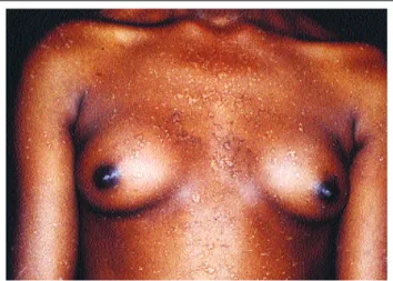
A forma maligna foi observada na maioria dos doen-tes (62%), que apresentou intenso polimorfismo clínico, com lesões tipo verruga plana, tipo PV, placas eritêmato-escamosas, queratose seborréica-símile, queratose actínica-símile e cânceres cutâneos (Figura 2).
A forma mista foi observada nos doentes 3 e 8 (15%) (Figura 3). A possível explicação para o desenvolvimento dessa forma em alguns doentes seria uma importante defi-ciência da imunidade celular causada pela infecção dissemi-nada e de longa duração pelo HPV-3, tornando-os susceptí-veis aos HPVEVs.17,24
Não existe, até o momento, explicação para o fato de a população estudada ter obedecido a essa freqüência de distribuição de suas formas clínicas, como também não existem relatos dessa freqüência nos trabalhos revisados.
A presença das lesões de queratose seborréica-sími-le apenas nas formas clínicas maligna e mista (Figura 4) sugere sua associação aos HPVEVs. Chama atenção a alta incidência (46% dos casos) dessas lesões nos doentes aqui analisados, pois não é freqüente tal descrição na literatu-ra.25,26
As mucosas e o couro cabeludo foram poupados. Encontraram-se lesões de verrugas palmoplantares em 38% dos doentes, fato divergente dos casos revisados, em que essa localização é excepcional.8,27,28
A doença cursa sem sintomas ou com discreto pruri-do.3,7Cerca de 38% dos doentes referiram discreto prurido, ocasionalmente, após exposição solar, o que leva os autores a acreditar que tenha pouca importância no quadro clínico da doença.
São relatados casos de regressão espontânea das lesões na forma benigna da EV.5,7,29No seguimento, obser-vou-se evolução progressiva das lesões em todas as formas
The malignant form was observed in most patients (62%), who demonstrated intense polymorphic clinical manifestation, with flat wart-like lesions, PV-type, erythe-matous-squamous plaques, seborrheic keratosis-like, kera-tosis actinic-like and cutaneous cancers. (Figure 2).
The mixed form was observed in patients 3 and 8 (15%) (Figure 3). The possible explanation for the develop-ment of this form in some patients would be an important deficiency in cellular immunity caused by disseminated and long-term infection due to HPV-3, which becomes suscepti-ble to HPVEVs.17,24
For the time being, there is no explanation for the fact that the population studied has obtained this frequency of distribution of its clinical forms. Nor do any reports exist of this frequency in revised works.
The presence of these seborrheic keratosis-like lesions only of malignant and mixed clinical forms (Figure 4) suggests their association with HPVEVs. It calls attention to the high incidence (46% of cases) of these lesions in the patients analyzed in this paper, because such a description is not frequent in the literature.25,26
The mucoses and scalp were preserved. Palmo-plantar wart lesions were found in 38% of patients, a fact diverging from the cases reviewed in which this localization is exceptional.8,27,28
The disease evolves without symptoms or with dis-crete pruritus. Roughly 38% of patients referred to discreet pruritus occasionally after sun-exposure, leading the authors to believe little importance is attached to the clini-cal condition of the disease.
Cases have been reported of spontaneous regres-sions in benign leregres-sions of EV.5,7,29
In what follows, progres-sive evolution of the disease was observed in all of the disease forms, without a tendency to remission. The patients did not demonstrate diseases either, (or were receiving
Figura 1: lesões de verrugas planas com isomorfismo no tronco e membros superiores – forma benigna
Figure 1: Flat “verruca” (wart-like) lesions with isomorphism on the trunk and upper limbs – benign form
Figura 2: placas eritematosas disseminadas e lesões de queratose actínica no dorso – forma maligna
da doença, sem tendência à remissão. Os enfermos também não apresentavam doenças consuntivas (ou estavam rece-bendo terapia imunossupressora) que justificasse uma infecção generalizada pelo HPVe câncer cutâneo (síndro-me se(síndro-melhante à EV).12,13,14,27
Em três irmãos (23%) foram observados grau varia-dos de retardo mental, apresentando índice maior do que os 10% referidos na literatura.9
O exame histopatológico é importante método diag-nóstico da doença, sendo possível individualizar nas lesões o efeito citopático da infecção viral,1,7,16,30que foi demonstrado em todas as lesões de verrugas, independente da forma clíni-ca.
Nas lesões de verrugas planas-símiles da forma maligna, o efeito citopático viral foi observado desde a camada suprabasal até as camadas superiores da epiderme, o que é característico das lesões causadas pelo HPVEV
(principalmente o HPV5 e 8).12Nas lesões da forma benig-na, o efeito citopático viral só foi observado nas camadas superiores do epitélio, conferindo o aspecto de “olhos de pássaros”, característico dessa forma da doença.8,13
Os resultados imuno-histoquímicos positivos nas amostras de verrugas de todos os doentes de EVindicam a presença do antígeno do HPV. Os resultados negativos nas lesões do tipo queratose seborréica e actínica e nos tumores cutâneos poderiam ser justificado pela diminuição da sen-sibilidade do método empregado nos tecidos displásicos.1,13 A EVé aceita como condição pré-maligna. A trans-formação maligna ocorre em torno de 30 a 50% dos casos e está associada aos HPVEVs oncogênicos, fatores genéti-cos do hospedeiro e à ação de co-carcinógenos extrínsegenéti-cos, principalmente UVBe radioterapia.1,5,8,12,28,31,32
Os doentes podem apresentar, precocemente, elasto-se solar e numerosas lesões de queratoelasto-se actínica-símile nas áreas de maior fotoexposição.8,28,33 Essas lesões
immunosuppressor therapy) which can justify a generali-zed infection by HPV and cutaneous cancer (syndrome similar to EV).12,13,14,27
In three siblings (23%), variable degrees of mental retardation were observed, showing a rate higher than the 10% referred to in the literature.9
The histopathological test is an important diagnos-tic method of the disease. It is possible to individualize the cytopathic effect of the viral infection in the lesions,1,7,16,30
which were demonstrated in all wart lesions, independent of clinical manifestation.
In the flat wart-like lesions of malignant form, the viral cytopathic effect was observed from the suprabasal layer until the upper layers of the epidermis, which is cha-racteristic in lesions caused by HPVEV(mainly HPV 5and
8).12
In benign lesions, the viral cytopathic effect was only observed in the upper layers of the epithelium, granted the aspect of “bird eyes”, characteristic of this kind of disea-se.8,13
The positive immunohistochemical results in the wart samples of all EVpatients indicate the presence of the
HPVantigen. The negative results in seborrheic and actinic keratosis-type lesions, and cutaneous tumors could be jus-tified by the reduced sensitivity of the method applied in dysplasic tissues.1,13
EV is accepted as a pre-malignant condition. The malignant transformation occurs in about 30 to 50% of cases and is associated with oncogenic HPVEVs, genetic factors of the host cell, and actions of the extrinsic co-car-cinogens, mainly UVBand radiotherapy.1,5,8,12,28,31,32
Patients can prematurely show solar elastosis and numerous actinic keratosis-like lesions in the areas of grea-test photoexhibition.8,28,33
These lesions demonstrate clinical characteristics equal to those observed population in gene-ral, but with more malignization power.8,12,13,28
There was no
Figura 3: verrugas planas no dorso das mãos e lesões semelhantes a PV no tronco e membros superiores – forma mista
Figure 3: Flat “Verruca” (wart-like) lesions on the back of the hands and lesions similar to PV on the trunk and upper
limbs- mixed form
Figura 4: lesão de queratose seborréica na região cervical – forma maligna
demonstram características clínicas iguais às observadas na população geral, porém com maior poder de maligniza-ção.8,12,13,28Não foi observada elastose solar nesses doentes, porém todos os que desenvolveram tumores cutâneos apre-sentaram previamente lesões de queratose actínica-símile. Todos os doentes com a forma maligna desenvolve-ram tumores cutâneos, apresentando índice de maligniza-ção de 62%. Esse índice pode estar subestimado devido ao curto período de observação deste estudo (três anos) e pelo fato de os doentes 7 e 9, sem tumores, estarem em idade inferior à preconizada para o início de desenvolvimento de tumores cutâneos na EV(de 24 a 30 anos).6,23
Os doentes com a forma mista, um pardo e um negro, não desenvolveram tumores, apesar do comprometi-mento por HPVEVs oncogênicos e intensa exposição solar. A ausência de tumores deve-se provavelmente ao papel protetor da melanina e ao menor tempo de exposição aos
HPVEVs nessa forma clínica da doença.
O desenvolvimento de tumores na EVé processo de múltiplas etapas, iniciando-se principalmente na pele exposta ao sol.13,30Nesta casuística, o menor período obser-vado, entre 11 e 25 anos, poderia ser justificado pela maior exposição solar a que esses doentes foram submetidos.
A maioria dos tumores malignos (em torno de 50%) desenvolveu-se na fronte dos doentes, o que poderia ser justificado pela ação conjunta ou isolada dos seguintes fatores: 1) área de grande exposição aos raios ultravioleta; 2) presença das células de longa vida, steem cells, dos
folí-culos pilosos, prováveis reservatórios do HPVEV;34 3) ação
sinérgica dos carcinógenos químicos (esqualenos e ácidos graxos do sebo).8,28
Os enfermos desenvolveram múltiplas lesões tumorais, sendo a maioria do tipo carcinoma espinocelular e doença de Bowen, o que está de acordo com a literatura pesquisada.1,5,6,7
É descrito na EV o comportamento invasivo dos tumores malignos, porém de crescimento lento e bom prog-nóstico, exceto quando tratados com radioterapia, situação em que se associam a doença metástatica,30 usualmente invadindo os gânglios linfáticos regionais.8,9,28
Os autores observaram neste estudo um comporta-mento bastante agressivo por parte dos tumores cutâneos, que comprometeram tecidos muscular, cartilaginoso e ósseo, órbitas e meninges. A agressividade tumoral pela aplicação prévia de radioterapia poderia ser justificada nos doentes número 2, 6 e 13. A radioterapia provoca a libera-ção de grande quantidade da citocina imunossupressora
TNFα(fator αde necrose tumoral), o que determina o com-portamento tumoral mais agressivo, com formação de metástases.12,13,14,30Nos demais pacientes, o comportamento agressivo poderia ser explicado pelo prolongado tempo de exposição solar associado ao tipo da pele II de Fitzpatrick. Embora existam relatos de óbitos por EVdevidos a tumores malignos avançados, não há casos de morte de doentes jovens.2,23Nesta casuística, entretanto, os doentes 2 e 10, com 34 e 32 anos de idade, respectivamente, foram a óbito
solar elastosis observed in these patients, but all of those who went on to develop cutaneous tumors had previously shown actinic keratosis-like lesions.
All patients with malignant condition developed cutaneous tumors, and showed a 62% malignization rate. This rate may be underestimated due to the study’s short observation period (three years) and due to the fact that patients 7 and 9, without tumors, were younger than the age suggested as the beginning of cutaneous tumor deve-lopment in EV(from 24 to 30 years of age).6,23
Patients with mixed form, one pardo and one black patient, did not develop tumors, in spite of being compro-mised by oncogenic HPVEVs and intense sun-exposure. The absence of tumors is probably due to the protector role of melanin and to the short exposure time to HPVEVs in this clinical form of the disease.
Tumor development in EVis a multiple-step process, with onset mainly in sun-exposed skin.13,30
In this sampling, the shortest duration observed, from ages 11 to 25, could be justified by the greater degree of sun-exposure to which these patients were subjected.
Most malignant tumors (roughly 50%) developed on the forehead of patients, which may be justified by the joint or isolated action of the following factors: 1) greater area exposed to ultra-violet rays; 2) presence of long-life cells, stem cells, hair follicles, probable reserves of HPVEV; 3) synergic action of chemical carcinogens (squalus and fatty acids in sebum)
Patients developed multiple tumor lesions, the majority with spinocellular-type carcinoma and Bowen’s disease, which confers with the research literature.1,5,6,7
The invasive behavior of malignant tumors in EV
has been described, but it has slow growth and good prognosis, except when treated with radiotherapy. The latter situation is one with which metastatic disease, 30 usually invading the regional lymphatic ganglions,8,9,28
is associated.
In this study, the authors observed noticeably aggressive behavior by the cutaneous tumors, which compromised the muscular, cartilaginous and bone tis-sues, and orbits and meninges. Tumor aggressivity due to the planned application of radiotherapy might be justified in patient numbers 2, 6 and 13. Radiotherapy provokes the release of a large amount of immunosup-pressor cytokines TNF-alpha (tumor necrosis factor alpha), which determines more aggressive tumor beha-vior with formation of metastases.12,13,14,30
In the other patients, the aggressive behavior could be explained by prolonged sun-exposure associated with Fitzpatrick’s skin type-II.
Although there have been reports of death by EV
carcinoma of the face with subsequent affliction of the cerebral tissue.
CONCLUSIONS
Data analysis justified the conclusion that EV is a genodermatosis of high familial incidence, especially pre-sent in children from consanguine marriages. Transmission of EVis probably recessive autosomal. They begin as flat wart-like and/or erythematous macules on the face and cer-vical region in general. Intense polymorphism of cutaneous lesions develop with diffuse distribution, except for the scalp and mucosa. The subjective symptoms are infrequent and irrelevant. The most common clinical manifestation of the disease is the “malignant” form (62%), followed by “-benign” (23%) and “mixed” (15%) forms.
The seborrheic keratosis-like lesions are probably caused by HPVEVs, because only carriers with “malignant” and “mixed” forms of the disease were identified, and viral cytopathic effect of the disease was detected by histopatho-logical exam.
Malignant cutaneous tumors are not very frequent. Predominantly multiple (75% of diseases), they develop in the areas most exposed to the sun, especially on the fore-head. They are especially invasive spinocellular and in situ carcinoma types, followed by basocellular carcinoma. They demonstrated very aggressive behavior, invaded subjacent tissues (65%), generated metastases (38%) and induced death (25%). q
devido ao desenvolvimento de carcinoma espinocelular agres-sivo da face com comprometimento tardio de tecido cerebral.
CONCLUSÕES
A análise dos dados permitiu concluir que a EV é genodermatose de alta incidência familiar, presente sobretu-do em filhos de casamentos consangüíneos, e provavelmen-te de transmissão autossômica recessiva. Inicia-se como verruga plana e/ou mácula eritematosa, em geral na face e região cervical. Desenvolve intenso polimorfismo das lesões cutâneas com distribuição difusa, poupa o couro cabeludo e mucosas, e os sintomas subjetivos são infre-qüentes e irrelevantes. A apresentação clínica mais comum da doença é a forma “maligna” (62%), seguida pelas formas “benigna” (23%) e “mista” (15%).
As lesões de queratose seborréica-símile provavel-mente são causadas pelos HPVEVs, pois só foram identifi-cadas nos portadores das formas “maligna” e “mista” da doença e apresentaram ao exame histopatológico o efeito citopático viral da doença.
Os tumores cutâneos malignos são muito freqüentes. Predominantemente múltiplos (75% dos doentes), desen-volvem-se nas áreas de maior exposição solar, em especial na fronte. São sobretudo do tipo carcinoma espinocelular invasivo e in situ, seguido pelo carcinoma basocelular.
Apresentam comportamento muito agressivo, invadem teci-dos subjacentes (65%), geram metástases (38%) e provo-cam morte (25%). q
skin. Philadelphia: WB Saunders, 1991:101-13.
9. Lutzner ME, Blanchet-Bardon C. Epidermodisplasia Verruciforme (síndrome de Lewandosky-Lutz) In: Fitzpatrick TB, Wolff K, Freedberg IN, Austen KF eds. Dermatologia en Medicina general. Buenos Aires: Panamericana,1984;3:2621-34.
10. Haftek M, Jablonska S, Orth G. Specific cell-mediated immu-nity in patients with epidermodysplasia veruciformis and plane warts. Dermatologica 1985;170:213-20.
11. Majewski S, Jablonska S. Epidermodysplasia verruciformis as a model of human papillomavirus – induced genetic cancers: the role of local immunosurveilance. Am J Med Sci 1992;304:174-9. 12. Jablonska S, Majewski S. Epidermodysplasia verruciformis: Immunological and clinical aspects. Curr Top Microbiol Immunol 1994;186:157-75.
13. Majewski S, Jablonska S. Epidermodysplasia verruciformis as a model of human papillomavirus - induced genetic cancer of the skin. Arch Dermatol 1995;131:1312-8.
14. Majewski S, Jablonska S. Human papillomavirus - associated tumors of the skin and mucosa. J Am Acad Dermatol 1997;36:659-87.
15. Majewski S, Jablonska S. Immunology of HPV infection and HPV-associated tumors. J Invest Dematol 1998;37:81-95. 16. Prawer SE, Pass F, Vance JC, Greenberg LJ, Yunis EJ, Zelickson AS. Depresssed immune function in epidermodysplasia
REFERÊNCIAS / REFERENCES
1. Jablonska S, Dabrowski J, Jakubowicz K. Epidermodysplasia verruciformis as a model in studies on the role of papovaviruses in oncogenesis. Cancer Res 1972;2:583-9.
2. Glisnki W, Jablonska S, Langner A, Obalek S, Haftek M, Proniewska M. Cell-mediated immunity in epidermodysplasia verruciformis. Dermatologica 1976;153:218-27.
3. Jablonska S, Orth G, Jarzabek-Chorzelska M, “et al”.. Immunological studies in epidermodysplasia verruciformis. Bull Cancer 1978;65:183-90.
4. Orth G, Jablonska S, Favre M, Croissant O, Jarbazek-Chorzelska M, Rzesa G. Characterization of two types of human papillomaviruses in lesions of epidermodysplasia verrucifomis. Proc Natl Acad Sci USA 1978;75:1537-41.
5. Orth G, Jablonska S, Jarzabeck-Chorzelska M, “et al”.Cha-racteristics of the lesions and risk of malignant conversion asso-ciated with the type of human papillomavirus involved in epider-modysplasia verruciformis. Cancer Res 1979;39:1074-89. 6. Lutzner MA. Epidermodysplasia verruciformis. Bull Cancer 1978;65:169-82.
7. Jablonska S, Orth G, Jarbazek-Chorzelska M “et al”. Twenty-one years of follow-up studies of familial epidermodysplasia ver-ruciformis. Dermatologica 1979;158:309-27.
verruciformis. Arch Dermatol 1977;113:495-9.
17. Majewski S, Skopinska-Rosewska E, Jablonska S, Wasik M, Misiewick J, Orth G. Partial defects of cell-mediated immunity in patients with epidermodysplasia verruciformis. J Am Acad Dermatol 1986;15:966-73.
18. Kurman RJ, Jenson B, Sinclair CF, Lancaster WD. Detection of human papillomavirus by imunocytochemistry. In: De Lellis eds. Advances in imunnohistochemistry. Washington: Masson, 1984:201-21.
19. Kawashima M. Epidemodysplasia verruciformis. J Dermatol 1992;19:707-9.
20. Rajagopalan K, Bahru J, Loo DSC, Tay CH, Chin KN, Tan KK. Familial epidermodysplasia verruciformis of Lewandowski and Lutz. Arch Dermatol 1972;105:73-8.
21. Androphy EJ, Dvoretzky I, Lowy DR. X-Linked inheritance of epidermodysplasia verruciformis. Arch Dermatol 1985;121:864-8. 22. Aizawa H, Abo T, Aiba S, Sugawara S, Kumagai K, Tagami H. Epidermodysplasia verruciformis accompanied by large granu-lar lymphocytosis. Arch Dermatol 1989;125:660-5.
23. Kanda R, Tanigaki T, Kitano Y, Yoshikawa L, Yutsudo M, Hakura A. Types of human papillomavirus isolated from japanese patients with epidermodysplasia verruciformis. Br J Dermatol 1989;121:463-9.
24. Majewski S, Malejczyk J, Jablonska S “et al.”. Natural cell-mediated citotoxicity against various target cells in patients with epidermodysplasia verruciformis. J Am Acad Dermatol 1990;22:423-7.
25. Tomasini C, Aloi F. Pippione M. Sborrheic keratosis-like lesions in epidermodysplasia verruciformis. J Cutan pathol 1993;20:237-41.
26. Jacyk WK, Dreyer L, Villiers EM. Seborrheic keratoses of black patients with epidermoysplasia verruciformis contain human papillomavirus DNA. Am J Dermatophatol 1993;15:1-6.
27. Pfister H. Human papillomaviruses and impaired immunity vs epidermodysplasia verruciformis. Arch Dermatol 1987;123:1469-70. 28. Jablonska S. Epidermodysplasia verruciformis. In: Fitzpatrick TB, Eisen AW, Wolff K, Freedberg IN, Austen KF eds. Dermatology in general medicine. New York: McGraw-Hill,1993;2:2621-67.
29. Jablonska S, Obalek S, Orth G, Haftek M, Jarzabek-Chorzelska, M. Regression of the lesions of epidermodysplasia verruciformis. Br J Dermatol 1982;107:109-16.
30. Cobb MW. Human papillomavirus infection. J Am Acad Dermatol 1990;22:547-65.
31. Ostrow RS, Bender M, Niimura M “et al”. Human papilloma-virus DNA in cutaneous primary and metastasized squamous cells carcinomas from patients with epidermodysplasia verruciformis. Proc Natl Acad Sci USA 1982;79:1634-8.
32. Haftek M, Jablonska S, Szymanczyk J, Jarzabek-Chorzelska. Langerhans cells in epidermodysplasia verruciformis. Dermatologica 1987;174:173-9.
33. Ruiter M, Van Mullem PJ. Behavior of virus in malignant degeneration skin lesion in epidermodysplasia verruciformis. J Invest Dermatol 1970;4:324-31.
34. Boxmann ILA, Berkhout RJM, Mulder LHC “et al”. Detection of human papillomavirus DNA in plucked hairs from renal trans-plant recipients and healthy volunteers. J Invest Dermatol 1997;108:712-5.
ENDEREÇO PARA CORRESPONDÊNCIA: / MAILINGADDRESS:
Walmar Roncalli Pereira de Oliveira
Rua Capote Valente, 640 apto 154 - Pinheiros São Paulo SP 05409 000