R e v i s t a d a S o c i e d a d e B r a s i l e i r a d e M e d i c i n a T r o p ic a l 2 1 ( 4 ) : 1 6 5 - 1 7 2 , O u t- D e z , 1 9 8 8
A R T I G O S
A M E R I C A N C U T A N E O U S L E I S H M A N I A S I S : P R E S E N T A T I O N A N D
P R O B L E M S O F P A T I E N T M A N A G E M E N T
Jeffrey D. Chulay, Charles N. Oster, Patrick B. McGreevy, Larry D. Hendricks and Richard D. Kreutzer
W e repo rt o u r e x pe rie n ce w ith th e d ia g n o s is a n d tr e a tm e n t o f 6 0 p a tie n ts w ith
A m e r ic a n c u ta n e o u s le is h m a n ia s is . T h e y were in fe c te d in P a n a m a (5 5), B r a z il (4 ) o r C o lo m b ia ( I ) . A m o n g 3 5 p a tie n ts w ith a 3 w e e k e x p o s u r e in P a n a m a , th e m e a n m a x im u m in c u b a tio n p e r io d w a s 3 3 d a y s ( ran g e 4 -8 1 da ys). D ia g n o s is w a s d e la y e d a n
a v e ra g e o f 93 d a y s a fte r o n s e t o f s k in lesions, d u e to th e p a t i e n t ’s d e la y in se e k in g m e d ic a l a tte n tio n (3 1 da ys), m e d ic a l p e r s o n n e l’s d e la y in c o n sid e rin g the d ia g n o s is (4 5 d a y s), a n d th e la b o r a to r y ’s d e la y in c o n fir m in g th e d ia g n o s is ( 1 7 da ys). F o r ty -fo u r
p a tie n ts (7 3 % ) d e v e lo p e d ulcers ty p ic a l o f c u ta n e o u s le is h m a n ia s is . S ix te e n a d d itio n a l p a tie n ts (2 7 % ) h a d a ty p ic a l m a cu la r, p a p u la r , s q u a m o u s , verru co us o r a c n e ifo rm s k in
le s io n s th a t were d ia g n o s e d o n ly b e c a u se le is h m a n ia l c u ltu re s were o b ta in ed . O f th e 59 p a tie n ts tr e a te d w ith p e n ta v a le n t a n tim o n ia l drugs, o n ly 3 4 (5 8 % ) were cu r e d a fte r th e f i r s t cou rse o f tre a tm e n t. L e s io n s w hic h were a t le a s t 2 cm in d ia m eter, ulcerated , or
c a u s e d by Leishmania braziliensis were less lik e ly to be c u re d a fte r a s in g le co u rse o f tr e a tm e n t th a n were le s io n s s m a lle r th a n 2 cm , n o n u lc e r a te d o r c a u s e d b y Leishmania mexicana o r Leishmania donovani.
Key words: American cutaneous leishmaniasis. Diagnosis. Treatment. Prognosis. Clinical Pathology.
Leishmaniasis is a group of diseases caused by parasitic protozoa of the genus L e is h m a n ia . Different species of L e is h m a n ia cause different disease syndro mes. Visceral leishmaniasis is caused by infection with L e is h m a n ia d o n o v a n i. Cutaneous leishmaniasis in the Old World is caused by L e is h m a n ia tropica, L e is h m a n ia m a jo r or L e is h m a n ia a e th io p ic a , while in the New World it is caused by L e is h m a n ia m e x ic a n a or L e is h m a n ia b r a z ilie n sis . L . b r a z ilie n s is can also cause mucosal disease.
Between 1977 and 1982, 60 patients with American cutaneous leishmaniasis were treated at the W alter Reed Army Medical Center. The management of these patients was often difficult because: 1) many presented with atypical lesions; 2) confirmation of the diagnosis often required several weeks; and 3) only 5 8% of patients were cured after a single 10 day course of treatment with pentavalent antimonial drugs. We report here our experience with these 60 patients.
W alter R eed A rray Institute of R esearch, W ashington, D C 20307-5100 and Youngstown State University, Youngstown, O H 44555 U S A .
A ddress correspondence to Dr. C hulay, D epartm ent of Im munology, W alter R eed A rm y Institute o f Research, W a shington, D C 20307-5100.
R ecebido para publicação em 9 /9 /8 8 .
M ATERIALS A N D M ETH O D S
W e obtained a general medical history, a detail ed travel history, and a complete physical examination on each patient. Serum was examined for antibodies to L e is h m a n ia by the direct agglutination test (one patient) performed at the Centers for Disease Control, Atlanta, G A 1, or by the indirect fluorescent antibody (IF A) test (44 patients) performed at the W alter Reed Army Institute of Research^3. Tissue was aspirated from all cutaneous lesions and cultured at 24 C in Schneider’s Drosophila Medium with 30% fetal bovi ne serum13. Skin biopsies performed on 31 patients were examined histologically and cultured for L e is h m a n ia , bacteria, fungi and mycobacteria. Leishmanial isolates which grew sufficiently in culture were specia- ted by isoenzyme analysis16 17. In brief, lysates of promastigotes were electrophoresed on cellulose ace tate to separate enzymes into discrete bands which were visualized by staining for specific enzyme activity. Species identification was made by comparing the isoenzyme profiles of the patient isolates with profiles from World Health Organization reference strains run in parallel.
M ost patients were treated with sodium stibo gluconate according to the manufacturer’s recommen dations (10 patients)** or under an Investigational New Drug Protocol of the W alter Reed Army Institute of
C h u l a y J D , O s te r C N , M c G r e e v y P B , H e n d r i c k s L D , K r e u t z e r R D . A m e r i c a n c u ta n e o u s le i s h m a n ia s is : p r e s e n t a ti o n a n d p r o b l e m s o f p a t i e n t m a n a g e m e n t. R e v i s t a d a S o c ie d a d e B r a s i l e i r a d e M e d i c i n a T r o p ic a l 2 1 : 1 6 5 - 1 7 2 , O u t- D e z , 1 9 8 8
Research (45 p a t i e n t s ) 2 2 . The drug was given at a dose
of 10 mg antimony (Sb)/kg body weight/day (maxi mum 600mg Sb/d) for 10 day. Patients who were not cured were given additional 10 day courses at this same dose. Three patients were treated with meglumi ne antimoniate, 850 mg Sb/day for 20 day, as initial therapy. Three patients were treated with amphoteri cin B, and one patient with trimethoprim-sulfame- thoxazole, after failure of antimony therapy.
All patients were examined daily for signs of response to therapy and questioned about the occur rence of symptoms related to therapy. Before, during, and after each treatment course, the following labora
to ry te s ts w e re p e rfo rm e d : serum N a + , K + , C1-, H C 03-, C a + + , P 0 4 = , glucose, blood urea nitrogen, creatinine, alanine aminotransferase (ALT), aspartate aminotransferase (AST), alkaline phosphatase, lactic dehydrogenase (LDH), creatine phosphokinase (CPK), bilirubin, albumin, total protein, cholesterol, triglycerides and uric acid; routine urinalysis with microscopic examination of the sediment; and com plete blood count (hemoglobin, hematocrit, and erythrocyte, leukocyte, platelet, and differential leukocyte counts). Electrocardiograms (ECG ) were performed on admission and daily during therapy.
Lesion aspirate cultures were obtained 2 days after completion of therapy. Patients were asked to return 1 ,3 ,6 and 12 months after apparent cure, at which time an interval history, physical examination, and lesion aspiration for culture were obtained.
RESULTS
Clinical Histories: During 1977-1982 we diag nosed American cutaneous leishmaniasis in 60 pati ents. The mean age of the patients was 24 (range 2-41) years, and 57 of the 60 were men in active military service. The ethnic background and details of exposure are given in Table 1. Ten of the patients were diagnosed during a prospective study of an Army battalion participating in jungle warfare training at F ort Sherman in P a n a m a ^ . Two patients acquired
their disease in Brazil and presented themselves to the medical field team evaluating the battalion from Panama. The remaining 48 patients presented them selves or were referred to the W alter Reed Army Medical Center with documented or suspected cuta neous leishmaniasis.
Among 35 patients with a 3 week exposure to infection in Panama, the maximum possible incubat ion period ranged from 4 to 81 days, with a mean of 3 3 days. Ten of these 35 patients developed their first lesion during the period of exposure (mean of 13 days after arrival). The other 25 developed a lesion 2 to 60 days after leaving the endemic area (mean of 42 days after first exposure and 21 days after last exposure).
T a b le 1 - Demographic and epidemiologic data on 60 patients with American cutaneous leishmaniasis.
E th n ic b a c k g r o u n d
White 37
Black 14
Hispanic 9
C o u n tr y o f E x p o s u r e
Panama 55
Brazil 4
Colombia 1
D u r a tio n o f E x p o s u r e
2 weeks 1
3 weeks 35
2-9 months 7 1-4 years 16
22 years 1
One Panamanian patient developed a cutaneous lesion caused by L . m e x ic a n a a m a z o n e n s is seven months after leaving her native country; the onset of her disease coincided with the beginning of pregnancy.
Clinical Features: M ost patients had no symp toms other than awareness of their skin lesions. However, the lesions were pruritic in 19 and painful in eight. While the patients usually had a single or a few lesions located on the exposed body surfaces, 10% of them had more than five lesions (Table 2). A few patients had multiple clustered lesions, suggesting repeated probing by an infected sand fly6. Lesions varied in size from 0.2 to 6.5 cm maximum diameter
(Table 3).
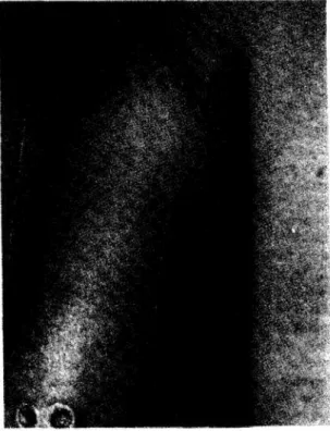
The most common type of lesion, seen in 44 patients, was an ulcer which often had elevated, indurated margins (Table 2; Figs. 1, 2).The center of the ulcer was often purulent, but occasionally it was heavily crusted. Satellite lesions around a central ulcer were seen in 15 patients. Small papular lesions were also common and occurred in 11 patients (Fig. 3). Squamous lesions (Fig. 4) were seen in three patients, an exophytic, verrucous lesion in one (Fig. 5), and acneiform lesions in one (Fig. 6). One patient develop ed a leishmanial ulcer at the site of an old surgical scar.
Sixteen patients had subcutaneous nodules pro ximal to an ulcerative skin lesion, occasionally with multiple nodules in a linear distribution. One patient with two ulcers on his arm developed a third ulcer where a subcutaneous nodule had been biops ied. Regional lymphadenopathy was present in 20 patients, and 10 of these also had subcutaneous nodules.
C h u l a y J D , O s te r C N , M c G r e e v y P B , H e n d r i c k s L D , K r e u t z e r R D . A m e r i c a n c u ta n e o u s le i s h m a n ia s is : p r e s e n t a ti o n a n d p r o b l e m s o f p a t i e n t m a n a g e m e n t. R e v is t a d a S o c ie d a d e B r a s i l e i r a d e M e d i c i n a T r o p ic a l 2 1 : 1 6 5 - 1 7 2 , O u t-D e z , 1 9 8 8
Diagnosis: There was a long delay (mean 93 days) between the onset of disease and confirmation of the diagnosis. For 51 patients, data were available to assess the factors responsible for this delay. There was a mean interval of 31 days (range 0-365 days) between appearance of a skin lesion and medical consultation, of 45 days (range 0-425 days) between medical consultation and initial consideration of cutaneous leishmaniasis, and of 17 days (range 2-120 days) until laboratory confirmation of the disease.
Twenty-three of the 60 patients were referred to
u s a f t e r t h e d i a g n o s i s h a d b e e n e s t a b l i s h e d b y f i n d in g
T a b le 2 - Number, location and type of lesions in 60 patients with American cutaneous leish maniasis.
N o . o f le sio n s N o. o f p a tie n ts
1 26
2 14
3 7
4-5 7
6-10 4
> 1 0 2
L o c a tio n o f le sio n s
Arm 29
Hand or wrist 18
Leg 16
Face or neck 15
Trunk 6
T y p e o f lesion
Ulcerated 44
Papular 11
Squamous 3
Verrucous 1
Acneiform 1
Figure 1 - A 2 8 x 3 5 m m u lc e r in a c a s e o f Leishm ania braziliensis panam anensis in fe c tio n .
amastigotes in histologic sections of skin biopsies. In another 35 patients the diagnosis of leishmaniasis was first confirmed by finding promastigotes in cultures from lesion aspirates (32), lesion biopsies (2), and a lymph node biopsy (1). The final two patients were diagnosed on the basis of exposure history, clinical appearance, and a positive serological test (one had a direct agglutination titer of 4096 and the other had an IF A titer of 32).
Overall, cultures of lesion aspirates were positi ve in 44/60 (7 3%) patients (including 12 of the 23 first
Figure 2 - M u l t i p l e u lc e r s o f c u ta n e o u s l e is h m a n ia s is .
Figure 3 - A 4 x 8 m m p a p u l e in a c a s e o f L eishm ania m exicana in fe c tio n .
C h u l a y J D , O s t e r C N , M c G r e e v y P B , H e n d r i c k s L D , K r e u t z e r R D . A m e r i c a n c u t a n e o u s l e i s h m a n i a s i s : p r e s e n t a t i o n a n d p r o b l e m s o f p a t i e n t m a n a g e m e n t . R e v i s t a d a S o c i e d a d e B r a s i l e i r a d e M e d i c i n a T r o p i c a l 2 1 : 1 6 5 - 1 7 2 , O u t - D e z , 1 9 8 8
F i g u r e 4 - A 3 0 x 4 0 m m s q u a m o u s l e s i o n f r o m w h i c h
L e ish m a n ia w e r e c u l t u r e d .
diagnosed histologically). The initial aspirate culture was positive in 37 of these 44, while six patients required two cultures and one required four cultures before organisms were isolated. Isolates from 28 patients grew sufficiently for isoenzyme analysis. L e i s h m a n i a b r a z i l i e n s i s p a n a m e n s i s was isolated from 13 patients, L . b r a z i l i e n s i s b r a z i l i e n s i s from 1, L . m e x i c a n a a m a z o n e n s i s from 9, L . m e x i c a n a m e x i c a n a from 1 ,L . d o n o v a n i c h a g a s i from 3, and a L e i s h m a n i a species with an unknown isoenzyme pattern.
The isolation of L . d . c h a g a s i from three patients with cutaneous lesions is of special interest because it is usually a viscerotropic species. One of these patients had a single small (13 mm) ulcer and another had five ulcers, including a verrucose lesion on the cheek (Fig. 5). The third patient had organisms cultured from a variety of lesions, including small papules on each elbow, excoriated papules on his thighs, an excoriation on his shoulder, and pustular acneiform lesions on his neck (Fig. 6). Because of the unusual appearance of these lesions, repeated cultures were obtained. All of six lesion cultures were positive, while two control cultures inoculated with the normal saline used to obtain aspirate cultures were negative. None of these patients had evidence of visceral leishmaniasis.
The isolation ofL. m . a m a z o n e n s i s from simple cutaneous lesions was also interesting because this species is rarely found in man (18). Eight of these patients had papular lesions (one lesion in four and two, three or more than 10 lesions in one each) with a mean maximum diameter of 13 + /- 8 mm. The other patient had a small (5 mm) ulcer. None of these patients had evidence of diffuse cutaneous leishmaniasis.
Thirty-one patients had portions of skin biops
F i g u r e 6 - A c n e i f o r m l e s i o n s o n t h e n e c k f r o m w h i c h L e i s h m a n i a d o n o v a n i c h a g a s i w a s c u l t u r e d .
ies cultured for L e i s h m a n i a , and 12 (39% ) were positive. L e i s h m a n i a were cultured from lymph node aspirates or biopsies in three patients.
IF A tests for leishmanial antibodies were posi tive in serum from 36 (82%) of the 44 patients tested. The titers ranged from 8 to 64 (geometric mean titer = 1 6 ).
Many patients were diagnosed early in the course of their disease because of our increased awareness of cutaneous leishmaniasis. Among the 12 patients who were diagnosed in connection with the prospective study duringjungle warfare training, nine had small papular or squamous lesions not usually considered suggestive of leishmaniasis, and the lesions of five of these had improved spontaneously prior to diagnosis. Isolates from 7 of these papular lesions were identified as L . m e x i c a n a ( L . m . a m a z o n e n s i s in six and L . m . m e x i c a n a in one). Another patient diagnosed early in his illness was an Army parasitologist studying l e i s h m a n i a in his laboratory who developed a small, culture-positive papule on his thumb caused by L . m . a m a z o n e n s i s . The individual with excoriated papular and acneiform lesions was cultured only because he had resided in Panam a for 2 years.
C h u l a y J D , O s te r C N , M c G r e e v y P B , H e n d r i c k s L D , K r e u t z e r R D . A m e r i c a n c u ta n e o u s le i s h m a n ia s is : p r e s e n t a ti o n a n d p r o b l e m s o f p a t i e n t m a n a g e m e n t. R e v is t a d a S o c ie d a d e B r a s i l e i r a d e M e d ic in a T r o p ic a l 2 1 : 1 6 5 - 1 7 2 , O u t- D e z , 1 9 8 8
Treatment. Fifty-six patients, including 36 who participated in a prospective randomized clinical trial (22), were treated with sodium stibogluconate intrave nously (54 patients) or intramuscularly (2 patients). Only 32 (57% ) were cured (healed lesions and negative aspiration cultures) after a single 10 day course of treatment. Sixteen patients required a se cond, two a third, and one a fourth 10 day course of sodium stibogluconate to achieve clinical and parasi tological cure.
The clinical response to sodium stibogluconate treatment was usually prompt. Within 7 to 10 days after starting treatment, all patients showed signs of healing (reduced drainage and crusting, less induration of the lesion margin, and epithelialization of the ulcer base). H o w e v e r, 24 patients required additional the rapy because of the persistance of the lesions or the relapse of healed lesions (11 patients), a positive culture (3), or both a positive culture and persistent lesions (10). Complete healing often required several months.
Five patients were not cured after multiple 10 day courses of sodium stibogluconate. One of these patients had healing of his lesions after treatment with trimethoprim sulfamethoxazole. In a second patient, ulcers on his arm healed after three courses of sodium stibogluconate but he had recurrent, nonulcerating subcutaneous nodules. Repeated cultures of the nodu les were negative, and he was not given additional therapy. Three of these antimony failures were treated with amphotericin B, but none had a favorable respon se. The first had persistent positive cultures despite clinical healing of his lesions after three courses of sodium stibogluconate and treatment with amphoteri cin (which was limited to 0.28 gm total dose because of rapid onset of azotemia). The second and third patients received larger doses of amphotericin (1.0 and 2.9 gm). Their shallow ulcers healed slowly after the end of treatment, but recurred intermittently for more than 2 years.
Meglumine antimonate was given intramuscu larly to three patients. Two achieved clinical and parasitological cure. The third healed clinically, but remained culture positive. His cultures became nega tive after retreatment with one course of sodium stibogluconate.
Twenty-five patients were seen at least 9 months after completion of treatment. Twenty-one of these remained clinically and parasitologically cured. Four patients had clinically healed lesions eventhough parasites were cultured from the lesion site on at least one occasion after treatment; lesions in all four remained healed without further treatment. Among the 18 patients followed for 2-8 months, 17 appeared cured and one had mild clinically active disease despite negative cultures. Sixteen patients who ap
peared cured at discharge 2 weeks after completing therapy failed to return for follow-up examination. Six of these patients responded to a mailed questionaire, and each denied any activity of his disease since treatment. One patient had spontaneous healing without therapy.
We identified three factors which appeared to influence prognosis: lesion type, lesion size, and the species of L e is h m a n ia causing the infection. Ulcerat ed lesions were less likely to be cured after a single course of treatment than were nonulcerated lesions (19/43 vs 15/16, x2 = 11.73, p < 0.001). Large lesions were less likely to be cured after a single course of treatment than were small lesions (Table 3). Lesions caused by L . b r a z ilie n s is were less likely to be cured after a single course of treatment than were lesions caused by other species (4/14 vs 12/14, x2 = 9.33. p < 0.005). However, these three factors are interrelat ed, because lesions caused by L . b r a z ilie n s is were larger (diameter 34 + /- 13 mm) than those caused by other species (diameter 16 + /- 10 mm. p < 0.001. Student's t-test), and lesions caused by L . m e x ic a n a were less likely to ulcerate (1/10) than those caused by L . d o n o v a n i c h a g a s i (2/3) or L . b r a z ilie n s is (14/14) (X2 = 20.28, 2 df, p > 0 .0 0 1 ).
The duration of infection did not appear to influence either the type or size of the lesions or the response to treatment. The median duration of infect ion before treatment was 106 days for ulcerative lesions and 105 days for papular lesions; 107 days for lesions < 2 cm in diameter and 111 days for lesions > 2 cm; and 111 days for lesions which where cured after
T a b le 3 - Relationship between size of lesion and _ clinical response to initial treatment with pentavalent antimonial drugs in 59 patients with American cutaneous leishmaniasis.*
D ia m e te r N u m b e r o f N u m b e r o f p a tie n ts o f la rg est p a tie n ts c u re d w ith in itia l le sio n (c m ) tr e a te d tr e a tm e n t co u rs e ( % )
< 1.0 6 5 (8 3 )
1.0-1.9 14 10(71)
2.0-2.9 12 6 (5 0 )
3.0-3.9 14 6 (4 3 )
> 4.0 13 7 (5 3 )
Total 59 34(58)
* C ure rate for lesions < 2 cm diam eter tended to be better th a n for lesions > 2 cm (75% vs 46% , x2 = 3.74, p < 0.06).
C h u l a y J D , O s te r C N , M c G r e e v y P B , H e n d r i c k s L D , K r e u t z e r R D . A m e r i c a n c u ta n e o u s le i s h m a n ia s is : p r e s e n t a ti o n a n d p r o b l e m s o f p a t i e n t m a n a g e m e n t. R e v i s t a d a S o c ie d a d e B r a s i l e i r a d e M e d i c i n a T r o p ic a l 2 1 : 1 6 5 - 1 7 2 , O u t- D e z , 1 9 8 8
one course of treatment and 106 days for lesions
re q u irin g m o re th a n o n e t r e a tm e n t c o u rs e .
Side Effects: Treatment with antimonials did not produce serious side effects. All five patients who received intramuscular therapy complained of pain at the injection site. Other symptoms possibly related to sodium stibogluconate treatment developed in 13 patients. These included headache (8 patients), blurred vision ( 3 patients), and mild abdominal pain, diarrhea, pruritic rash, tinnitus, myalgias, anorexia, depression and palpitations (1 each). Twenty-one patients developed objective clinical or laboratory abnormalities during treatment. Four developed su perficial thrombophlebitis at the site of infusion and one developed a painful, indurated, violaceous lesion at an infiltrated intravenous injection site. Transient elevations of serum ALT, LD H or A ST occurred in eight, four and three patients, respectively. Transient elevations of serum triglycerides, CPK, or alkaline phosphatase occurred in three, two and one patient, respectively. Three patients had nonspecific ST and T wave changes on E C G . One young man had an intermittent low atrial pacemaker during treatment. This was considered to be a normal v a r i a n t 1 ^ and
unrelated to the sodium stibogluconate therapy. Thirty-five patients had no side effects from treatment.
D ISC U SSIO N
Our patients with American cutaneous leish maniasis presented with a variety of skin lesions. Although ulcerated lesions were most common, 27% of the patients had atypical papular, macular, squam ous, verrucous or acneiform lesions which were diagnosed because of a high index of suspicion and the ability to obtain leishmanial cultures. Cutaneous leish maniasis should be considered in the differential diagnosis of any chronic skin lesion in a patient who has spent time in an endemic area. In the Americas these areas include parts of Texas and every country south of the United States, with the exception of Uruguay. The incubation period for 35 soldiers who had a brief exposure to infection in Panama was always less than 90 days; therefore, leishmaniasis should be strongly considered when chronic skin lesions develop during residence in an endemic area, or within 3 months of leaving the area. Infrequently, skin lesions due to leishmaniasis may appear later, as in the patient who developed her first lesion 7 months after arriving in the U.S. Consequently, a prolonged incub ation period should not be used as grounds to exclude the possibility of leishmaniasis.
After experimental infections with L . tro p ic a in man, only 50% of lesions ulcerate; the rest become papular, often with scaling, and then heal spontane
o u s l y ^ . Our experience with 10 patients diagnosed in
conjunction with a prospective study during jungle warfare training suggests that spontaneous healing, often without progression to ulceration, can also occur in American cutaneous leishmaniasis caused by L . m. a m a z o n e n s is . This is in sharp contrast to persistant diffuse cutaneous leishmaniasis and mucosal leishma niasis caused by L . m . a m a z o n e n s is. 1 ® ^4
The diagnosis of cutaneous leishmaniasis was delayed an average of 3 months in our patients due to three factors: patient delay in seeking medical advice, physician delay in considering the diagnosis, and laboratory delay in confirming the diagnosis. The first two factors might be addressed by educating people at risk and the physicians likely to provide them health care.
Confirming the diagnosis of cutaneous leishma niasis is often difficult. Serogical tests are non-speci- fic 23 and relatively insensitive1 31, especially early in the course of disease2**. Moreover, they are available at only a few reference laboratories. Histopathologic changes are said to be highly suggestive of the correct diagnosis3^ but, in our experience, histopathology often revealed only non-specific inflammatory chan ges without identifying the causative organism2**. The Montenegro skin test has not been standardized or adequately evaluated and it is not generally available. Antigen preparations vary among investigators. A number of publications indicate that the sensitivity of these preparations is > 80% , but there is only meagre data on their specificity and cross-reactivity. Slit skin smears that are performed the same way as in cases of suspected leprosy and stained with Giemsa may demonstrate amastigotes, but they are often negative in infections caused by L . b r a z ilie n s is . Culturing tissues from lesion aspirates and biopsies in appropriate media at room temperature allows conversion of amastigotes into promastigotes which subsequently multiply. Performed carefully, cultures are the most sensitive and practical method currently available to establish the diagnosis33. Various culture media have been used for this purpose13 2^ 33, but none is univer sally successful^. There is a need for new diagnostic methods that are more sensitive and rapid. The recent development of L e is h m a n ia subspecies-specific mo noclonal antibodies21 22 and kinetoplast D N A pro- bes^ hold forth the promise of rapid, definitive diagno sis by visualizing amastigotes in diseased tissue by fluorescent microscopy or D N A hybridization techni ques3 5.
The management of cutaneous leishmaniasis is often difficult because of the varied natural history of the disease and the suboptimal response to current treatment regimens. Skin lesions are usually self limited, but they often require 6-18 months to heal spontaneously 30 and may cause considerable cosmetic disfigurement. Mucosal disease occurs rarely after
C h u l a y J D, O s te r C N , M c G r e e v y P B , H e n d r i c k s L D , K r e u t z e r R D . A m e r i c a n c u ta n e o u s le i s h m a n ia s is : p r e s e n t a ti o n a n d p r o b l e m s o f p a t i e n t m a n a g e m e n t. R e v is t a d a S o c ie d a d e B r a s i l e i r a d e M e d i c i n a T r o p ic a l 2 1 : 1 6 5 - 1 7 2 , O u t- D e z , 1 9 8 8
cutaneous infection with L . b. p a n a m e n s is . Ho wever, 3% of patients with simple cutaneous leish maniasis caused by L . b. b r a z ilie n s is develop mucosal disease from months to years after an inapparent or self-healing infection19 25 32 Treatment with penta- valent antimonial drugs appears to shorten the time required for healing of skin lesions15 30. Our expe rience supports this impression as clinically active lesions improved within 10 days of starting therapy in all of our patients. Although data are limited, it is likely that systemic treatment with antimonial drugs also decreases the risk of late mucosal disease19. F or these reasons, we recommend treatment for all patients with active lesions, and for patients with confirmed or presumed L . b r a z ilie n s is infections, even if their lesions have healed spontaneously.
Forty-two percent of the patients we treated required more than one course of therapy, and 9% were not cured after multiple courses of pentavalent antimonial drugs. It is likely that the dose in conventio nal treatment regimens of sodium stibogluconate is too small and the duration of treatment too short. The levels of sodium stibogluconate required to inhibit L e is h m a n ia in vitro are frequently equal to or higher than the peak blood concentration after standard therapy with 600 mg Sb^. Due to rapid urinary excretion these peak blood levels are maintained for only a brief period of tim e12. Studies of visceral and cutaneous leishmaniasis in Kenya and India have demonstrated increased efficacy without serious toxi city when higher daily doses of sodium stibogluconate were used for up to 40 days2 10 11 29. Based on this information, we recently conducted a controlled trial in 40 patients with cutaneous ulcerative lesions caused by L . b r a z ilie n s is p a n a m e n s is in which we compared treatment with sodium stibogluconate at 10 and 20 mg Sb/kg daily for 20 days. The lower dose cured only 76% of 21 patients, but the higher dose cured all of 19 patients^. Because the prognosis of American cutan eous leishmaniasis is influenced by the type and size of ths lesion as well as the species of L e is h m a n ia , similar studies on the treatment of cutaneous leishmaniasis caused by other species of L e is h m a n ia or other types of lesions would be useful.
RESUM O
Relatamos nossa experiência em 60 pacientes com leishmaniose tegumentar americana diagnostica da e tratada entre 1977 e 1982. Cinqüenta e cinco pacientes foram infectados no Panamá, 4 no Brasil, e 1 na Colômbia. Entre 35 pacientes com uma exposição de 3 semanas no Panamá, a média do período de incubação foi 33 dias (limite sobre 4 e 81 dias). O diagnóstico foi feito, em média, 93 dias depois do início das lesões de pele, devido a demora do paciente
em procurar o serviço médico (31 dias), a demora do médico em considerar o diagnóstico (45 dias), e a demora do laboratório em confirmar o diagnóstico (17 dias). Quarenta e quatro pacientes (73% ) desenvolve ram úlceras típicas de leishmaniose cutânea. Porém,
16 pacientes (27% ) tiveram lesões de pele atípicas maculares, papulares, escamosas, verrucosas ou ac- neiformes que foram diagnosticadas somente através de culturas de leishmania. De 59 pacientes tratados com o antimonial pentavalente, só 34 (58%) obtive ram cura depois da primeira série de tratamento. Em pacientes com lesões maiores do que 2 cm de diâme tro, ou causada por L e is h m a n ia b r a z ilie n s is , ocorrem menos índice de cura depois da primeira série de tratamento do que naqueles com lesões menores do que 2 cm, fechadas, ou causadas porL. m e x ic a n a ou L . d o n o v a n i.
Palavras-chaves: Leishmaniose tegumentar america na. Diagnóstico. Tratamento. Prognóstico. Patologia Clínica.
A C K N O W L E D G E M E N T S
We thank B. Stoltman, N. Wright, J. Andujar, E. Jenkins, and A. W olf for assistance in culturing and typing L e is h m a n ia , and M. Pappas and R. Hajkowski for performing the IF A tests.
The views of the authors do not purport to reflect the position of the Department of the Army or the Department of Defense, USA.
R E FE R E N C E S
1. A llain D S , K agan IG . A direct agglutination test for leishm aniasis. A m erican Journal of T ropical M edicine and Hygiene 24: 2 32-236, 1975.
2. A nabw ani G M , N gira JA , D im iti G , Bryceson A D M . C om parison o f two dosage schedules of sodium stiboglu conate in the treatm ent o f visceral leishm aniasis in Kenya. L ancet I: 2 10-212, 1983.
3. Anthony RL, G rogl M, Sacci JB , Ballou RW . Rapid detection of L eishm ania am astigotes in fluid aspirates and biopsies o f hum an tissues. A m erican Journal of T ropical M edicine and Hygiene 37: 2 7 1 -2 7 6 , 1987. 4. Ballou W R ,M cC lain JB , G ordon D M , Shanks G D ,
A ndujar J, Berm an JD , C hulay JD . Safety and efficacy of high-dose sodium stibogluconate therapy of A m erican cutaneous leishm aniasis. L ancet II: 13-16, 1987. 5. B arker D C. D N A diagnosis of hum an leishm aniasis.
Parasitology T oday 3: 177-184, 1987.
6. Beach R, Kiilu G , H endricks L, O ste rC , L eeuw enburgJ. C utaneous leishm aniasis in K enya: transm ission of
L e i s h m a n i a m a j o r to m an by the bite of a naturally infected P h l e b o t o m u s d u b o s c q i. T ransactions o f the Royal Society of T ropical M edicine and Hygiene 78: 747-751, 1984.
C h u l a y J D , O s te r C N , M c G r e e v y P B , H e n d r i c k s L D , K r e u t z e r R D . A m e r i c a n c u ta n e o u s le i s h m a n ia s is : p r e s e n t a ti o n a n a p r o b l e m s o f p a t i e n t m a n a g e m e n t. R e v is t a d a S o c ie d a d e B r a s i l e i r a d e M e d i c i n a T r o p ic a l 2 1 : 1 6 5 - 1 7 2 , O u t- D e z , 1 9 8 8
7. B erm an J D , C hulay J D , H endricks L D , O ster C N . Susceptibility of clinically sensitive and resistant L e i s h -m a n i a to pentavalent antim ony i n v itr o . A m erican Journal of T ropical M edicine and H ygiene 31: 459-465, 1982.
8. Burroughs W ellcom e Co. Pentostam package insert. 1976.
9. C haves F , Silva S, Z eledón R, C om parison o f two culture m edia for the isolation of L e i s h m a n i a strains. Journal o f Parasitology 68: 346-347, 1982.
10. C hulay JD , A nzeze E M , K oech D K , B ryceson A D . High-dose sodium stibogluconate treatm ent of cutaneous leishm aniasis in K enya. T ransactions o f the Royal Socie ty of T ropical M edicine and Hygiene 77: 7 1 7 -7 2 1 ,1 9 8 3 . 11. Chulay JD , Bhatt SM , Muigai R, H o M , G achihi,G , W ere
JB, Chunge C, Bryceson A D . A com parison o f three dosage regimens of sodium stibogluconate in the treat m ent of visceral leishm aniasis in K enya. Journal of Infectious D iseases 148: 148-155, 1983.
12. Chulay JD , Fleckenstein L, Smith D H . Pharmacokinetics of antim ony during treatm ent o f visceral leishm aniasis ■with sodium stibogluconate or m eglumine antim oniate. T ransactions of the Royal Society o f T ropical M edicine and Hygiene 82: 6 9-72, 1988.
13. Hendricks LD , W right N , D iagnosis of cutaneous leish m aniasis by in v itr o cultivation of saline aspirates in Schneider’s D rosophila M edium . A m erican Journal of T ropical M edicine and Hygiene 28: 962-964, 1979. 14. Hiss R G , L am b LE. E lectrocardiographic findings in
122, 043 individuals. Circulation 25: 947-961, 1962. 15. K ern F , Pedersen JK . Leishm aniasis in the U nited
States. A report of ten cases in m ilitary personnel. Journal of the A m erican M edical A ssociation 226: 872-874, 1973.
16. K reutzer R D , Sem ko M E , Hendricks L D , W right N. Identification o f L e i s h m a n i a spp. by m ultiple isozyme analysis. A m erican Journal of T ropical M edicine and Hygiene 32: 7 03-715, 1983.
17. K reutzer R D , Souraty N , Sem ko M E . Biochemical identities and differences a m o n g L e i s h m a n i a species and subspecies. A m erican Journal of T ropical M edicine and Hygiene 36: 22-32, 1987.
18. Lainson R. T he A m erican leishm aniases: some observa tions on their ecology and epidemiology. T ransactions of the Royal Society o f T ropical M edicine and H ygiene 77: 569-596, 1983.
19. M arsden P D , Llanos-C uentos E A , Lago E L , C u b a C C , Barreto A C , C osta JM , Jones T C . H um an m ucocutan eous leishm aniasis in T res B raços, Bahia - Brazil. A n area o f L e i s h m a n i a b r a z il i e n s is b r a z il i e n s is transm is sion. III. M ucosal disease presentation and initial evolut ion. R evista da Sociedade B rasileira de M edicina T ropi cal 17: 179-186, 1984.
20. M cM ahon-P ratt D , Bennett E , D avid JR . M onoclonal antibodies that distinguish subspecies o f L e i s h m a n i a b r a z il i e n s is . Journal of Im munology 129: 926-927,
1982.
21. M cM ah o n -P ratt D , B ennett E , G rim aldi G , Jaffe CL. Subspecies - and species-specific antigens of L e i s h m a
n ia m e x i c a n a characterized by m onoclonal antibodies. Journal of Im m unology 134: 1935-1940, 1985. 22. O ster C N , C hulay JD , H endricks L D , Pam plin III CL,
Ballou W R, B erm an J D , Takafuji E T , T ram ont EC , Canfield C J. A m erican cutaneous leishm aniasis: a com parison of three sodium stibogluconate treatm ent sche dules. A m erican Journal o f T ropical M edicine and Hygiene 34: 856-860, 1985.
23. P appas M G , M cG reev y PB, Hajkowski R, H endricks L D , O ster C N , H ockm eyer W T . E valuation of prom as- tigote and am astigote antigens in the indirect fluorescent antibody test for A m erican cutaneous Aeishmaniasis. A m erican Journal of T ropical M edicine and Hygiene 32: 1260-1267, 1983.
24. Sam paio R N , M arsden P D , Llanos C uentas E A , C uba C A C , Grim aldi G . L e i s h m a n i a m e x i c a n a a m a z o n e n s i s
isolated from a p atient with fatal m ucosal leishm aniasis. R evista da Sociedade Brasileira de M edicina Tropical
18: 273-274, 1985.
25. Saravia N G , H olguin A F , M cM ah o n -P ratt D , D ’A les sandro A . M ucocutaneous leishm anisis in Colombia:
L e i s h m a n i a b r a z il i e n s is subspecies diversity. A m erican Journal of T ropical M edicine and H ygiene 34: 714-720,
1985.
26. Senekji H A , B eattie C P. A rtificial infection and imm uni zation of m an with cultures of L e i s h m a n i a tr o p ic a .
T ransactions o f the Royal Society o f T ropical M edicine and Hygiene 34: 4 15-419, 1941.
27. Shaw JJ , L ainson R. T he in v itr o cultivation o f m em bers of the L e i s h m a n i a b r a z il i e n s is complex. Transactions of the Royal Society o f T ropical M edicine and H ygiene 75: 127, 1981.
28. Takafuji E T , H endricks L D , D aubek JL , M cN eil KM , Scagliola H M , Diggs C L . C utaneous leishm aniasis as sociated with jungle training. A m erican Journal of T ropi cal M edicine and Hygiene 29: 5 16-520, 1980. 29. T hakur C P , K u m ar M , K u m ar P , M ish ra B N , Pandey
A K . R ationalisation o f regim ens o f treatm ent of K ala-azar with sodium stibogluconate in India: a ran dom ised study. British M edical Journal 2 9 6 :1 5 5 7 -1 5 6 1 ,
1988.
30. T hornburgh D B, Johnson C M , E lton N W . H istopatho- logy of cutaneous leishm aniasis in Panam a. T ransactions of the R oyal Society o f T ropical M edicine and Hygiene 46: 550-554, 1952.
31. W alton BC, Brooks W H , A rjona I. Serodiagnosis of A m erican leishm aniasis by indirect fluorescent antibody test. A m erican Journal o fT ro p ic al M edicine and H ygie ne 21:296-299, 1972.
32. W alton BC, Chinel LV, E guia O E . O nset o f espundia after m any years of occult infection with L e i s h m a n i a b r a z il i e n s is . A m erican Journal o f T ropical M edicine and Hygiene 22: 6 96-698, 1973.
33. W eigle K A , de D âvalos M , H eredia P, M olineros R, Saravia N G , D ’A lessandro A. D iagnosis of cutaneous and m ucocutaneous leishm aniasis in Colombia: A com parison o f seven m ethods. A m erican Journal ofT ro p ical M edicine and Hygiene 36: 4 89-496, 1987.