Abstract
Staphylococcus aureus (S. aureus) osteomyelitis is a significant complication for orthopaedic patients undergoing surgery, particularly with fracture ixation and arthroplasty. Given the dificulty in studying S. aureus infections in human subjects, animal models serve an integral role in exploring the pathogenesis of osteomyelitis, and aid in determining the eficacy of prophylactic and therapeutic treatments. Animal models should mimic the clinical scenarios seen in patients as closely as possible to permit the experimental results to be translated to the corresponding clinical care. To help understand existing animal models of S. aureus, we conducted a systematic search of PubMed and Ovid MEDLINE to identify in vivo animal experiments that have investigated the management of S. aureus osteomyelitis in the context of fractures and metallic implants. In this review, experimental studies are categorised by animal species and are further classiied by the setting of the infection. Study methods are summarised and the relevant advantages and disadvantages of each species and model are discussed. While no ideal animal model exists, the understanding of a model’s strengths and limitations should assist clinicians and researchers to appropriately select an animal model to translate the conclusions to the clinical setting.
Keywords: Osteomyelitis; animal models,
methicillin-resistant Staphylococcus aureus (MRSA), infection; fracture, implant.
*Address for correspondence: Stephen L. Kates
601 Elmwood Ave, Box 665 Rochester, NY 14642, USA
Email: [email protected]
Introduction
Infection and osteomyelitis represent significant complications of orthopaedic surgery. Infection rates associated with total hip arthroplasty and total knee arthroplasty have been historically reported to be 0.88 % and 0.92 %, respectively, and result in substantial morbidity (Kurtz et al., 2008). As the number of total hip and knee replacements are projected to grow between 174 % and 673 %, during the next 20 years in the United States alone (Kurtz et al., 2007), controlling these rates is of great concern. A recent report by Cram et al. has shown the infection rate after total knee replacement to be increasing with time to 3 % (Cram et al., 2012). In contrast to the rates associated with the controlled environment of arthroplasty, the infection rates of open fractures range from several percentage points to a staggering 50 % (Gustilo et al., 1984; Zalavras et al., 2005).Implant associated osteomyelitis inlicts signiicant morbidity and mortality to the patient and proves a notable challenge for the orthopaedic surgeon.
The increase in antibiotic resistant pathogens has further complicated the management of osteomyelitis (Cardo et al., 2004). S. aureus and coagulase-negative Staphylococci account for 65-80 % of infections (Schmidt and Swiontkowski, 2000); Darouiche, 2004 and a steep rise in the incidence of methicillin-resistant S. aureus and the ongoing emergence of vancomycin-resistant S. aureus is well documented (Parvizi et al., 2009; Loomba et al., 2010). Recent studies have shown that patients with a conirmed methicillin-resistant Staphylococcus aureus (MRSA) infection experience longer hospital stays, require prolonged antibiotic therapy (usually intravenous) and experience a 2.7 fold increase in mortality compared to non-infected inpatients (Nixon et al., 2006; Lee et al., 2010). One estimate of the mean cost attributable to an MRSA infection is $35,367 (Stone et al., 2002); and, in a 2011 APIC (Association for Professionals in Infection Control and Epidemiology) publication (Rebmann and Aureden, 2011), the annual cost to treat MRSA in hospitalised patients in the United States was quoted between $3.2 billion to $4.2 billion. Accordingly, the successful development of novel therapies to prevent and manage MRSA infections promises to deliver vast beneits in patient care and for the healthcare community.
In the face of such infections, infectious disease and orthopaedic researchers collaborate to reine the management of osteomyelitis. Human clinical trials are inherently difficult to conduct secondary to the low incidence of implant-associated osteomyelitis, the heterogeneous population, various treatment modalities
A SYSTEMATIC REVIEW OF ANIMAL MODELS FOR
STAPHYLOCOCCUS AUREUS OSTEOMYELITIS
W. Reizner, J.G. Hunter, N.T. O’Malley, R.D. Southgate, E.M. Schwarz and S.L. Kates*
and the broad range of causative pathogens and associated virulence patterns (Lazzarini et al., 2006). Experimental studies in animal models serve to ill this void. While it is dificult to precisely replicate disease onset and progression characteristics of human infection, animal models facilitate our understanding of osteomyelitis and often inform clinical practice (Norden, 1988). In order to address implant associated osteomyelitis, investigators must ask disease speciic questions and build on established animal models capable of answering these questions. For this purpose, we have systematically reviewed experimental studies in animals that explore S. aureus osteomyelitis to provide both clinicians and basic scientists with an understanding of current animal models that address the prevention and management of osteomyelitis.
Methods
Search strategy
Two databases, PubMed and Ovid MEDLINE, were used to systematically identify studies exploring osteomyelitis in S. aureus animal models. The PubMed database was systematically searched with the following search string and included articles through December 31st, 2012: ‘(infection) AND (animal model) AND (arthroplasty OR fracture OR internal ixation OR prosthesis).’ The search strategy for Ovid MEDLINE (1902 – December 31st, 2012) is described in Table 1. Three reviewers participated in screening and study selection. Two authors (NO’M and WR) individually reviewed all abstracts of identified articles. Studies that met eligibility criteria during this initial screening were retrieved and reviewed in full. A third reviewer (JH) resolved any discrepancies. Additional publications were identiied within the reference section of acquired studies, and additional publications of interest were included to supplement those identiied by means of the systematic search.
Inclusion/exclusion criteria
Inclusion criteria included: (1) in vivo experimental study, (2) investigation of the management of S. aureus osteomyelitis, and (3) an experimental model based on an animal species. Exclusion criteria included (1) vertebral osteomyelitis, (2) craniofacial osteomyelitis, and (3) infections not clearly deined as osteomyelitis. Furthermore, letters, abstracts, cases and reviews were excluded. Non-English reports that were unavailable in an English transcript were excluded.
Summary of the literature search
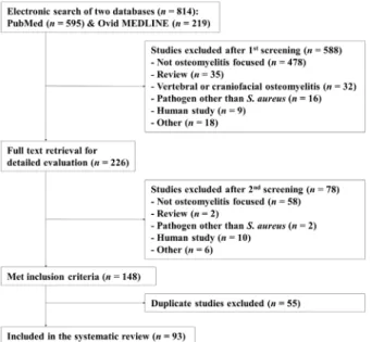
Fig. 1 depicts the results of our systematic search. A total of 814 articles were identiied. 588 articles were excluded during the initial screening of titles and abstracts and seventy-eight articles were excluded during the second screening. Of the 148 articles that met eligibility criteria, fifty-five were duplicates. Ultimately, ninety-three experimental studies of S. aureus were included to be discussed in this systematic review.
Within the initial screening, 478 of the 588 excluded studies did not explore osteomyelitis. The majority of these articles investigated non-orthopaedic infections (i.e. cardiovascular infections, gastrointestinal infections, etc.). Additionally, any orthopaedic study that did not include an infection-related endpoint did not meet eligibility criteria. Thirty-ive articles were reviews, thirty-two studies investigated vertebral or craniofacial osteomyelitis, sixteen studies explored non-S. aureus osteomyelitis (pathogens included S. epidermidis, Pseudomonas aeruginosa, Escherichia coli and Acinetobacter baumannii), and nine articles were human studies. The remaining eighteen excluded studies consist of seventeen non-English articles that could not be reviewed and one case study.
Once the remaining 226 articles were retrieved in full, the reviewers excluded ifty-eight studies which did not directly address osteomyelitis. Ten experimental studies were primarily in vivo human studies with a minor animal species component, two manuscripts were classiied as
# Search Words
1 exp Infection/
2 exp Models, Animal/
3 exp Arthroplasty
4 exp Fracture Fixation, Internal/ or exp Fracture Fixation/ or exp Fracture Healing/ or exp Fracture Fixation, Intramedullary/
5 exp Bone Screws/ or exp Femoral Fractures/ or exp Fractures, Bone/ or exp Fracture Fixation, Internal/ or exp Bone Plates
6 exp “Prostheses and Implants”/
7 1 and 2 and (3 or 4 or 5 or 6)
8 Limit 7 to yr = “1902-2012”
Table1. Ovid MEDLINE Advanced Search Strategy
reviews, and two studies did not focus on S. aureus. Of the six additional excluded articles, three were non-English studies, two articles were letters, and one was a detailed study proposal that did not possess outcomes. Of note, the number of excluded articles is overestimated as the screening of the PubMed and Ovid MEDLINE searches were done in parallel. Duplicates were calculated only for included studies following the second screening.
Model design characteristics
The experimental studies obtained using the systematic search utilise various animal species, study varying strains of Staphylococcus aureus, and explore distinct aspects of osteomyelitis including diagnostic, prophylactic, and therapeutic measures. Table 2 highlights the major elements of each of the ninety-three experimental studies that met our inclusion criteria. Tables 2-5 provide speciic details: (1) the animal species utilised, (2) the pathogenic strain studied, (3) the amount of inoculum and the method of inoculation, (4) the study question/purpose, and (5) the principal indings for each reference. Experimental studies are organised according to the use of an internal ixation, open fracture, periprosthetic, external ixation, or haematogenous model. Of note, this systematic review did not identify any rabbit or murine studies employing an external ixation model.
Animal species
Thirty-six references utilised the rabbit as the animal model (Tables 2a, and 2b), thirty-three of which employed the New Zealand white rabbit (NZW). Twenty-nine references utilised a rat model (Tables 3a and 3b), the majority of which employed either the Wistar rat or the Sprague Dawley rat. Seven references utilised a mouse model (Table 4): either the BALB/c or C57BL/6 strain. Seven references utilised an ovine model, six employed a canine model, four a goat model, two a porcine model, one a guinea pig model, and one a hamster model (Table 5).
Staphylococcus aureus strain
Researchers utilised a wide range of Staphylococcus aureus strains to investigate osteomyelitis. Of note, twenty-eight references possessed a clinical isolate or unspeciied strain. The sensitivities of clinical isolates are often detailed in the accompanying reference. Common strains include ATCC 25923, ATCC 29213, ATCC 49230 as well as bioluminescent strains (i.e. ATCC12600). Thirteen experiments explored osteomyelitis secondary to methicillin-resistant Staphylococcus aureus. Nine of these utilised a clinical isolate. MRSA strains included ATCC 33591, ST-021 (a clinical isolate from University of Maryland, Shock Trauma) (Craig et al., 2005), an MLST-80 clone (Poultsides et al., 2008), and the bioluminescent USA300 LAC:;lux strain (Niska et al., 2012).
Methods of evaluation
Animal experiments exploring osteomyelitis aim to either investigate the pathogenesis of osteomyelitis, examine diagnostic tools, study the efficacy of prophylactic modalities and consider varying therapeutic options. These studies typically conirmed the presence of osteomyelitis,
and those studies exploring the natural course of osteomyelitis or examining the eficacy of prophylactic and therapeutic protocols monitored the progression of the infection in a reproducible and an informative manner. Studies commonly deined the primary endpoint as rate of clinical infection. This was commonly documented by one or more of the following criteria: gross infection (i.e., purulent drainage from the infection site), histologic analysis, X-ray, serum markers of inlammation or capture of photons using bioluminescent imaging. Nonetheless, the manner in which a study establishes the presence of infection varies. Beyond noting local (erythema, swelling or abscess formation) or systemic (fever or lethargy) clinical signs of infection, studies often conducted advanced imaging (i.e., X-ray, computed tomography (CT) scan or magnetic resonance imaging (MRI)), as well as microbiologic and histologic evaluations. Additionally, researchers cultured intracardial blood samples, bone samples, or swabs from pins sites or hardware. Cultures were grown over nutrient agar or in tryptic soy broth. Growth was often considered a binary result (i.e., with growth or without growth). Pin site swabs were assessed in a similar manner. When quantifying the magnitude of infection in bone, researchers pulverised bone samples, suspended the samples in sterile phosphate buffered saline solution and determined colony forming units per gram of bone. To expand on the colony forming units per gram of bone measurement, select studies performed polymerase chain reaction (PCR) assays to assess the presence of bacterial DNA and potentially quantify the bacterial load (Nijhof et al., 2001).
Infections were also qualitatively assessed by radiologic, macroscopic and histologic evaluation. Radiographic evaluation of infected bone is commonly performed according to a score by An et al. (An and Friedman, 1998; Lucke et al., 2003a), grading the involved bone on periosteal reaction, osteolysis, soft-tissue swelling, deformity, sequestrum formation, spontaneous fracture and general impression. One macroscopic approach or lesion scoring employs the Rissing scale (Rissing et al., 1985b; Shandley et al., 2012), converting the appearance of infected bone into a quantitative scale: 0 = no visible evidence of infection; 1 = minimal erythema without bone destruction, without abscess; 2 = erythema with bone formation and minimal bone destruction; 3 = abscess with new bone formation, bone destruction and with purulent exudates; 4 = severe bone resorption, abscess, and total bone involvement. Studies performing a histologic examination of bone samples, such as Huneault et al., analysed periosteal proliferation, cortex remodelling, endosteal proliferation, periosteal neutrophilic inlammation, periosteal lymphoplasmocytic inlammation, marrow lymphoplasmocytic inlammation, sequestrum and bacteria ( Petty et al., 1985; Huneault et al., 2004).
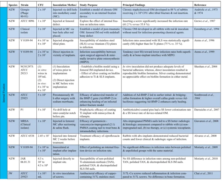
Table 2a. Experimental rabbit studies of Staphylococcus aureus osteomyelitis
as to the degree and extent of infection and offers valuable insight to the metabolic status of the bacteria. These quantitative measurements are made over time in the same animal, in contrast to gross and histologic evaluations that require sacriice of the test subject. Consequently, one is able to observe the temporal response of infection to an intervention in real-time.
The following section summarises many of the articles that met inclusion criteria and references additional experiments that inform and supplement those found by our systematic approach.
Literature Review of Animal Models
Rabbit models of osteomyelitis
Internal ixation models: acute and chronic osteomyelitis
The original tibial rabbit model was described by Schemen and then later modiied by Norden (Norden, 1970), and involved inoculation of a sclerosing agent (5 % sodium morrhuate) via an 18 gauge needle followed with S. aureus to produce chronic osteomyelitis of the tibia. Insertion of a foreign body (i.e. metallic implants) was introduced by Andriole et al. (Andriole et al., 1973). They fractured the tibia using a simple three-pronged clamp and then used a stainless steel pin for ixation, and were able to monitor
infection for up to 18 months. Other authors have used the same technique without fracture (Moriarty et al., 2010). Arens et al. created a model using plate ixation for the tibiae using 6-hole dynamic compression plating (DCP), and in one study, performed it using minimally invasive plate osteosynthesis (MIPO) (Arens et al., 1999). However, no fracture was created and only unicortical screws were inserted, limiting its applications.
With respect to the femur, investigators historically accessed the femoral canal through the greater trochanter (Eerenberg et al., 1994); however, recently published models are accessing the canal through the intercondylar notch using a parapatellar incision (Castro et al., 2003; Sheehan et al., 2004; Darouiche et al., 2007; Giavaresi et al., 2008). Authors report ease of exposure and access to the canal, though this technique compromises the knee joint and thus infection may not be limited to the femoral shaft. Once access is obtained after reaming, the investigator can perform the bacterial inoculation, place the cement (Castro et al., 2003) or screw (Darouiche et al., 2007), and inally, seal the intercondylar notch with bone wax. As such, these models provide multiple options for treatment and testing techniques. These models could be improved upon by creating a closed fracture to more closely mimic the clinical scenario. Using a simple lateral approach to the femur, Southwood et al. dissected through the soft tissue
Species Strain CFU Inoculation Method Study Purpose Principal Findings Reference
Internal Fixation
NZW Giorgio (Phage 80-81)
2 x 10⁸ Injected via drill hole
into medullary canal (MC).
Establish a model of chronic OM
secondary to an internal ixation
device.
Chronic staphylococcal OM developed in 88 % of rabbits
receiving 2 x 10⁸ CFU after tibial fracture & rod insertion.
Andriole et al., 1973 & 1974
NZW ATCC 8096 1 x 10⁸ Injected at femoral
defect.
Explore the effect of internal ixa -tion on infec-tion rates.
Inserting a screw signiicantly increased the infection rate
(61.2 % versus 35.8 %).
Grewe et al., 1987
NZW Clinical isolate
3 x 10⁵ Injected in MC via
burr hole after rod placed.
Establish a model of post-traumatic OM: femoral IM rod with midshaft bony defect
Chronic OM developed in all rabbits with rod & inoculum without need for infection-promoting chemical agents.
Eerenberg et al., 1994
NZW V 8189-94 4 x 10³ to 4 x 10⁶
Injected alongside tibial plate.
Susceptibility of stainless steel (SLS) versus titanium (Ti) plates to infection.
Infection rates associated with SLS was statistically signii
-cantly (SS) higher than for Ti plates (75 % vs. 35 %).
Arens et al., 1996
NZW V 8189-94 2 x 10³ to 2 x 10⁷
Direct injection in vicinity of implants.
Infection susceptibility between open surgical approach & mini-mally invasive plate osteosynthesis (MIPO).
Tendency (not SS) toward lower infection rates both superi -cially & at bone-implant interface with MIPO.
Arens et al., 1999
NZW NC012973: ATCC 29213
(1)
in vitro: wires in 108/mL
(2)
in vivo:
4 x 10⁵ to 4 x 10⁶
(1) Inoculation of wires prior to implantation
(2) Direct injection in MC before wire implantation
- Establish a bioilm model using a
femoral IM-implanted device.
- Effect of silver coating on bioilm
adhesion to Ti & SLS implants.
In vitro inoculation did not produce adequate levels of bacterial adhesion; whereas, direct inoculation resulted in
reproducible bioilm formation. Silver coating demonstrated no appreciable effect on bioilm formation in either metal.
Sheehan et al., 2004
NZW ATCC
25923 5 x 10
6 Percutaneously 48
h after surgery with sodium morrhuate.
Eficacy of adenoviral transfer of
the BMP-2 gene (Ad-BMP-2) in enhancing healing of an infected defect fracture model
Addition of Ad-BMP-2 led to earlier initial- & bridging-callus formation & higher overall bridging-callus grade versus Ad-luciferase suggesting Ad-BMP-2 enhances early healing.
Southwood et al., 2004
NZW P1 5 x 10² Via drill hole at
inter-condylar notch before pin.
Prophylactic eficacy of coating
Ti implant with minocycline & rifampin.
Antibiotic(abx)-coated pins had a SS lower colonisation rate
& a SS lower rate of device-related OM.
Darouiche et al., 2007
NZW MRSA (Clinical isolate)
1 x 10⁶ Injected in femoral
MC after suctioning
& saline lush.
Eficacy of gentamicin -
vancomycin-impregnated (2:1) PMMA coating nail to treat bone & intramedullary infections.
Abx-impregnated PMMA nails led to a SS better radiologic
& histologic assessment compared to rabbits with (a) non-impregnated nail, (b) no therapy, or (c) systemic teicoplanin.
Giavaresi et al., 2008
NZW ATCC 6538 2.45 x 10⁷ Injected into femoral
MC 2 weeks before treatment.
Treatment eficacy of ciproloxacin
implants.
Rabbits with abx-implants demonstrated reduced bacterial
counts and fewer clinical & radiologic signs of infection.
Alvarez et al., 2008
NZW V 8189-94 2 x 10⁴ to 1 x 10⁸
Innoculated at implant after implan-tation.
Effect of polishing an internal ixa -tion device on infec-tion rate.
No signiicant difference in infection rates between polished
& unpolished groups with the same material.
Moriarty et al., 2009
NZW JAR
06.01.31
4.3 x
101 to 4.3 x 10⁴
Injected directly to implant site.
Susceptibility of non-polished Ti-aluminium-niobium (TAN), polished TAN, & electropolished SLS IM nails.
No SS difference in infection rates among non-polished TAN, polished TAN, & electropolished SLS IM nails.
Moriarty et al., 2010
JW ATCC
25923 1 x 10 5/
mL
In vitro inoculation of screws.
Antibacterial eficacy of copper-
containing 317L stainless steel.
317L-Cu screws reduced inlammation & infection com -pared to 317L screws. No difference in bone formation.
and periosteum of the femoral diaphysis where a 10 mm defect could be made with a burr (Southwood et al., 2004). Fixation followed with stacked bone plates, 2.0 mm cortical screws and cerclage wire. While these authors used sodium morrhuate to aid in creating osteomyelitis, this is somewhat controversial and may not be needed. Many investigators referenced in this article have shown reproducible models without the aid of a sclerosing agent, and it may also skew outcomes if host response is being studied, as the infection cannot all be attributed to the bacteria.
Open fracture models
Open fracture rabbit models primarily utilise the tibia. Ashhurst et al. irst described a fracture model; fractures
were created by a saw and were stabilised with plates (Ashhurst et al., 1982). Worlock et al. modiied this scheme to generate a model with ixation of an intramedullary rod (Worlock et al., 1988); this method has in turn been employed by others (Kraft et al., 2001). An essential feature of this model is creating a wound over the fracture site where the contamination can take place. Worlock et al. report 107 colony forming units (CFU) as the minimal dose to produce a consistent infection (> 80 %). Subsequently, ixation can take place at the discretion of the investigator to appropriately address the experimental question. The intramedullary (IM) nail described by Worlock lacked rotational stability, differing from human cases where interlocking bolts are used to provide stability.
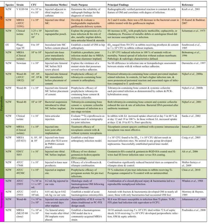
Table 2b. Experimental rabbit studies of Staphylococcus aureus osteomyelitis (...continued)
Species Strain CFU Inoculation Method Study Purpose Principal Findings Reference
OF
NZW V 8189-94 4 x 10³ to 4 x 10⁷
Injected adjacent to tibial implant via
catheter.
Determine the reliability of
radiograph indings for the
diagnosis of osteitis.
Radiographically veriied periosteal reaction is constant & early
feature of OM and correlates with degree of infection.
Kraft et al., 2001
NZW MRSA (ATCC
33591)
1 x 10⁶ Injected into tibial
MC.
Develop & evaluate a biodegradable implantable
gatiloxacin delivery system.
At 2 and 4 weeks, there was a SS decrease in the bacterial count in
rabbits treated with the gatiloxacin implant.
El-Kamel & Baddour, 2007
Periprosthetic
NZW Clinical isolate
3.25 x 101 to 3.9 x
104
Injected into suprapatellar pouch.
Explore the progression of a
knee infection & the role of
abx, metallic implant debris,
intra-articular blood & steroids.
SS increase in ID50 with prophylactic methicillin, cephacetrile, or clindamycin. Presence of metallic debris or autologous blood did not increase susceptibility to infection.
Schurman et al., 1975
ES Phage
type 95 0 to 10
8 Inoculated into MC
before cement placed.
Establish a model of OM in hip arthroplasty.
ID50 ranged from 50 CFU in rabbits receiving prosthesis & cement
to 1 x 10⁴ CFU in rabbits without implants.
Southwood et al.,
1985
NZW MRSA (Clinical isolate)
105 to 108 Injected into knee
joint immediately after implantation.
Establish a prosthetic joint infection model due to MRSA. [Silicone elastomer implant]
5 x 10⁶ CFU induced infection in 100 % of animals with no
mortality. Natural spread of infection without sodium morrhuate.
Pathologic & radiologic characteristics deined.
Belmatoug et al., 1996
NZW Newman 1 x 10⁴ Injected into femoral
MC before SLS screw placed.
Explore the virulence of a
mutant strain that possesses several surface adhesins.
No SS difference in infection rate or histopathologic assessment between strains with & without surface adhesins.
Darouiche et al., 1997
NZW Wood-46 (ATCC 10832)
103, 104,
105, 106 &
107
Injected into femoral MC immediately before placement of cement.
Prophylactic eficacy of
tobramycin-containing bone cement in arthroplasty.
Premixed tobramycin-containing bone cement prevented
implant-related infection. In controls, SA had a higher infection rate, & more pronounced periosteal reaction and associated signs of infec-tion compared to S. epidermidis.
Nijhof et al., 2000a
NZW Wood-46 1 x 10⁶ Injected into femoral
MC before placement of cement.
Prophylactic eficacy of
prophylactic tobramycin-containing bone cement & systemic cefazolin.
Tobramycin-containing bone cement & systemic cefazolin each prevented infection as demonstrated by culture & PCR-hybridisation assay.
Nijhof et al., 2000b
NZW Wood-46 105 or 106 Bacterial suspension
introduced to tibial MC before implant.
Tobramycin-containing bone cement vs. systemic cefazolin for treatment of infection in a one-stage revision.
Both tobramycin-containing bone cement and systemic cefazolin reduced the size & rate of infection. Bacterial DNA persisted after antibiotic treatment.
Nijhof et al., 2001
NZW Clinical isolate
1 x 10⁷ Intra-articular
injection.
Evaluate 99mTc-ciproloxacin as
a marker used in scintigraphy for joint infection.
In rabbits with SA, increased uptake observed at day 5 in 60 % &
at day 12 and 19 in 100 %. In those without SA, increased uptake
at days 12 & 19 in 83 %. Poor speciicity.
Sarda et al., 2002
NZW MRSA (Clinical isolate)
1 x 10⁸ Injected into
knee joint after implantation.
Treatment eficacy of
teicoplanin cement with & without systemic teicoplanin.
Teicoplanin cement spacers combined with systemic intramuscular teicoplanin was most effective.
Ismael et al., 2003
NZW MRSA (ST-021)
0, 102, 103
& 104
Injected into knee joint after SLS screw & PMMA cement placed.
Establish a total knee arthroplasty infection model.
1 x 10³ CFU found to be ID70. 1 x 10⁴ CFU did not result in
increased infection rates. No animal in any group developed septicaemia. Successfully established paired knee model.
Craig et al., 2005
NZW EDCC
5055 1 x 10
7 Injected into tibial
MC before implant.
Eficacy of two distinct gentamicin-hydroxyapatite
(HA) coatings.
Gentamicin-HA-coated & gentamicin-RGD-HA-coated steel K-wires had SS lower infection rates versus HA-coating.
Alt et al., 2006
NZW ATCC
17848
1 x 10⁷ Injected in knee near
prosthesis.
Eficacy of a levoloxacin &
rifampin combination.
Combination signiicantly reduced bacterial titers as compared to
rifampin alone & control.
Muller-Serieys et al., 2009
NZW ATCC
49230C
1 x 10⁸ Injected at implant
site.
Prophylactic eficacy of pexiganan acetate for pin tract
infections.
75 % reduction of pin tract infections in rabbits receiving Ti-Pexiganan compared to Ti-control with no antimicrobial.
Chou et al., 2010
KEY: NZW – New Zealand White; JW – Japanese White; ES – English Shorthair; SA – S. aureus; SS – signiicantly, statistically signiicant; OM – osteomyelitis; MC – medullary canal; SLS – stainless steel; Ti – titanium
Hematogenous
NZW ATCC
25953 7 x 10 7 to 1.1 x 10⁸/
mL
0.2 mL/kg injected in
ear vein.
Histologic study of haematogenous OM following physeal fracture.
Combination of a closed physeal injury & bacteraemia led to a reproducible metaphyseal infection.
Whalen et al., 1988
NZW ATCC
25953 4.65 x 10⁸/mL
0.01 mL/kg or 0.02 mL/kg via ear vein.
Establish a model of acute haematogenous osteomyelitis.
Animals with fracture & bacteraemia developed OM in nearly all cases; those with only bacteraemia had occasional OM foci
Morrissy & Haynes, 1989
NZW Wood-46 1 x 10⁸ to 2 x 10⁹
Injected into auricular vein several days after implant.
Susceptibility of SLS versus Ti plates (traditional or PC-FIX design used).
SLS was SS more susceptible to infection than Ti plates. Ti-PC-FIX plate had infection risk equivalent to 0 CFU.
Johansson et al., 1999
NZW MRSA
(MLST-80 clone)
5 x 10⁸ or 3 x 10⁸
Via femoral artery four weeks after tibial IM implants were placed.
Establish a haematogenous OM model due to a community-acquired MRSA strain.
10/10 animals receiving 5 x 10⁸ CFU died within 72 h of septic shock. 8/10 receiving 3 x 10⁸ CFU developed periprosthetic
infec-tion, OM & septic arthritis.
In a rabbit osteomyelitis model that involved inoculation of bacteria into devascularised bone, Smeltzer et al. demonstrated the importance of virulence factors in establishing osteomyelitis (Smeltzer et al., 1997). Interestingly, the strain Smith diffuse, a strain that had demonstrated enhanced virulence in a murine peritonitis model, exhibited relatively little evidence of osteomyelitis compared to that of the strain UAMS-1. UAMS-1 achieved an infection rate of 75 % with an inoculum of 2 x 10³ CFU.
Periprosthetic models
Rabbits are the smallest animals in which true models of prosthesis related osteomyelitis are described in the literature. Knee arthroplasty components exist with modiication of existing joint replacements for humans. Belmatoug et al. used a human irst metatarsophalangeal (MTP) silicone-elastomer implant for the tibial tray of a knee arthroplasty (Belmatoug et al., 1996). Inoculants of 5 x 10⁶ resulted in consistent infections. Of note, a
silicone implant allows for MRI and micro-computed tomography (μCT) without artefact distortion, a signiicant advantage over most implants. Craig et al. produced a novel arthroplasty model in which a metal screw and ultra-high molecular weight polyethylene (UHMWPE) washer was secured to the non-articulating surface of the lateral femoral condyle (Craig et al., 2005). All the various materials seen in a typical arthroplasty are present; however, the same stresses and strains placed on a weight bearing total joint arthroplasty (TJA) are not recreated with this model, which may bias results of therapeutic studies. A one-stage revision model has been described by inserting a standardised cement plug in to the tibial metaphysis, followed by contamination of 105 or 106 S. aureus directly anterior to the insertion of the anterior cruciate ligament of the knee (Nijhof et al., 2001). This plug has a hook, thereby making a single exchange reproducible, even after several weeks of incorporation in the host.
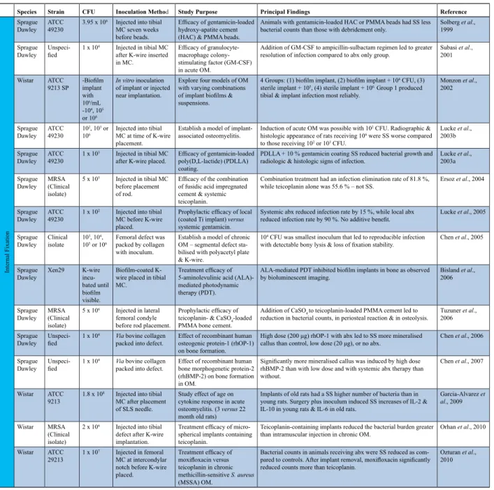
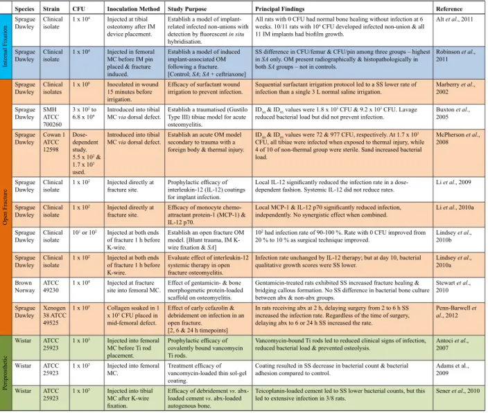
Table 3a. Experimental rat studies of Staphylococcus aureus osteomyelitis
Species Strain CFU Inoculation Method Study Purpose Principal Findings Reference
Internal Fixation
Sprague Dawley
ATCC 49230
3.95 x 10⁶ Injected into tibial
MC seven weeks before beads.
Eficacy of gentamicin-loaded hydroxy-apatite cement
(HAC) & PMMA beads.
Animals with gentamicin-loaded HAC or PMMA beads had SS less bacterial counts than those with debridement only.
Solberg et al., 1999
Sprague Dawley
Unspeci-ied 1 x 10
4 Injected in tibial MC
after K-wire inserted in MC.
Eficacy of
granulocyte-macrophage colony-stimulating factor (GM-CSF) in acute OM.
Addition of GM-CSF to ampicillin-sulbactam regimen led to greater
resolution of infection compared to abx only group.
Subasi et al., 2001 Wistar ATCC 9213 SP -Bioilm implant with 106/mL
-104, 105
or 106
In vitro inoculation of implant or injected near implantation.
Explore four models of OM
with varying combinations
of implant bioilms &
suspensions.
4 Groups: (1) bioilm implant, (2) bioilm implant + 104 CFU, (3) sterile implant + 105, (4) sterile implant + 106. Group 1 produced
tibial & implant infection most reliably.
Monzon et al., 2002
Sprague Dawley
ATCC 49230
102, 103 or
106
Injected into tibial MC at time of K-wire placement.
Establish a model of implant-associated osteomyelitis.
Induction of acute OM was possible with 102 CFU. Radiographic &
histologic appearance of rats receiving 106 were SS worse compared
to those receiving 102 or 103 CFU.
Lucke et al., 2003b
Sprague Dawley
ATCC 49230
1 x 10³ Injected in tibial MC
after K-wire placed.
Eficacy of gentamicin-loaded
poly(D,L-lactide) (PDLLA) coating.
PDLLA + 10 % gentamicin coating SS reduced bacterial growth and
radiologic & histologic signs of infection.
Lucke et al., 2003a Sprague Dawley MRSA (Clinical isolate)
5 x 10⁵ Injected in tibial MC
before placement of rod.
Eficacy of the combination
of fusidic acid impregnated cement & systemic teicoplanin.
Combination treatment had an infection elimination rate of 81.8 %,
while teicoplanin alone was 55.6 % – not SS. Ersoz et al., 2004
Sprague Dawley
ATCC 49230
1 x 10² Injected into tibial
MC before K-wire placed.
Prophylactic eficacy of local
(coated Ti implant) versus
systemic gentamicin.
Systemic abx reduced infection rate by 15 %, while local abx
reduced infection rate by 90 %. No additive beneit. Lucke et al., 2005
Sprague Dawley
Clinical isolate
103, 104,
105 or 106
Femoral defect was packed by collagen with inoculum.
Establish a model of chronic OM – segmental defect sta-bilised with polyacetyl plate & K-wire.
104 CFU was smallest inoculum that led to reproducible infection
with detectable bony lysis & loss of ixation stability. Chen et al., 2005
Sprague Dawley Xen29 K-wire incu-bated until bioilm visible. Bioilm-coated
K-wire placed in tibial MC.
Treatment eficacy of 5-aminolevulinic acid
(ALA)-mediated photodynamic therapy (PDT).
ALA-mediated PDT inhibited bioilm implants in bone as observed
by bioluminescent imaging.
Bisland et al., 2006 Sprague Dawley MRSA (Clinical isolate)
5 x 10⁶ Injected in lateral
femoral condyle before rod placement.
Prophylactic eficacy of
teicoplanin- & CaSO4-loaded PMMA bone cement.
Addition of CaSO4 to teicoplanin-loaded PMMA cement led to
reduction in bacterial counts, in periosteal reaction & in osteolysis.
Tuzuner et al., 2006
Sprague Dawley
Unspeci-ied 1 x 10
4 Via bovine collagen
packed into defect.
Effect of recombinant human osteogenic protein-1 (rhOP-1) on bone formation.
High dose (200 μg) rhOP-1 with abx led to SS more mineralised callus than control, low dose (20 μg), or no abx.
Chen et al., 2006
Sprague Dawley
Unspeci-ied 1 x 10
4 Via bovine collagen
packed into defect.
Effect of recombinant human bone morphogenetic protein-2 (rhBMP-2) on bone formation in OM.
Signiicantly more mineralised callus was induced by high dose rhBMP-2 than with low dose and with systemic abx therapy than
without.
Chen et al., 2007
Wistar ATCC 9213
1.8 x 10⁸ Injected into tibial
MC after placement of SLS needle.
Study effect of age on cytokine response in acute osteomyelitis. (3 versus 22 month old rats)
Implants of old rats had a SS higher number of bacteria than in young rats. Surgery plus inoculum induced SS increases of IL-2 & IL-10 in young rats & IL-6 in old rats.
Garcia-Alvarez et al., 2009
Wistar MRSA (Clinical isolate)
2 x 10⁶ Injected into tibial
defect after K-wire implantation.
Treatment eficacy of micro -spherical implants containing teicoplanin.
Teicoplanin-containing implants reduced the bacterial burden greater than intramuscular injection in chronic OM.
Orhan et al., 2010
Wistar ATCC 29213
1 x 10⁷ Injected in femoral
MC at intercondylar notch before K-wire placed.
Treatment eficacy of moxiloxacin versus
teicoplanin in chronic methicillin-sensitive S. aureus
(MSSA) OM.
Bacterial counts in animals receiving abx were SS reduced as com
-pared to controls. After implant removal, moxiloxacin signiicantly
reduced counts more than teicoplanin.
Table 3b. Experimental rat studies of Staphylococcus aureus osteomyelitis (…continued)
Table 4. Experimental mouse studies of Staphylococcus aureus osteomyelitis
Species Strain CFU Inoculation Method Study Purpose Principal Findings Reference
Internal Fixation
Balb/c FDA209P 5 x 10³ Injected at fracture
site before K-wire placement.
Treatment eficacy of
hyaluronic acid gel as a carrier of gentamicin.
Gentamicin-containing hyaluronic gel suppressed bacterial growth without interfering with bone growth.
Matsuno et al., 2006
Balb/c Clinical isolate
1 x 10⁶ Injected into the
osseous cavity of the tibiae.
Study the expression of human β-defensin-2 during
osteomyelitis.
The murine homologue, murine β-defensin-3, was upregulated
following tibial contamination.
Varoga et al., 2008
C57BL/6 ATCC 49230 Xen29
9.5 x 10⁵
ATCC 49230 &
4.2 x 10⁵
Xen29 per pin
In vitro inoculation of SLS pin before implantation.
Establish a quantitative model of implant-associated OM utilising bioluminescent imaging (BLI).
In vivo BLI of Xen29 combined with nuc gene real-time quantitative
PCR demonstrated the initial exponential growth phase of bacteria that peaks on day 4, followed by the bioilm growth phase at a lower
metabolic rate.
Li et al., 2008
C57BL/6 MRSA (Clinical isolate)
2.7 x 10⁴ In vitro inoculation
of SLS pin.
Eficacy of hyperbaric oxygen (HBO) therapy in the prophylaxis & treatment of
osteomyelitis.
HBO therapy did not decrease bacterial burden. Of note, a positive correlation was found between the receptor activator of NF-κB
ligand concentration and lesion score.
Shandley et al., 2012
Periprosthetic
C57BL/6 ALC2906 5 x 10², 5 x 10³, & 5 x 10⁴
Injected into joint space after K-wire implantation.
Explore a treatment modality
for a post-arthroplasty infection of bioluminescent S. aureus.
Ex vivo bacterial counts highly correlated with in vivo
bioluminescent signals. Minocycline/rifampin-loaded implants reduced infection, inlammation & bioilm formation.
Bernthal et al., 2010 C57BL/6 MSSA (Xen29) MRSA (USA300 LAC:lux)
1 x 10⁴ Injected into knee
joint after device implantation.
Prophylactic eficacy of low-
versus high-dose daptomycin, tigecycline, & vancomycin.
Low- & high-dose daptomycin & tigecycline and high-dose
vancomycin resulted in signiicantly fewer bacterial counts as
compared to control.
Niska et al., 2012
C57BL/6 ALC2906, Xen29, Xen40 & Xen36
102, 103 or
104
Injected into joint space after K-wire implantation.
Establish a chronic post-arthroplasty infection with a bioluminescent S. aureus strain.
Xen29, Xen40 & Xen36 had increased bioluminescent signal through day 42; ALC2906 became undetectable at day 10. Xen36
induced the least inlammation.
Pribaz et al., 2012
Species Strain CFU Inoculation Method Study Purpose Principal Findings Reference
Internal Fixation
Sprague Dawley
Clinical isolate
1 x 10⁴ Injected at tibial
osteotomy after IM device placement.
Establish a model of implant-related infected non-unions with
detection by luorescent in situ
hybridisation.
All rats with 0 CFU had normal bone healing without infection at 6
weeks. 10/11 rats with 104 CFU developed infected non-union & all 11 IM implants had bioilm growth.
Alt et al., 2011
Sprague Dawley
Clinical isolate
1 x 10⁴ Injected in femoral
MC before IM pin placed & fracture induced.
Establish a model of induced implant-associated OM following a fracture. [Control; SA; SA + ceftriaxone]
SS difference in CFU/femur & CFU/pin among three groups – highest
in SA only. OM present radiographically & histopathologically in both SA groups – not in controls.
Robinson et al., 2011 Open Fracture Sprague Dawley Clinical isolates
1 x 10⁸ Inoculated in wound 15 minutes before
irrigation.
Eficacy of surfactant wound
irrigation to prevent infection.
Sequential surfactant irrigation protocol led to a SS lower rate of infection than a single 3 L normal saline irrigation.
Marberry et al., 2002 Sprague Dawley SMH ATCC 700260
3 x 10² to 6.8 x 10⁴
Introduced into tibial MC via dorsal defect.
Establish a traumatised (Gustilo Type III) tibiae model for acute osteomyelitis.
ID50 & ID95 values were 1.8 x 10³ CFU & 9.2 x 10³ CFU. Lavage
reduced bacterial load but did not prevent infection.
Buxton et al.,
2005 Sprague Dawley Cowan 1 ATCC 12598 Dose-dependent study.
5.5 x 10² & 1.7 x 10³
used.
Introduced into tibial MC via dorsal defect.
Establish an acute OM model secondary to trauma with a foreign body & thermal injury.
ID50 & ID95 values were 72 & 977 CFU, respectively. At 1.7 x 10³ CFU, all tibiae were infected when exposed to thermal injury, while
4 of 10 of non-thermal group were sterile. Sand increased bacterial load.
McPherson et al., 2008
Sprague Dawley
Clinical isolate
1 x 10² Injected directly at
fracture site.
Prophylactic eficacy of
interleukin-12 (IL-12) coatings for implant infection.
Local IL-12 signiicantly reduced the infection rate in a
dose-dependent fashion. Systemic IL-12 did not reduce rates.
Li et al., 2009
Sprague Dawley
Clinical isolate
1 x 10² Injected directly at
fracture site.
Eficacy of monocyte
chemo-attractant protein-1 (MCP-1) & IL-12 p70.
Local MCP-1 & IL-12 p70 signiicantly reducedinfection, independently. No synergistic effect when combined.
Li et al., 2010a
Sprague Dawley
Clinical isolate
101 or 102 Injected at both ends
of fracture 1 h before K-wire.
Establish an open fracture OM model. [Blunt trauma, IM
K-wire ixation & SA]
102 had infection rate of 90-100 %. Rate with 0 CFU improved from
20 % to 10 % as surgical technique improved.
Lindsey et al., 2010b
Sprague Dawley
Clinical isolate
1 x 10² Injected at both ends
of fracture 1 h before K-wire.
Evaluate effect of interleukin-12 systemic therapy in open fracture osteomyelitis.
Infection rate unchanged by IL-12 therapy; but at day 10, bacterial qualitative growth scores were SS lower.
Lindsey et al., 2010a
Brown Norway
ATCC 49230
1 x 10⁴ Injected at fracture
site into femoral MC.
Effect of gentamicin- & bone morphogenetic protein-loaded scaffold on osteomyelitis.
Gentamicin-treated rats exhibited SS increased fracture healing &
bridging callous formation. No SS difference in bacterial bone culture
between abx & non-abx groups.
Stewart et al., 2010 Sprague Dawley Xenogen 38 ATCC 49525
1 x 10⁵ Collagen soaked in 1 x 10⁵ CFU placed in
mid-femoral defect.
Effect of early cefazolin & debridement on infection in an open fracture.
[2, 6 & 24 h timepoints]
In rats receiving abx at 2 h, delaying surgery from 2 to 6 h SS increased the infection rate. Regardless of the time of surgery,
delaying abx to 6 or 24 h SS increased the rate.
Penn-Barwell et al., 2012
Periprosthetic
Wistar ATCC
25923 1 x 10
3 Injected into femoral
MC before Ti rod placement.
Prophylactic eficacy of
covalently bound vancomycin Ti rods.
Vancomycin-bound Ti rods led to reduced clinical signs of infection, reduced bacterial load & prevented osteolysis.
Antoci et al., 2007
Wistar ATCC
25923 1 x 10
3 Injected into femoral
MC.
Treatment eficacy of
vancomycin-loaded thin sol-gel coating.
Coating resulted in SS decrease in bacterial count & bacterial adhesion compared to control.
Adams et al., 2009
Wistar ATCC
25923 1 x 10
3 Injected into tibial
MC after K-wire
ixation.
Eficacy of debridement vs.
abx-loaded cement vs. abx-loaded
autogenous bone.
Teicoplanin-loaded cement led to SS lower bacterial counts, but this
led to extensive infection in 3/8 rats.
Several authors have investigated the role of cement with various mixtures of antibiotics. Nijhof et al. describe a method of gaining access to the femoral canal by the trochanter tertius (Nijhof et al., 2000a; Nijhof et al., 2000b). Once the bone is exposed and the canal accessed, a silicone sleeve is inserted into the shaft. This sleeve will house both the inoculum and the cement which serves to seal off the bony defect and prevents subsequent leakage of the inoculum into the surrounding soft tissues.
Haematogenous models
Morrisy et al. and Whalen et al. first published a haematogenous model involving trauma to the proximal epiphysis of the tibia (Whalen et al., 1988; Morrissy and Haynes, 1989). A three point bending force over the proximal part of the tibia creates a reproducible shearing injury to the physis. This is followed by intravenous (IV) injection of a high bacterial load (108 CFU) and provides good analysis of histologic parameters. This model was adapted by Johansson et al. (Johansson et al., 1999) who implemented the presence of metallic hardware in the distal part of the tibia, although no fracture was created prior to ixation. Exposure to the distal diaphysis was made through a lateral approach, and a 5 x 35 mm dynamic compression plate (DCP) was applied followed by skin closure. Three to ive days later, after the incision had healed, 108 S. aureus were injected into the auricular vein. This model had a relatively poor infection rate with a high mortality rate for the animals. Again, the addition of an injury or
fracture may create an area of weakness to the host immune system, theoretically allowing greater infection rates as irst published by Morrissy et al. (1989).
Poultsides et al. produced a haematogenous infection after insertion of a porous tantalum intramedullary implant press-it into the proximal part of the tibia, which was capped by a silicone cup to permit load transmission within the knee joint (Poultsides et al., 2008). Four weeks later, the femoral artery was cannulated and a catheter was advanced to 20 mm proximal to the knee joint where 1 mL of bacterial inoculum was injected to maximise the implant exposure to bacteria. While a higher inoculum (5 x 10⁸) resulted in 100 % mortality due to septic shock, lower inoculates did not consistently produce infection reliably. This reinforces the dificulty of injecting the smallest load to ensure infection without overwhelming the animal immune system.
Advantages and disadvantages
Rabbits are employed in a large portion of the reviewed experiments. As an intermediate-sized animal, they possess distinct versatility and are relatively easy to handle, manipulate and maintain. Consequently, rabbits are relatively inexpensive. While their size lends itself to easy maintenance, their bones remain large enough to perform plate and screw ixation. The medullary canal of both the tibia and femur can easily accommodate a modiied nail and are sizeable enough to house implants, in which an investigator can remove and replace in a reproducible
Table 5. Other experimental animal studies of Staphylococcus aureus osteomyelitis
Animal Strain CFU Inoculation Method Study Purpose Principal Findings Reference
Internal Fixation
Canine Unspeciied 103, 108 or
109
Injected into tibial MC via cortical window.
Establish a model of
subacute OM & explore the
role of gentamicin using this model.
Tibial OM was induced with 109 CFU. Gentamicin-loaded cement
prevented sepsis in dogs with 103 to 109 CFU; but it was ineffective in
treatment of established infection.
Fitzgerald, 1983
Canine ATCC 29213 6 x 10⁵ to
8 x 10⁵
Directly dropped on 2.0 mm screw, then inserted in femoral diaphysis.
Eficacy of
ciproloxacin-loaded crosslinked high amylose starch implants as
prophylaxis & treatment.
Preventative group had absence of radiographic, macroscopic & histologic signs of infection. In established OM, systemic & local
ciproloxacin had equivalent curative eficacy.
Huneault et al., 2004
Guinea Pig
879R4S &
879R4S/1536 4 x 10
6/mL Explanted
miniplates & mini-screws bathed for 1 h.
Explore role of host proteins
absorbed on implant in adhesion & colonisation.
SS reduction in adhesion of the ibronectin adhesin-defective mutant
of SAversus wild-type strain on explanted implants.
Fischer et al., 1996
Ovine ATCC 25923 2.5 x 10⁶ Inoculated at
osteotomy site after surgical closure.
Effect of
vancomycin-modiied implant on bioilm
formation & bone-healing.
Vancomycin-derivatised plate surface inhibited colonisation & supported bone-healing (homogenous remodelling).
Stewart et al., 2012
Ovine ATCC 25923 2.5 x 10⁶ Injected via catheter
into osteotomy site.
Effect of hydrophobic polycationic implant
coatings on bioilm
formation & bone-healing.
Both Ti & SLS implants coated by N,N-dodecyl,methyl-PEI exhibited SS less bioilm formation & greater bone healing compared to
uncoated implants.
Schaer et al., 2012
External Fixation
Goat ATCC 25923 7.6 x 10⁵ Inoculated on pin
after insertion.
Prophylactic eficacy of
tobramycin-loaded PMMA pin sleeve.
Abx-loaded sleeves had no gross evidence of infection. At 48 h, all
untreated pins were colonised; treated had no growth.
Voos et al., 1999
Goat ATCC 29213 3 x 10⁴ Inoculated on pin
before insertion.
Eficacy of hydroxyapatite- chlorhexadine coating on
SLS & Ti pins.
Coating SS decreased pin tract infections from 100 % in uncoated pins to 83.3 % with no growth in coated pins.
Dejong et al., 2001
Ovine ATCC 29213 5 x 10⁷ Inoculated in
wound around pin.
Explore pathogenesis of
pin infections: spread
of bacteria & luid
accumulation.
When a luid reservoir was maintained around the pin, the infection rate was signiicantly greater (7 of 9 versus 0 of 9).
Clasper et al., 2001a
Ovine ATCC 29213 2.4 x 10⁷ Inoculated in
wound at pin two weeks before IM nailing.
Explore the outcome of
secondary IM nailing following pin track infection.
IM nailing in the setting of no abx led to sepsis in all sheep. Local debridement, MC lavage, and systemic and local abx controlled
spread of infection; did not prevent chronic OM.
Clasper et al., 2001b
Hematogenous
Canine Phage types
52, 80, & 81 1 x 10
5 Injected into tibial
nutrient artery.
Establish a canine model of acute haematogenous OM.
At 48 h, 10 % died of septicaemia. Surviving dogs developed medullary destruction, spontaneous fractures, & notable periosteal bone formation.
Deysine et al., 1976
Canine Phage type
80/81 5 x 10
5 Injected into tibial
nutrient artery.
Explore progression of
acute haematogenous OM into chronic infection.
At 2 years, dogs exhibited clinical, histologic, radiologic &
microbiologic changes associated with chronic OM.
multi-staged fashion. Based on the included rabbit studies, the typical inoculation dose ranges from 103-108. Higher doses are typically required when manually coating implants with bacteria in contrast to direct inoculation in order to successfully initiate an infection.
Rat models of osteomyelitis: Internal ixation models: acute and chronic osteomyelitis
There are few studies designed to model acute osteomyelitis. The rat is known to possess a strong immune system that at times can complicate infection models in this species. Nonetheless, Subasi et al. explored acute osteomyelitis after pre-drilling the medial cortex of the proximal part of the tibia, directly inoculating bacteria and sealing the hole with bone cement in the absence of a sclerosing agent (Subasi et al., 2001). A similar method was used by a group that used photodynamic therapy to eradicate acute S. aureus tibial osteomyelitis (Burch et al., 2005).
Chronic tibial osteomyelitis was initially described by Zak et al. (Zak et al., 1982) and has been modiied by several investigators since that time. Zak et al. used sodium morrhuate as a sclerosing agent to help establish an infection; arachidonic acid has also been described (Rissing et al., 1985a; Mendel et al., 1999). More recent models, however, do not rely upon a sclerosing agent, which may cause variability in the inlammatory pathology of the infection. They describe a craniomedial approach to the proximal tibia. This is ideal, as there is minimal soft tissue for dissection and for placement of desired instrumentation. Groups have used either a short segment of wire or needle tip as the metallic implant into the medullary canal (Hamblen, 1968; Korkusuz et al., 1993; Solberg et al., 1999; Ersoz et al., 2004). While these are adequate models of developing osteomyelitis, they are not directly comparable to a human subject. A model designed by Lucke et al. (Lucke et al., 2003b), though technically more challenging, mimics the human condition more accurately. It requires placement of a Kirschner wire (K-wire) or needle down the length of the medullary canal of the tibia with contamination prior to implanting the metal implant, which has been reproduced by other investigators (Lucke et al., 2005; Bisland et al., 2006; Garcia-Alvarez et al., 2009). Others have modiied this technique to include internal ixation of the tibia after performing an osteotomy of the tibial diaphyseal area (Alt et al., 2011).
Several rat models designed to recreate femoral implant associated chronic osteomyelitis. Skott et al. (Skott et al., 2006) originally described a medial parapatellar approach, which allowed reaming of the distal femur generating easy access to the canal for intramedullary fracture ixation. This has been modiied to implant a bacterial inoculum and steel pin with the hole subsequently sealed by bone wax or cement to develop intramedullary osteomyelitis (Tuzuner et al., 2006; Ozturan et al., 2010; Robinson et al., 2011). A novel model of extramedullary ixation was irst described by Chen et al. They used a 6-hole polyacetyl plate with threaded K-wires to secure the femur after creation of a 6 mm defect (Chen et al., 2005). A disadvantage to this model is the need to expose the entire femur laterally for application of the hardware.
Open fracture osteomyelitis
The majority of open rat models involved the femur, in which a comminuted midshaft femur fracture was created by dropping a weighted blade onto the hind leg. Lateral exposure of the fracture ensued and the ixation took place by intramedullary K-wire placement. The fracture site is then contaminated and left open to air for 1 hour to mimic environmental exposure of the trauma patient prior to treatment (Li et al., 2009; Li et al., 2010a; Lindsey et al., 2010a). Stewart et al. used this same technique to generate the femur fracture, though they removed small pieces of bone to create a 5.0 mm gap and implemented a polypropylene fumarate scaffold, to serve as a carrier for their treatment (Stewart et al., 2010). Similarly, an open femur fracture model with polymethylene plate and threaded K-wires has been performed (Penn-Barwell et al., 2012). A 6.0 mm defect is created after ixation and the bone is contaminated and closed prior to treatment. Two studies identiied described an open tibial fracture model. Both studies used a 1.0 mm burr to create a 10 mm longitudinal trough in the cranial tibial cortex to expose the medullary canal. Using cautery, the endosteal blood supply was subsequently disrupted and bacterial contamination performed (Buxton et al., 2005). No instrumentation was used. This technique was later modiied with the addition of sand as the foreign body and Escherichia coli and S. aureus as bacterial contaminants (McPherson et al., 2008).
Periprosthetic osteomyelitis
Given the small size of the rat, it is dificult to generate a true model for total joint arthroplasty. Antoci et al. provide a limited model of arthroplasty-related osteomyelitis (Antoci et al., 2007), which is similar to the chronic femoral model by Skott et al. The notable difference is that after bacterial inoculation, a titanium rod is press-itted into the femoral notch, effectively sealing the canal. Consequently, the titanium implant still has direct exposure to the joint, in contrast to being sealed off by bone wax, effectively simulating the pathogenesis of arthroplasty-related osteomyelitis. It should be stated that models in larger animals, where joint replacement is possible, is preferred as it clinically replicates the infection.
External-ixation osteomyelitis-related model
There are few rat studies specifically addressing the question of external ixation induced osteomyelitis despite being a recognised risk factor for infection. One citation identiied independently of the systematic search described a model that inserted three 2.0 mm diameter pins into the third, fourth and ifth tail vertebrae (Holt et al., 2011); however, no direct inoculation took place at the time of implantation. No additional models of external ixation of the rat were identiied.
Haematogenous models
for haematogenous spread, effectively generating acute osteomyelitis.
Advantages and disadvantages
Rats possess a distinct set of pros and cons. Rat bones are of suficient size to reproduce fracture patterns, and to perform drilling and ixation, as well as intramedullary nailing. The medullary canal is large enough to implant foreign objects and despite their small size, screw and plate ixation has been well documented (Histing et al., 2011). As an alternative to larger animals, such as the rabbit or sheep, rats are inexpensive, abundant in supply, and are easily housed and maintained for prolonged periods of time during an experiment. Rats require inoculation doses ranging from 103-106 CFU, with several open fracture models reporting inoculation doses as low as 102 CFU.
Mouse models of osteomyelitis
Acute and chronic osteomyelitis
The work by Funao et al. (Funao et al., 2012) utilised a femoral implant and monitored the infection with bioluminescence imaging. A midline lower limb arthrotomy was performed to allow introduction of an intramedullary inoculate of S. aureus through the distal part of the femur, without contaminating the actual joint space. Bone wax was used to seal the access burr hole and sequential study of the infectious process and establishment of osteomyelitis was observed. Both Sottnik et al. (Sottnik et al., 2010) and Yoshii et al. (Yoshii et al., 2002a; Yoshii et al., 2002b) utilise a transcortical hole in the proximal part of the tibia with implanted S. aureus seeded suture material as the source of the osteomyelitis. Varoga et al. (Varoga et al., 2008; Varoga et al., 2009) modiied a rat model initially described by Lucke et al. (Lucke et al., 2003b), in which an access hole in the proximal part of the tibia allowed access to the medullary canal for the introduction of an inoculum. Mariott et al. (Marriott et al., 2004; Marriott et al., 2005) accessed the femur with a burr for inoculation with S. aureus, based on original work by Spagnolo et al. (Spagnolo et al., 1993). In 2006, Matsuno and colleagues (Matsuno et al., 2006) described a mouse model in which the femur was surgically exposed and fractured with surgical scissors, followed by placement of S. aureus and an intramedullary Kirschner wire. They do not specify whether the fracture site is closed at the supericial level or left open, both methods would be suitable depending on the desired clinical scenario being modelled.
Li et al. described a reproducible murine model with the use of bioluminescent S. aureus (Xen29) for an implant-associated osteomyelitis in which a stainless steel pin is coated with S. aureus and implanted transcortically, medial to lateral, through the tibial metaphysis (Li et al., 2008; Li et al., 2010b). This has the advantage of not involving the knee joint. This led to a highly reproducible localised abscess in greater than 90 % of the mice, without any detectable haematogenous spread, sepsis or mortality. The method was subsequently reproduced by Shandley et al. (Shandley et al., 2012). Johansson et al. (Johansson et al., 2001) similarly inserted 0.4 mm cerclage wire through a 0.6 mm drill hole in the medial tibial metaphysis,
before an inoculation suspension was injected close to the metaphyseal drill hole.
Periprosthetic models
Bernthal et al. (Bernthal et al., 2010) used a model of periprosthetic infection established through a medial parapatellar approach and then introduced a Kirschner wire into the murine femur with 1.0 mm left protruding into the knee joint before inoculation with S. aureus. This model is also similar to an arthroplasty infection and could be used to assess synovial response, but fails to adequately recreate the environment of a total joint arthroplasty. Other authors have published long term (42 days) evaluation with this model (Pribaz et al., 2012).
Advantages and disadvantages
In recent years, mice have served as the animal of choice to establish models utilising bioluminescent imaging. Given the superior knowledge into the function and regulation of their immune system, the choice of utilising a mouse model is not surprising (Patel et al., 2009). Supplement this advantage with their small size, ease of handling, and overall lower cost, and it becomes evident why mouse models are quickly becoming a frequently used model for the study of osteomyelitis. While the size of mice offers many advantages, it is noteworthy that the smaller size makes two-stage revisions and multiple procedures in a single mouse more challenging.
Nonetheless, with the advent of bioluminescent imaging, the advantages of mouse models abound. As imaging photon emissions through thicker tissues skews the ability of researchers to interpret the metabolic activity of bacteria, small animals, such as mice and rats, prove valuable. As bioluminescent imaging permits real-time assessment of the degree of infection in a single animal longitudinally without sacriicing the animal, the sheer number of animals needed in an experiment is reduced; and more importantly, researchers are able to examine the response of bacteria to an intervention in a single animal over time. Researchers need not compare the degree of infection in mouse ‘a’ at time ‘t’ to the degree of infection in mouse ‘b’ at time ‘t + 1.’ Consequently, animals can serve as their own control and experimental variability is greatly reduced. No models of external ixation for the mouse were identiied in this review.
Other animal models of osteomyelitis
Internal ixation models: acute and chronic osteomyelitis