Submitted 24 July 2015 Accepted 3 September 2015 Published22 September 2015
Corresponding author Vladimir N. Uversky, [email protected]
Academic editor Alla Kostyukova
Additional Information and Declarations can be found on page 21
DOI10.7717/peerj.1265
Copyright 2015 Permyakov et al.
Distributed under
Creative Commons CC-BY 4.0
OPEN ACCESS
Intrinsically disordered caldesmon binds
calmodulin via the “buttons on a string”
mechanism
Sergei E. Permyakov1, Eugene A. Permyakov1and Vladimir N. Uversky1,2 1Protein Research Group, Institute for Biological Instrumentation, Russian Academy of Sciences,
Pushchino, Moscow Region, Russia
2Department of Molecular Medicine, University of South Florida, Tampa, FL, USA
ABSTRACT
We show here that chicken gizzard caldesmon (CaD) and its C-terminal domain (residues 636–771, CaD136) are intrinsically disordered proteins. The computational
and experimental analyses of the wild type CaD136and series of its single tryptophan
mutants (W674A, W707A, and W737A) and a double tryptophan mutant
(W674A/W707A) suggested that although the interaction of CaD136with calmodulin
(CaM) can be driven by the non-specific electrostatic attraction between these oppositely charged molecules, the specificity of CaD136-CaM binding is likely to be
determined by the specific packing of important CaD136tryptophan residues at the
CaD136-CaM interface. It is suggested that this interaction can be described as the
“buttons on a charged string” model, where the electrostatic attraction between the intrinsically disordered CaD136and the CaM is solidified in a “snapping buttons”
manner by specific packing of the CaD136“pliable buttons” (which are the short
segments of fluctuating local structure condensed around the tryptophan residues) at the CaD136-CaM interface. Our data also show that all three “buttons” are important
for binding, since mutation of any of the tryptophans affects CaD136-CaM binding
and since CaD136remains CaM-buttoned even when two of the three tryptophans are
mutated to alanines.
Subjects Biochemistry, Bioinformatics, Biophysics, Computational Biology
Keywords Intrinsically disordered protein, Caldesmon, Calmodulin, Protein–protein interaction, Molecular Recognition Feature (MoRF).
INTRODUCTION
Caldesmon (CaD) is a ubiquitous actin-binding protein of∼770 residues with the molec-ular mass of 88.75 kDa andpIof 5.56 (Mabuchi et al., 1996). CaD is involved in the reg-ulation of smooth muscle contraction, non-muscle motility, and cytoskeleton formation (Czurylo & Kulikova, 2012;Gusev, 2001;Marston & Redwood, 1991;Martson & Huber, 1996;
& Filipek, 1987). The functional activity of CaD is further regulated by phosphorylation at multiple sites (Shirinsky, Vorotnikov & Gusev, 1999). CaD is also engaged in the interaction with F-actin (Adelstein & Eisenberg, 1980;Gusev, 2001). These thin filament-based modulatory effects provide additional “fine-tuning” to the well-established, myosin light chain phosphorylation-dependent, thick filament-based regulation of smooth muscle con-traction (Adelstein & Eisenberg, 1980). CaD is found to form tight complexes with several proteins, such as myosin, actin, CaM (Marston & Redwood, 1991), caltropin (Gusev, 2001;
Mani & Kay, 1996), calcyclin (Kuznicki & Filipek, 1987), S100ao, S100a and S100b proteins
(Polyakov et al., 1998), and non-muscle tropomyosin (Gusev, 2001). It also possesses distinctive phospholipid-binding properties (Czurylo, Zborowski & Dabrowska, 1993;
Makowski et al., 1997;Vorotnikov, Bogatcheva & Gusev, 1992;Vorotnikov & Gusev, 1990). Sequence of CaD can be divided to four independent functional domains. The first N-terminal domain interacts with myosin and tropomyosin. The second domain is characteristic for smooth muscle CaD and also participates in the tropomyosin binding. The third domain is involved in the CaD interaction of with myosin, tropomyosin, and actin. The fourth C-terminal domain plays the most important role in the function of CaD, interacting with actin, various Ca2+-binding proteins, myosin, tropomyosin, and
phospholipids (Gusev, 2001). Furthermore, interaction of CaD with actin, tropomyosin, and CaM involves multiple sites (Fraser et al., 1997;Gusev, 2001;Huber et al., 1996;
Medvedeva et al., 1997;Wang et al., 1997), with CaD being wrapped around its partners (Gusev, 2001;Permyakov et al., 2003).
CaD exists as two isoforms that are generated by alternative splicing of a single mRNA transcript. These CaD isoforms are differently distributed among tissues (Abrams et al., 2012;Kordowska, Huang & Wang, 2006). The light (or low molecular weight) isoform (l-CaD) is expressed in most cell types, including at low levels in smooth muscle, where it mediates actin and non-muscle myosin interaction in the cortical cytoskeleton (Helfman et al., 1999). The heavy (or high molecular weight) isoform (h-CaD) is expressed specifically in smooth muscle. It is believed that this isoform is capable of simultaneous binding to smooth muscle actin and myosin filaments due to the presence of a peptide spacer domain in the middle of the protein (Wang et al., 1991).
common in nature (Dunker et al., 2000;Tokuriki et al., 2009;Uversky, 2010;Ward et al., 2004;Xue, Dunker & Uversky, 2012;Xue et al., 2010b). They constitute significant fractions of all known proteomes, where the overall amount of disorder in proteins increases from bacteria to archaea to eukaryota, and over a half of the eukaryotic proteins are predicted to possess long IDP regions (IDPRs) (Dunker et al., 2000;Oldfield et al., 2005a;Uversky, 2010;
Ward et al., 2004;Xue, Dunker & Uversky, 2012). Due to the lack of unique 3D-structures, IDPs/IDPRs carry out numerous crucial biological functions (such as signaling, regulation, and recognition) (Daughdrill et al., 2005;Dunker et al., 2002a;Dunker, Brown & Obradovic, 2002;Dunker et al., 2005;Dunker et al., 1998;Dunker et al., 2001;Dyson & Wright, 2005;
Tompa, 2002;Tompa, 2005;Tompa & Csermely, 2004;Tompa, Szasz & Buday, 2005;Uversky, 2002a;Uversky, 2002b;Uversky, 2003;Uversky, 2010;Uversky, Gillespie & Fink, 2000;
Uversky, Oldfield & Dunker, 2005;Vucetic et al., 2007;Wright & Dyson, 1999;Xie et al., 2007a;Xie et al., 2007b) that complement functions of ordered proteins (Vucetic et al., 2007;Xie et al., 2007a;Xie et al., 2007b) Furthermore, many IDPs/IDPRs are associated with the variety of human diseases (Uversky et al., 2014;Uversky, Oldfield & Dunker, 2008).
In our previous study, we showed that the C-terminal domain of chicken gizzard CaD, CaD136(636–771 fragment), is a typical extended IDP characterized by the almost
complete lack of secondary structure, absence of a globular core, and a large hydrodynamic volume (Permyakov et al., 2003). Although CaD136can effectively bind to the Ca2+-loaded
CaM, this protein was shown to remain mostly unfolded within its complex with CaM (Permyakov et al., 2003). In this paper, we first performed comprehensive computational characterization of chicken gizzard CaD to confirm the overall disorder status of this protein. Then, we found that the CaD136has three major disorder-based potential binding
sites located around the tryptophan residues W674, W707, and W737. To verify the role of these sites in CaD136binding to CaM, we designed and characterized biophysically three
single tryptophan mutants (W674A, W707A, and W737A) and a double tryptophan mutant (W674A/W707A). This analysis suggests that CaD136 potentially binds CaM
via the “buttons on a charged string” mechanism. Some biological significance of these observations is discussed.
MATERIALS AND METHODS
MaterialsSamples of chicken gizzard CaM, CaD136, its single tryptophan mutants (W674A, W707A,
and W737A), and a double tryptophan mutant (W674A/W707A) were a kind gift from Dr. Yuji Kobajashi (Department of Physical Chemistry, Institute of Protein Research, Osaka University, Osaka 565, Japan).
All chemicals were of analytical grade from Fisher Chemicals. Concentrations of CaD and CaM were estimated spectrophotometrically. Molar extinction coefficient for CaM was calculated based upon amino acids content according toPace et al. (1995): ε280 nm=2,980 M−1cm−1. For the wild type CaDε280 nm=17,990 M−1cm−1was used,
Methods
Absorption Spectroscopy. Absorption spectra were measured on a spectrophotometer designed and manufactured in the Institute for Biological Instrumentation (Pushchino, Russia).
Circular Dichroism Measurements. Circular dichroism measurements were carried out by means of a AVIV 60DS spectropolarimeter (Lakewood, NJ., USA), using cells with a path length of 0.1 and 10.0 mm for far and near UV CD measurements, respectively. Protein concentration was kept at 0.6–0.8 mg/ml throughout all the experiments.
Fluorescence Measurements. Fluorescence measurements were carried out on a lab-made spectrofluorimeter main characteristics of which were described earlier (Permyakov et al., 1977). All spectra were corrected for spectral sensitivity of the instrument and fitted to log-normal curves (Burstein & Emelyanenko, 1996) using nonlinear regression analysis (Marquardt, 1963). The maximum positions of the spectra were obtained from the fits. The temperature inside the cell was monitored with a copper-constantan thermopile.
Parameters of CaD136 Binding to CaM. The apparent binding constants for complexes of calmodulin with the caldesmon mutants were evaluated from a fit of the fluorescence titration data to the specific binding scheme using nonlinear regression analysis ( Mar-quardt, 1963). The binding scheme was chosen on the “simplest best fit” basis. The quality of the fit was judged by a randomness of distribution of residuals. Temperature dependence of intrinsic fluorescence was analyzed according to (Permyakov & Burstein, 1984).
Differential Scanning Microcalorimetry. Scanning microcalorimetric measurements were carried out on a DASM-4M differential scanning microcalorimeter (Institute for Biological Instrumentation of the Russian Academy of Sciences, Pushchino, Russia) in 0.48 mL cells at a 1 K/min heating rate. An extra pressure of 1.5 atm was maintained in order to prevent possible degassing of the solutions on heating. Protein concentrations were in the 0.5 to 0.7 mg/mL range. The heat sorption curves were baseline corrected by heating the measurement cells filled by the solvent only. Specific heat capacities of the proteins were calculated according toPrivalov (1979)andPrivalov & Potekhin (1986).
Sequence Analyses. Amino acid sequences of human and chicken caldesmons (UniProt IDs: P12957 and Q05682, respectively) and human and chicken calmodulins (UniProt IDs: P62149 and P62158, respectively) were retrieved from UniProt (http://www.uniprot.org/).
and TopIDP (Campen et al., 2008). Disorder propensities of CaD and CaM were further analyzed using the MobiDB database (http://mobidb.bio.unipd.it/) (Di Domenico et al., 2012;Potenza et al., 2015) that generates consensus disorder scores based on the outputs of ten disorder predictors, such as ESpritz in its two flavors (Walsh et al., 2012), IUPred in its two flavors (Dosztanyi et al., 2005a), DisEMBL in two of its flavors (Linding et al., 2003a), GlobPlot (Linding et al., 2003b), PONDR®VSL2B (Obradovic et al., 2005;Peng et al., 2006), and JRONN (Yang et al., 2005).
For human CaM and CaD proteins, disorder evaluations together with the important disorder-related functional annotations were retrieved from D2P2database (http://d2p2. pro/) (Oates et al., 2013). D2P2is a database of predicted disorder that represents a community resource for pre-computed disorder predictions on a large library of proteins from completely sequenced genomes (Oates et al., 2013). D2P2database uses outputs of PONDR®VLXT (Dunker et al., 2001), IUPred (Dosztanyi et al., 2005a), PONDR®VSL2B (Obradovic et al., 2005;Peng et al., 2006), PrDOS (Ishida & Kinoshita, 2007), ESpritz (Walsh et al., 2012), and PV2 (Oates et al., 2013). This database is further enhanced by information on the curated sites of various posttranslational modifications and on the location of predicted disorder-based potential binding sites.
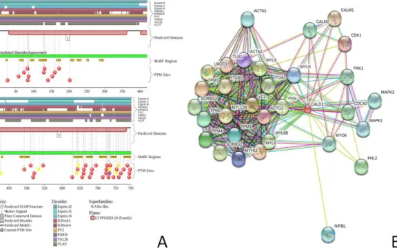
Interactability of chicken CaD and CaM was evaluated by STRING (Search Tool for the Retrieval of Interacting Genes,http://string-db.org/), which is the online database resource, that provides both experimental and predicted interaction information (Szklarczyk et al., 2011). STRING produces the network of predicted associations for a particular group of proteins. The network nodes are proteins, whereas the edges represent the predicted or known functional associations. An edge may be drawn with up to 7 differently colored lines that represent the existence of the seven types of evidence used in predicting the associations. A red line indicates the presence of fusion evidence; a green line, neighborhood evidence; a blue line, co-occurrence evidence; a purple line, experimental evidence; a yellow line, text mining evidence; a light blue line, database evidence; a black line, co-expression evidence (Szklarczyk et al., 2011).
Potential disorder-based binding sites in CaD136(which is the C-terminal domain
(636–771) of CaD) were found using three computational tools,α-MoRF identifier (Cheng et al., 2007;Oldfield et al., 2005b), ANCHOR (Dosztanyi, Meszaros & Simon, 2009;Meszaros, Simon & Dosztanyi, 2009), and MoRFpred (Disfani et al., 2012). Since IDPs/IDPRs are commonly involved in protein-protein interactions (Daughdrill et al., 2005;Dunker et al., 2002a;Dunker, Brown & Obradovic, 2002;Dunker et al., 2001;Dunker et al., 2008b;Dunker & Uversky, 2008;Oldfield et al., 2005b;Radivojac et al., 2007;
Tompa, 2002;Uversky, 2011b;Uversky, 2012;Uversky, 2013b;Uversky & Dunker, 2010;
regions. Such motifs are known as Molecular Recognition Feature (MoRF), they are able to undergo disorder-to-order transition during the binding to a specific partner, and can be identified computationally (Cheng et al., 2007;Oldfield et al., 2005b). For example, an α-MoRF predictor indicates the presence of a relatively short, loosely structured region within a largely disordered sequence (Oldfield et al., 2005b), which can gain functionality upon a disorder-to-order transition induced by binding to partners (Mohan et al., 2006;
Vacic et al., 2007a). In addition to MoRF identifiers, potential binding sites in disordered regions can be identified by the ANCHOR algorithm (Dosztanyi, Meszaros & Simon, 2009;Meszaros, Simon & Dosztanyi, 2009). This approach relies on the pairwise energy estimation approach developed for the general disorder prediction method IUPred (Dosztanyi et al., 2005a;Dosztanyi et al., 2005b). being based on the hypothesis that long regions of disorder contain localized potential binding sites that cannot form enough favorable intrachain interactions to fold on their own, but are likely to gain stabilizing energy by interacting with a globular protein partner (Dosztanyi, Meszaros & Simon, 2009;
Meszaros, Simon & Dosztanyi, 2009). Regions of a protein suggested by the ANCHOR
algorithm to have significant potential to be binding sites are the ANCHOR-indicated binding site (AIBS).
RESULTS AND DISCUSSION
Characterization of functional disorder in caldesmon and calmodulin
The amino acid sequences and compositions of IDPs/IDPRs are significantly different from those of ordered proteins and domains. For example, the amino acid compositions of extended IDPs/IDPRs (i.e., highly disordered proteins and regions lacking almost any residual structure (Dunker et al., 2001;Uversky, 2002a;Uversky, 2002b;Uversky, 2003; Uver-sky, 2013a;Uversky, 2013c;Uversky & Dunker, 2010;Uversky, Gillespie & Fink, 2000)) are characterized by high mean net charge and low mean hydropathy, being significantly depleted in order-promoting residues C, W, Y, F, H, I, L, V, and N and significantly enriched in disorder-promoting residues A, R, G, Q, S, P, E, and K (Dunker et al., 2001;Radivojac et al., 2007;Romero et al., 2001;Vacic et al., 2007b). The fractional difference in composition between CaD and a set of ordered proteins from PDB Select 25 (Berman et al., 2000) was calculated as(CCaD−Corder)/Corder, where CCaDis the content of a given amino acid in
CaD, and Corderis the corresponding value for the set of ordered proteins. This analysis
revealed that in comparison with typical ordered proteins, CaD is significantly depleted in major order-promoting residues (C, Y, F, H, V, L, and I) and is significantly enriched in major disorder-promoting residues, such as A, R, E, and K. This means that CaD might contain multiple structural and functional signatures typical for the IDPs.
Figure 1 Evaluating the intrinsic disorder propensities of chicken CaD (A), CaD136(B), and chicken
CaM (C) by the family of PONDR predictors.A disorder threshold is indicated as a thin line (at score =0.5) in all plots to show a boundary between disorder (>0.5) and order (<0.5). Plot (D) represents the amino acid sequences of CaD136and CaM, for which the positively and negatively charged residues are highlighted. The positions of tryptophan residues within the CaD136sequence are also indicated.
full-length protein ranges, depending on the predictor, from 0.69 to 0.93, this analysis clearly shows that CaD is expected to be mostly disordered. In agreement with this conclusion, the consensus MobiDB analysis (http://mobidb.bio.unipd.it/entries/P12957) revealed that chicken gizzard CaD contains 98.4% disordered residues. Curiously, the C-terminal domain of this protein, CaD136, is predicted to be a bit more predisposed for
order than the remaining protein (depending on the predictor, the mean disorder score for this 636–771 fragment of CaD ranges from 0.52 to 0.81). This observation is further illustrated byFig. 1Bwhich represents the PONDR-based disorder profiles of this region.
Curiously, although several X-ray crystal (PDB IDs: 1ahr, 1up5, 2bcx, 2bki, 2o5g, 2o60, 2vb6, 3gog, and 3gp2) and NMR solution structures (PDB IDs: 2kz2 and 2m3s) of CaM are known,Fig. 1Cshows that this protein is predicted to be rather disordered too. These findings are not too surprising, since it is known that the CaM structure and folding are strongly dependent on the metal ion binding (Li, Wang & Takada, 2014;
Sulmann et al., 2014), and that there is a great variability in the crystal structures of CaM in isolation (i.e., where it is not bound to its protein or peptide partners and exists in the unliganded form) which is considered as an illustration of CaM plasticity in solution
solution using small angle scattering and other methods have indicated the presence of a mixture of conformations (Bertini et al., 2010;Heller, 2005;Kursula, 2014;Yamada et al., 2012). Also in agreement with these predictions, the analysis of one of the NMR structures of CaM (PDB ID: 2m3s) revealed that this protein might contain up to 50.3% of disordered residues in solution (Moroz et al., 2013). Again, the results of the per-residue predictions by the members of the PONDR family are further supported by the results of the MobiDB analysis, according to which the consensus disorder content of CaM based on the outputs of ten disorder predictors is 18.1%. The corresponding values evaluated by the individual predictors (http://mobidb.bio.unipd.it/entries/P62149) are ranging from 6.0% and 13.4% for the ESpritz-XRay and DisEMBL-465, respectively to 41.6% and 69.1% for the IUPred-long and PONDR®VSL2, respectively. Note that both ESpritz-XRay and DisEMBL-465 are trained based on proteins with known crystal structures and containing regions of missing electron density, whereas IUPred-long and PONDR VSL2 use different criteria for training.
Further information on the functional disorder status of CaD and CaM was retrieved from D2P2portal, which represents a database of pre-computed disorder predictions for a large library of proteins from completely sequenced genomes (Oates et al., 2013), which in addition to outputs of nine disorder predictors provides information on the curated sites of various posttranslational modifications and on the location of predicted disorder-based potential binding sites. Since this database does not include data for chicken, the human homologues of CaD and CaM were used for this analysis. The validity of this approach is justified by the fact that sequences of human and chicken CaMs are identical (100% iden-tity), whereas sequences of human and chicken CaD are highly conserved (61% identity).
Figures 2Aand3Arepresents the results of this analysis of CaD and CaM, respectively, and provide further support for the abundance and functional importance of intrinsic disorder in these proteins, which are predicted to contain long disordered regions enriched in potential disorder-based binding motifs and containing numerous predicted sites of po-tential posttranslational modifications (PTMs). The fact that disordered domains/regions of the human CaD and CaM contain numerous PTM sites is in agreement with the well-known notion that phosphorylation (Iakoucheva et al., 2004) and many other enzy-matically catalyzed PTMs are preferentially located within the IDPRs (Pejaver et al., 2014).
Figure 2 Evaluation of the functional intrinsic disorder propensity of the human CaD (UniProt ID: Q05682) by the D2P2platform (http://d2p2.pro/) (Oates et al., 2013).In this plot, top nine colored bars represent locations of disordered regions predicted by different computational tools (Espritz-D, Espritz-N, Espritz-X, IUPred-L, IUPred-S, PV2, PrDOS, PONDR®VSL2b, and PONDR® VLXT, see keys for the corresponding color codes). Dark red bar shows the location of the functional domain found by the Pfam platform, which is a database of protein families that includes their annotations and multiple sequence alignments generated using hidden Markov models (Berman et al., 2000;Finn et al., 2006;Finn et al., 2008). The green-and-white bar in the middle of the plot shows the predicted disorder agreement between these nine predictors, with green parts corresponding to disordered regions by consensus. Red, yellow and purple circles at the bottom of the plot show the locations of phosphorylation, acetylation and ubiquitination sites, respectively. (B) Analysis of the interactivity of the chicken gizzard CaD (UniProt ID: P12957) by STRING (Szklarczyk et al., 2011). STRING produces the network of predicted associations for a particular group of proteins. The network nodes are proteins, whereas the edges represent the predicted or known functional associations. An edge may be drawn with up to 7 differently colored lines that represent the existence of the seven types of evidence used in predicting the associations. A red line indicates the presence of fusion evidence; a green line, neighborhood evidence; a blue line, co-occurrence evidence; a purple line, experimental evidence; a yellow line, text mining evidence; a light blue line, database evidence; a black line, co-expression evidence (Szklarczyk et al., 2011).
signaling) (Dunker et al., 1998). In line with these considerations, intrinsically disordered nature of chicken CaD and CaM provides a plausible explanation for their potential roles as hub proteins. Therefore, data reported inFigs. 1,2and3suggest that both CaD and CaM are expected to contain substantial amounts of functional disorder, which CaD being predicted to be mostly disordered.
Figure 1Dshows that the positively charged R and K residues are evenly distributed within the CaD136sequence and that the sequence of CaM contains evenly spread
negatively charged residues D and E. Since under the physiologic conditions of neutral pH, the C-terminal interacting domain of CaD and CaM possess charges of opposite sign (+9 for CaD136and -24 for CaM) it is likely that electrostatic interactions play important
Figure 3 Evaluation of the functional intrinsic disorder propensity of human CaM (UniProt ID: P62158) by D2P2database (http://d2p2.pro/) (Oates et al., 2013).In this plot, top dark blue bar with green stripes shows the localization of disordered region annotated in the IDEAL database (Fukuchi et al. 2012) for this protein. Next nine colored bars represent location of disordered regions predicted by different disorder predictors (Espritz-D, Espritz-N, Espritz-X, IUPred-L, IUPred-S, PV2, PrDOS, PONDR®VSL2b, and PONDR®VLXT, see keys for the corresponding color codes). Dark red bar shows the location of the functional domain found by the Pfam platform, which is a database of protein families that includes their annotations and multiple sequence alignments generated using hidden Markov models (Berman et al., 2000;Finn et al., 2006;Finn et al., 2008). Blue-and-white bar in the middle of the plot shows the predicted disorder agreement between these nine predictors, with green parts corresponding to disordered regions by consensus. Red, yellow, purple and blue circles at the bottom of the plot show the location of phosphorylation, acetylation, ubiquitination, and methylation sites, respectively. (B) Analysis of the interactivity of the chicken CaM (UniProt ID: P62149) by STRING (Szklarczyk et al., 2011).
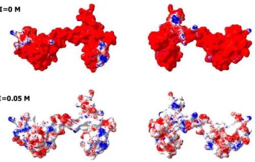
Fig. 4, which represents the charge distribution over the CaM surface and shows that negative charges are almost evenly distributed over the entire protein surface. What then defines the specificity of interaction between a highly positively charged IDP(CaD136)and
a highly negatively charged surface of CaM? Some answers to this important question can be obtained analyzing peculiarities of the disorder distribution in CaD136. In fact, many
Figure 4 Analysis of the charge distribution on the surface of CaM molecule.PDB file: 1CLM. Analyzed protein: calmodulin, Ca2+-form (1 chain, 4 Ca ions), without first 3 residues Ala, Gln, and Glu and without a last residue Lys. Ca2+ions and water molecules were removed, absent hydrogen atoms were added. Calculations were done using the Swiss-PdbViewer v3.7b2 program. Method of calculation: Poisson-Boltzmann, using partial atom charges, ionic strength 0M or 0.05M, dielectric constant of solvent 80, for protein—4. Colors: Red, potential value is NEGATIVE,−1.8 kT/e; White, potential value is ZERO; Blue, potential value is POSITIVE, 1.8 kT/e.
(MoRF), and they often can be found based on the peculiar shape of a disorder profile (sharp “dips” within the long IDPRs). These observations serve as a foundation for the corresponding computational tools, e.g.,α-MoRF-Pred (Cheng et al., 2007;Oldfield et al., 2005b) or MoRFpred (Disfani et al., 2012). Alternatively, the disorder-based binding sites can be identified by ANCHOR (Dosztanyi, Meszaros & Simon, 2009;Meszaros, Simon & Dosztanyi, 2009) (see Materials and Methods). There is generally a good agreement between the results of binding sites prediction by these two tools.
These analyses revealed that CaD136has several disorder-based potential binding
sites and three of them correspond to the major minima in the CaD136disorder plots
obtained by both PONDR®VLXT and PONDR-FIT (seeFig. 5). Since each of these three dip-centered potential binding sites include a tryptophan residue, we decided to mutate those tryptophans in order to evaluate their roles in the CaD136binding to CaM.
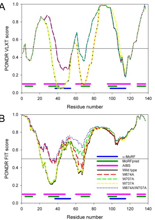
At the first stage, the disorder propensities of three single tryptophan mutants (W674A, W707A, and W737A) and a double tryptophan mutant (W674A/W707A) were compared using PONDR®VLXT and PONDR FIT algorithms.Figure 5represents the results of these analyses and shows that the local disorder propensities were noticeably affected by single mutations W674A and W707A and by the W674A/W707A double mutation, whereas W737A had a very minimal effect on the CaD136 disorder profile. Although
Figure 5 Computational analysis of the effect of tryptophan mutations on the disorder propensity of CaD136evaluated by PONDR®VLXT (A) and PONDR-FIT (B).Locations of the predicted
disorder-based binding sites are shown at the bottom of plots as pink (AIBSs), dark green (MoRFpreds), and dark blue (α-MoRFs) bars, respectively.
Table 1 Equilibrium association constants(KCaM)for complexes between CaM and wild type CaD136
and its mutants and their relative fluorescence quantum yields in the free and CaM-bound states.
Protein KCaM Q/Qtrp(in solution) Q/Qtrp(in complex
with calmodulin)
WT (6.5±1.6)×105 1.25 2.40
W674A (2.2±0.6)×105 1.25 2.72
W707A (3.0±0.8)×105 1.50 2.55
W737A (1.8±0.5)×106 1.49 2.95
Double mutant (4.4±1.1)×104 1.19 2.64
Effect of tryptophan substitutions on tryptophan fluorescence spectrum of the C-terminal CaD domain
Analysis of the normalized tryptophan fluorescence spectra of CD136and its mutants in
solution and in complex with CaM (which does not have tryptophan residues) revealed that the spectra of all the CD136proteins in their unbound forms are practically the same
(seeFig. S1). They have extremely long wavelength positions and are similar to spectrum of a free tryptophan in water, which shows that in all these proteins, the tryptophan residues are totally exposed to water. The spectra of the complexes with CaM are different. The CaM-complexes W737A mutant has the most blue-shifted spectrum, whereas the W707A mutant in its bound state has the least blue-shifted spectrum. TheTable 1represents the relative fluorescence quantum yields for CD136and its mutants in solution and in the
complex with CaM.
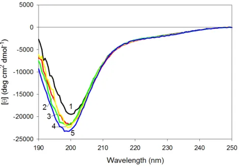
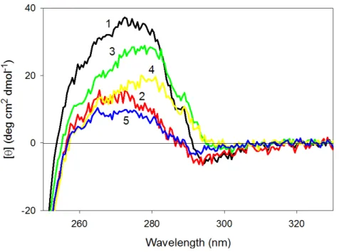
Effect of tryptophan substitutions on far-UV CD spectra of CaD136 mutants
Figure 6represents the far-UV CD spectra of wild type, W674A, W707A, W737A and W674A/W707A CaD136and shows that all these proteins have far-UV CD spectra typical
of the almost completely unfolded polypeptides. In other words, the data are consistent with the conclusion that at physiological conditions none of the CaD136domains has
considerable amount of ordered secondary structure; i.e., they belong to the family of so-called natively unfolded proteins, which are the most disordered members of the realm of intrinsically disordered proteins. On the other hand, more detailed analysis of the far-UV CD spectrum shows that the wild type CaD136, being mostly disordered, is still
far from to be completely unfolded and preserves some residual structure (e.g.,[θ]222 ∼−3,000 deg cm2dmol−1, the minimum is located at 200, rather than at 196–198 nm, see
Fig. 6).
Figure 6shows that all amino acid substitutions affect the far-UV CD spectrum of the C-terminal CaD domain in a similar manner, inducing considerable decrease in the spectrum intensity around 200 nm. This is further illustrated byFig. S2that represents the difference spectra between the wild type CaD136and mutated domains and clearly shows
Figure 6 Far-UV CD spectra of wild type (1), W674A (2), W707A (3), W737A (4) and W674A/W707A (5) CaD136. All measurements were carried out at a protein concentration of 0.6–0.8 mg/ml, cell
pathlength 0.1 mm, 15◦C.
Effect of tryptophan substitutions on the near-UV CD spectra of CaD136 mutants
Surprisingly,Fig. 7shows that wild type CaD136and all its mutants possess rather intensive
and pronounced near-UV CD spectra. This means that tryptophan residues of these proteins are in relatively asymmetric environment.Figure 7shows that any tryptophan substitution analyzed in this study has a considerable effect on the near-UV CD spectrum of CaD136, leading to the substantial decrease in the spectral intensity. It also can be seen
that different tryptophan residues have different contributions to the near-UV CD spec-trum of protein. In fact,Fig. 7shows that the effect of amino acid substitutions increases in the following order: W707A<W737A<W674A≤W674A/W707A. This conclusion is confirmed by the difference spectra shown inFig. S3. Therefore, these data suggest that tryptophan residues have noticeable contributions to the residual structure of CaD136,
likely serving as condensation centers around which the local dynamic structure is formed.
Conformational stability of CaD136 and its mutants analyzed by the
effect of temperature on their near- and far-UV CD spectra
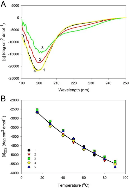
Figure 8represents near-UV CD spectra of the wild type and mutated CaD136measured
Figure 7 Near-UV CD spectra of wild type (1), W674A (2), W707A (3), W737A (4), and W674A/W707A (5) CaD136.All measurements were carried out at a protein concentration of 0.6–0.8
mg/ml, cell pathlength 10 mm, 15◦C.
the double W674A/W707A mutant is practically unaffected by temperature. Importantly,
Fig. 8shows that even at 90◦C all of the protein variants analyzed in this study show pronounced near-UV CD spectra, reflecting the fact that the temperature increase does not destroy completely the asymmetric environment of their aromatic residues.
Temperature had similar effect of the far-UV CD spectra of all the CaD136variants.
As an example,Fig. 9Arepresents the far-UV CD spectra of W674A mutant measured at different temperatures. It can be seen that shape and intensity of the spectrum undergo considerable changes with the increase in temperature, reflecting the temperature-induced formation of the more ordered secondary structure. Same spectral changes were observed for several other IDPs and were classified as the “turn-out” paradoxical response of extended IDPs (opposite to the response of ordered proteins) to changes in their environment (Uversky, 2002a;Uversky, 2002b;Uversky, 2011a;Uversky, 2013a;Uversky, 2013c;Uversky & Dunker, 2010).Figure 9Bsummarizes the data on the effect of heating on the secondary structure of the CaD136variants as corresponding[θ]222vs. temperature
dependences. One can see that in all cases studied temperature increase was accompanied by the steady increase in the negative ellipticity at 222 nm. It is necessary to emphasize here that this behavior is totally different from the conformational behavior of typical globular proteins, which show temperature-induced reduction in the content of ordered secondary structure.
Studying the CaD136 variants by scanning microcalorimetry
Figure S4represents the calorimetric scans obtained for the wild type CaD136and its
Figure 8 Near-UV CD spectra of the wild type (A), W674A (B), W707A (C), W737A (D) and W674A/W707A (E) CaD136measured at different temperatures.15◦C (1); 40◦C (2), 90◦C (3) and
Figure 9 Effect of temperature on far-UV CD spectra of CaD136.(A) Far-UV CD spectra of W674A
Figure 10 Spectrofluorimetric titration of the CaD136and its mutants by CaM.
and the absence of distinct heat absorption peaks within the temperature region from 10 to 100◦C for these proteins suggest that their structure is predominantly unfolded.
Interactions of the CaD136 and its tryptophan mutants with
calmodulin studied by intrinsic fluorescence
Figure 10represents the results of the spectrofluorimetric titration of CD136 and its
varying the binding constant. The values of the binding constants which give the best fits are collected inTable 1. This analysis revealed that the substitution of the tryptophan residues by alanines resulted in a decrease in the CaD136-CaM binding constant in all the
cases except W737A, where mutation caused an increase in the CaD136affinity for CaM. Table 1also shows that the double W674A/W707A mutation caused the largest reduction in the CaD136binding efficiency. The value of the association constant for wild type CaD136
in our work is in a good agreement with the literature data of another authors (Czurylo et al., 1991;Graether et al., 1997;Huber et al., 1996;Medvedeva et al., 1997;Shirinsky, Bushueva & Frolova, 1988;Wang et al., 1997).
The ability of the caldesmon and its C-terminal fragments to interact specifically with calmodulin has been established long ago (Shirinsky, Bushueva & Frolova, 1988), and several models of this complex have been suggested (reviewed inGusev, 2001). It is known that the C-terminal domain of CaD contains three CaM-binding sites, centers A (close to Trp674), B (close to Trp707), and B’ (close to Trp737). It has been shown that sites A and B interact with C-terminal lobe of CaM (this protein has dumbbell shape with twoα-helical Ca2+-binding globular domains, separated by an extended “handle” formed by a seven-turnα-helix), whereas center B forms complex with the N-terminal globular domain (Gusev, 2001;Marston et al., 1994;Mezgueldi et al., 1994;Zhan, Wong &
Wang, 1991). The idea of multiple-sited interaction of CaD and CaM and participation
of Trp residues in it was described earlier in a series of papers from different laboratories (for instance,Huber et al., 1996;Mezgueldi et al., 1994. For example, to determine the contribution of each of three Trp residues (659, 692, and 722, which are similar to 674, 707, and 737 in our protein) in the calmodulin-caldesmon interaction,Graether et al. (1997)
have mutated the Trp residues to Ala in the C-terminal domain of fibroblast caldesmon (CaD39) and studied the effects on calmodulin binding by fluorescence measurements and using immobilized calmodulin (Graether et al., 1997). All the mutations reduced the affinity of CaD to calmodulin, but mutation of Trp 722 at site B’ to Ala caused the smallest decrease in affinity. In our work similar mutation caused even an increase in affinity. The authors concluded that Trp 659 and Trp 692 are the major determinants in the fibroblast caldesmon-calmodulin interaction and that Trp 722 in site B’ plays a minor role (Graether et al., 1997). The results of our study show that in gizzard caldesmon the letter tryptophan seems to play more significant role in the interaction with calmodulin.
CONCLUSIONS
Altogether, data presented in our study suggest that CaD and its C-terminal domain, CaD136, are intrinsically disordered proteins. CaD potentially serves as a disordered hub
in several important protein-protein interaction networks. It is likely that CaD136-CaM
interaction is driven by the non-specific electrostatic attraction interactions due to the opposite charges of these two proteins. Specificity of CaD136-CaM binding is likely
to be determined by the definite packing of important tryptophan residues at the CaD136-CaM interface, which is manifested by the dramatic blue shift of the intrinsic
Figure 11 Schematic representation of the “buttons on a charge string” binding mode proposed in this study.Here, the CaD136is shown as a blue string containing three “buttons” (tryptophan-centric partially structured binding sites), whereas CaM is shown as mostly red surface. Note that positions of binding sites and length of the CaD136chain are arbitrary and used here only to illustrate an idea.
aforementioned tryptophan residues potentially serving as centers of local fluctuating structural elements. Therefore, our bioinformatics and experimental data suggest that the interaction between CaD136and CaM can be described within the “buttons on a charged
string” model, where the electrostatic attraction between the positively charged and highly disordered CaD136containing at least three segments of fluctuating local structure
(“pliable buttons”) and the negatively charged CaM is solidified by the specific packing of three short regions containing tryptophan residues in a “snapping a button” manner. This model is schematically represented inFig. 11. Curiously, it seems that all three “buttons” are important for binding, since mutation of any of the tryptophans affects CaD136-CaM
binding and since CaD136remains CaM-buttoned even when two of the three tryptophans
are mutated to alanines.
Abbreviations
AIBS disorder-based ANCHOR-identified binding site
CaD caldesmon
CaD136 C-terminal domain (636–771) of CaD
CaM calmodulin
CD circular dichroism
DSC differential scanning calorimetry
IDP intrinsically disordered protein
IDPR intrinsically disordered protein region
MoRF molecular recognition feature
PTM posttranslational modification
ADDITIONAL INFORMATION AND DECLARATIONS
Funding
This work was supported by grants from the Programs of the Russian Academy of Sciences “Molecular and Cellular Biology” (P.E.A.) and “Fundamental Science for Medicine” (P.S.E.). The funders had no role in study design, data collection and analysis, decision to publish, or preparation of the manuscript.
Grant Disclosures
The following grant information was disclosed by the authors: Programs of the Russian Academy of Sciences.
Competing Interests
Eugene A. Permyakov and Vladimir N. Uversky are Academic Editors for PeerJ.
Author Contributions
• Sergei E. Permyakov, Eugene A. Permyakov and Vladimir N. Uversky conceived and designed the experiments, performed the experiments, analyzed the data, wrote the paper, prepared figures and/or tables, reviewed drafts of the paper.
Supplemental Information
Supplemental information for this article can be found online athttp://dx.doi.org/ 10.7717/peerj.1265#supplemental-information.
REFERENCES
Abrams J, Davuluri G, Seiler C, Pack M. 2012.Smooth muscle caldesmon modulates peristalsis in the wild type and non-innervated zebrafish intestine.Neurogastroenterology & Motility 24:288–299DOI 10.1111/j.1365-2982.2011.01844.x.
Adelstein RS, Eisenberg E. 1980.Regulation and kinetics of the actin–myosin-ATP interaction.
Annual Review of Biochemistry49:921–956DOI 10.1146/annurev.bi.49.070180.004421.
Berman HM, Westbrook J, Feng Z, Gilliland G, Bhat TN, Weissig H, Shindyalov IN, Bourne PE. 2000.The protein data bank.Nucleic Acids Research28:235–242DOI 10.1093/nar/28.1.235.
Bertini I, Giachetti A, Luchinat C, Parigi G, Petoukhov MV, Pierattelli R, Ravera E, Svergun DI. 2010.Conformational space of flexible biological macromolecules from average data.Journal of the American Chemical Society132:13553–13558DOI 10.1021/ja1063923.
Burstein EA, Emelyanenko VI. 1996.Log-normal description of fluorescence spectra of organic fluorophores.Photochemistry and Photobiology64:316–320
DOI 10.1111/j.1751-1097.1996.tb02464.x.
Campen A, Williams RM, Brown CJ, Meng J, Uversky VN, Dunker AK. 2008.TOP-IDP-scale: a new amino acid scale measuring propensity for intrinsic disorder.Protein & Peptide Letters 15:956–963DOI 10.2174/092986608785849164.
Cheng Y, Oldfield CJ, Meng J, Romero P, Uversky VN, Dunker AK. 2007. Mining
alpha-helix-forming molecular recognition features with cross species sequence alignments.
Czurylo EA, Emelyanenko VI, Permyakov EA, Dabrowska R. 1991.Spectrofluorimetric studies on C-terminal 34 kDa fragment of caldesmon.Biophysical Chemistry40:181–188 DOI 10.1016/0301-4622(91)87007-R.
Czurylo EA, Kulikova N. 2012.Anatromy and physiology of proteins: caldesmon. New York: Nova Science Publishers.
Czurylo EA, Zborowski J, Dabrowska R. 1993.Interaction of caldesmon with phospholipids.
Biochemical Journal291:403–408DOI 10.1042/bj2910403.
Daughdrill GW, Pielak GJ, Uversky VN, Cortese MS, Dunker AK. 2005.Natively disordered proteins. In: Buchner J, Kiefhaber T, eds.Handbook of protein folding. Weinheim: Wiley-VCH, Verlag GmbH & Co, 271–353.
Di Domenico T, Walsh I, Martin AJ, Tosatto SC. 2012. MobiDB: a comprehensive database of intrinsic protein disorder annotations. Bioinformatics28:2080–2081 DOI 10.1093/bioinformatics/bts327.
Disfani FM, Hsu WL, Mizianty MJ, Oldfield CJ, Xue B, Dunker AK, Uversky VN, Kurgan L. 2012.MoRFpred, a computational tool for sequence-based prediction and characterization of short disorder-to-order transitioning binding regions in proteins.Bioinformatics28:i75–i83 DOI 10.1093/bioinformatics/bts209.
Dosztanyi Z, Chen J, Dunker AK, Simon I, Tompa P. 2006.Disorder and sequence repeats in hub proteins and their implications for network evolution.Journal of Proteome Research 5:2985–2995DOI 10.1021/pr060171o.
Dosztanyi Z, Csizmok V, Tompa P, Simon I. 2005a.IUPred: web server for the prediction of intrinsically unstructured regions of proteins based on estimated energy content.Bioinformatics 21:3433–3434DOI 10.1093/bioinformatics/bti541.
Dosztanyi Z, Csizmok V, Tompa P, Simon I. 2005b.The pairwise energy content estimated from amino acid composition discriminates between folded and intrinsically unstructured proteins.
Journal of Molecular Biology347:827–839DOI 10.1016/j.jmb.2005.01.071.
Dosztanyi Z, Meszaros B, Simon I. 2009.ANCHOR: web server for predicting protein binding re-gions in disordered proteins.Bioinformatics25:2745–2746DOI 10.1093/bioinformatics/btp518.
Dunker AK, Babu M, Barbar E, Blackledge M, Bondos SE, Doszt´anyi Z, Dyson HJ, Forman-Kay J, Fuxreiter M, Gsponer J, Han K-H, Jones DT, Longhi S, Metallo SJ, Nishikawa K, Nussinov R, Obradovic Z, Pappu R, Rost B, Selenko P, Subramaniam V, Sussman JL, Tompa P, Uversky VN. 2013.What’s in a name? Why these proteins are intrinsically disordered.Intrinsically Disordered Proteins1:e24157DOI 10.4161/idp.24157.
Dunker AK, Brown CJ, Lawson JD, Iakoucheva LM, Obradovic Z. 2002a.Intrinsic disorder and protein function.Biochemistry41:6573–6582DOI 10.1021/bi012159+.
Dunker AK, Brown CJ, Obradovic Z. 2002.Identification and functions of usefully disordered proteins.Advances in Protein Chemistry62:25–49.
Dunker AK, Cortese MS, Romero P, Iakoucheva LM, Uversky VN. 2005.Flexible nets. The roles of intrinsic disorder in protein interaction networks.FEBS Journal272:5129–5148 DOI 10.1111/j.1742-4658.2005.04948.x.
Dunker AK, Garner E, Guilliot S, Romero P, Albrecht K, Hart J, Obradovic Z, Kissinger C, Villafranca JE. 1998.Protein disorder and the evolution of molecular recognition: theory, predictions and observations.Pacific Symposium on Biocomputing473–484.
Dunker AK, Lawson JD, Brown CJ, Williams RM, Romero P, Oh JS, Oldfield CJ, Campen AM, RatliffCM, Hipps KW, Ausio J, Nissen MS, Reeves R, Kang C, Kissinger CR, Bailey RW, Griswold MD, Chiu W, Garner EC, Obradovic Z. 2001.Intrinsically disordered protein.
Dunker AK, Obradovic Z, Romero P, Garner EC, Brown CJ. 2000.Intrinsic protein disorder in complete genomes.Genome informatics. Workshop on Genome Informatics11:161–171.
Dunker AK, Oldfield CJ, Meng J, Romero P, Yang JY, Chen JW, Vacic V, Obradovic Z,
Uversky VN. 2008a.The unfoldomics decade: an update on intrinsically disordered proteins.
BMC Genomics9(Suppl 2):S1DOI 10.1186/1471-2164-9-S2-S1.
Dunker AK, Silman I, Uversky VN, Sussman JL. 2008b. Function and structure of inherently disordered proteins. Current Opinion in Structural Biology 18:756–764 DOI 10.1016/j.sbi.2008.10.002.
Dunker AK, Uversky VN. 2008.Signal transduction via unstructured protein conduits.Nature Chemical Biology4:229–230DOI 10.1038/nchembio0408-229.
Dyson HJ. 2011.Expanding the proteome: disordered and alternatively folded proteins.Quarterly Review of Biophysics44:467–518DOI 10.1017/S0033583511000060.
Dyson HJ, Wright PE. 2002.Coupling of folding and binding for unstructured proteins.Current Opinion in Structural Biology12:54–60DOI 10.1016/S0959-440X(02)00289-0.
Dyson HJ, Wright PE. 2005.Intrinsically unstructured proteins and their functions.Nature Reviews Molecular Cell Biology6:197–208DOI 10.1038/nrm1589.
Ekman D, Light S, Bjorklund AK, Elofsson A. 2006.What properties characterize the hub proteins of the protein-protein interaction network of Saccharomyces cerevisiae?Genome Biology7:R45DOI 10.1186/gb-2006-7-6-r45.
Fan X, Kurgan L. 2014.Accurate prediction of disorder in protein chains with a comprehensive and empirically designed consensus.Journal of Biomolecular Structure and Dynamics 32:448–464DOI 10.1080/07391102.2013.775969.
Finn RD, Mistry J, Schuster-Bockler B, Griffiths-Jones S, Hollich V, Lassmann T, Moxon S, Marshall M, Khanna A, Durbin R, Eddy SR, Sonnhammer EL, Bateman A. 2006.Pfam: clans, web tools and services.Nucleic Acids Research34:D247–D251DOI 10.1093/nar/gkj149.
Finn RD, Tate J, Mistry J, Coggill PC, Sammut SJ, Hotz HR, Ceric G, Forslund K, Eddy SR, Sonnhammer EL, Bateman A. 2008.The Pfam protein families database.Nucleic Acids Research 36:D281–D288DOI 10.1093/nar/gkm960.
Fraser IDC, Copeland O, Wu B, Marston SB. 1997.The inhibitory complex of smooth muscle caldesmon with actin and tropomyosin involves three interacting segments of the C-terminal domain 4.Biochemistry36:5483–5492DOI 10.1021/bi962969z.
Graether SP, Heinonen TY, Raharjo WH, Jin JP, Mak AS. 1997.Tryptophan residues in caldesmon are major determinants for calmodulin binding.Biochemistry 36:364–369 DOI 10.1021/bi962008k.
Gusev NB. 2001.Some properties of caldesmon and calponin and the participation of these proteins in regulation of smooth muscle contraction and cytoskeleton formation.Biochemistry (Moscow)66:1112–1121DOI 10.1023/A:1012480829618.
Haynes C, Oldfield CJ, Ji F, Klitgord N, Cusick ME, Radivojac P, Uversky VN, Vidal M, Iak-oucheva LM. 2006.Intrinsic disorder is a common feature of hub proteins from four eukaryotic interactomes.PLoS Computational Biology2:e100DOI 10.1371/journal.pcbi.0020100.
Heller WT. 2005.Influence of multiple well defined conformations on small-angle scattering of proteins in solution.Acta Crystallographica Section D-Biological Crystallography61:33–44 DOI 10.1107/S0907444904025855.
Huber PAJ, ElMezgueldi M, Grabarek Z, Slatter DA, Levine BA, Marston SB. 1996.
Multiple-sited interaction of caldesmon with Ca2(+)-calmodulin.Biochemical Journal 316:413–420DOI 10.1042/bj3160413.
Iakoucheva LM, Radivojac P, Brown CJ, O’Connor TR, Sikes JG, Obradovic Z, Dunker AK. 2004.The importance of intrinsic disorder for protein phosphorylation.Nucleic Acids Research 32:1037–1049DOI 10.1093/nar/gkh253.
Ishida T, Kinoshita K. 2007.PrDOS: prediction of disordered protein regions from amino acid sequence.Nucleic Acids Research35:W460–W464DOI 10.1093/nar/gkm363.
Kordowska J, Huang R, Wang CL. 2006.Phosphorylation of caldesmon during smooth muscle contraction and cell migration or proliferation.Journal of Biomedical Science13:159–172 DOI 10.1007/s11373-005-9060-8.
Kursula P. 2014.The many structural faces of calmodulin: a multitasking molecular jackknife.
Amino Acids46:2295–2304DOI 10.1007/s00726-014-1795-y.
Kuznicki J, Filipek A. 1987.Purification and Properties of a Novel Ca2+-Binding Protein (10.5 Kda) from Ehrlich-Ascites-Tumor Cells. Biochemical Journal247:663–667 DOI 10.1042/bj2470663.
Li W, Wang W, Takada S. 2014.Energy landscape views for interplays among folding, binding, and allostery of calmodulin domains.Proceedings of the National Academy of Sciences of the United States of America111:10550–10555DOI 10.1073/pnas.1402768111.
Linding R, Jensen LJ, Diella F, Bork P, Gibson TJ, Russell RB. 2003a.Protein disorder prediction: implications for structural proteomics.Structure11:1453–1459DOI 10.1016/j.str.2003.10.002.
Linding R, Russell RB, Neduva V, Gibson TJ. 2003b.GlobPlot: exploring protein sequences for globularity and disorder.Nucleic Acids Research31:3701–3708DOI 10.1093/nar/gkg519.
Mabuchi K, Li Y, Tao T, Wang CL. 1996.Immunocytochemical localization of caldesmon and calponin in chicken gizzard smooth muscle.Journal of Muscle Research and Cell Motility 17:243–260DOI 10.1007/BF00124246.
Makowski P, Makuch R, Sikorski AF, Jezierski A, Pikula S, Dabrowska R. 1997.Interaction of caldesmon with endoplasmic reticulum membrane: effects on the mobility of phospholipids in the membrane and on the phosphatidylserine base-exchange reaction.Biochemical Journal 328:505–509DOI 10.1042/bj3280505.
Mani RS, Kay CM. 1996.Calcium binding proteins. In: Barany M, ed.Biochemistry of smooth muscle contraction. New York: Academic Press, 105–116.
Marquardt DW. 1963.An algorithm for least-squares estimation of nonlinear parameters.Journal of the Society for Industrial and Applied Mathematics11:431–441DOI 10.1137/0111030.
Marston SB, Fraser ID, Huber PA, Pritchard K, Gusev NB, Torok K. 1994.Location of two contact sites between human smooth muscle caldesmon and Ca(2+)-calmodulin.Journal of Biological Chemistry269:8134–8139.
Marston SB, Redwood CS. 1991.The molecular anatomy of caldesmon.Biochemical Journal 279(Pt 1):1–16DOI 10.1042/bj2790001.
Martson SB, Huber PAJ. 1996.Caldesmon. In: Barany M, ed.Biochemistry of smooth muscle contraction. New York: Academic Press, 70–90.
Medvedeva MV, Kolobova EA, Huber PAJ, Fraser IDC, Marston SB, Gusev NB. 1997.Mapping of contact sites in the caldesmon-calmodulin complex.Biochemical Journal324:255–262 DOI 10.1042/bj3240255.
Meszaros B, Simon I, Dosztanyi Z. 2009.Prediction of protein binding regions in disordered proteins.PLoS Computational Biology5:e1000376DOI 10.1371/journal.pcbi.1000376.
Mezgueldi M, Derancourt J, Calas B, Kassab R, Fattoum A. 1994.Precise identification of the regulatory F-actin- and calmodulin-binding sequences in the 10-kDa carboxyl-terminal domain of caldesmon.Journal of Biological Chemistry269:12824–12832.
Mohan A, Oldfield CJ, Radivojac P, Vacic V, Cortese MS, Dunker AK, Uversky VN. 2006.
Analysis of molecular recognition features (MoRFs). Journal of Molecular Biology 362:1043–1059DOI 10.1016/j.jmb.2006.07.087.
Moroz OV, Moroz YS, Wu Y, Olsen AB, Cheng H, Mack KL, McLaughlin JM, Raymond EA, Zhezherya K, Roder H, Korendovych IV. 2013.A single mutation in a regulatory protein produces evolvable allosterically regulated catalyst of nonnatural reaction.Angewandte Chemie International Edition52:6246–6249DOI 10.1002/anie.201302339.
Oates ME, Romero P, Ishida T, Ghalwash M, Mizianty MJ, Xue B, Dosztanyi Z, Uversky VN, Obradovic Z, Kurgan L, Dunker AK, Gough J. 2013.D(2)P(2): database of disordered protein predictions.Nucleic Acids Research41:D508–D516DOI 10.1093/nar/gks1226.
Obradovic Z, Peng K, Vucetic S, Radivojac P, Dunker AK. 2005.Exploiting heterogeneous sequence properties improves prediction of protein disorder.Proteins61(Suppl 7):176–182 DOI 10.1002/prot.20735.
Oldfield CJ, Cheng Y, Cortese MS, Brown CJ, Uversky VN, Dunker AK. 2005a.Comparing and combining predictors of mostly disordered proteins.Biochemistry 44:1989–2000 DOI 10.1021/bi047993o.
Oldfield CJ, Cheng Y, Cortese MS, Romero P, Uversky VN, Dunker AK. 2005b.Coupled folding and binding with alpha-helix-forming molecular recognition elements.Biochemistry 44:12454–12470DOI 10.1021/bi050736e.
Pace CN, Vajdos F, Fee L, Grimsley G, Gray T. 1995.How to measure and predict the molar absorption-coefficient of a protein.Protein Science4:2411–2423DOI 10.1002/pro.5560041120.
Patil A, Nakamura H. 2006.Disordered domains and high surface charge confer hubs with the ability to interact with multiple proteins in interaction networks.FEBS Letters580:2041–2045 DOI 10.1016/j.febslet.2006.03.003.
Pejaver V, Hsu WL, Xin F, Dunker AK, Uversky VN, Radivojac P. 2014.The structural and functional signatures of proteins that undergo multiple events of post-translational modification.Protein Science23:1077–1093DOI 10.1002/pro.2494.
Peng K, Radivojac P, Vucetic S, Dunker AK, Obradovic Z. 2006.Length-dependent prediction of protein intrinsic disorder.BMC Bioinformatics7:208DOI 10.1186/1471-2105-7-208.
Peng K, Vucetic S, Radivojac P, Brown CJ, Dunker AK, Obradovic Z. 2005.Optimizing long intrinsic disorder predictors with protein evolutionary information.Journal of Bioinformatics and Computational Biology3:35–60DOI 10.1142/S0219720005000886.
Peng ZL, Kurgan L. 2012.Comprehensive comparative assessment of in-silico predictors of disor-dered regions.Current Protein & Peptide Science13:6–18DOI 10.2174/138920312799277938.
Permyakov EA, Burstein EA, Sawada Y, Yamazaki I. 1977.Luminescence of phenylalamine residues in superoxide dismutase from green pea.Biochimica et Biophysica ACTA/General Subjects491:149–154DOI 10.1016/0005-2795(77)90050-2.
Permyakov SE, Millett IS, Doniach S, Permyakov EA, Uversky VN. 2003.Natively unfolded C-terminal domain of caldesmon remains substantially unstructured after the effective binding to calmodulin.Proteins53:855–862DOI 10.1002/prot.10481.
Polyakov AA, Huber PAJ, Marston SB, Gusev NB. 1998.Interaction of isoforms of S100 protein with smooth muscle caldesmon.FEBS Letters422:235–239
DOI 10.1016/S0014-5793(98)00014-3.
Potenza E, Domenico TD, Walsh I, Tosatto SC. 2015.MobiDB 2.0: an improved database of intrinsically disordered and mobile proteins.Nucleic Acids Research43:D315–D320 DOI 10.1093/nar/gku982.
Prilusky J, Felder CE, Zeev-Ben-Mordehai T, Rydberg EH, Man O, Beckmann JS, Silman I, Sussman JL. 2005.FoldIndex: a simple tool to predict whether a given protein sequence is intrinsically unfolded.Bioinformatics21:3435–3438DOI 10.1093/bioinformatics/bti537.
Privalov PL. 1979.Stability of proteins: small globular proteins.Advances in Protein Chemistry 33:167–241.
Privalov PL, Potekhin SA. 1986.Scanning microcalorimetry in studying temperature-induced changes in proteins.Methods in Enzymology131:4–51.
Radivojac P, Iakoucheva LM, Oldfield CJ, Obradovic Z, Uversky VN, Dunker AK. 2007.
Intrinsic disorder and functional proteomics. Biophysical Journal 92:1439–1456 DOI 10.1529/biophysj.106.094045.
Romero P, Obradovic Z, Li X, Garner EC, Brown CJ, Dunker AK. 2001.Sequence complexity of disordered protein.Proteins42:38–48DOI 10.1002/1097-0134(20010101)42:1< 38::AID-PROT50>3.0.CO;2-3.
Shirinsky VP, Bushueva TL, Frolova SI. 1988.Caldesmon-calmodulin interaction. Study by the method of protein intrinsic tryptophan fluorescence.Biochemical Journal255:203–208.
Shirinsky VP, Vorotnikov AV, Gusev NB. 1999.Caldesmon phosphorylation and smooth muscle contraction. In: Kohama K, Sasaki Y, eds.Molecular biology of smooth muscle contraction. Austin: R.G. Landes Company, 59–79.
Singh GP, Ganapathi M, Sandhu KS, Dash D. 2006.Intrinsic unstructuredness and abundance of PEST motifs in eukaryotic proteomes.Proteins62:309–315DOI 10.1002/prot.20746.
Sobue K, Sellers JR. 1991.Caldesmon, a novel regulatory protein in smooth muscle and nonmuscle actomyosin systems.Journal of Biological Chemistry266:12115–12118.
Sulmann S, Dell’Orco D, Marino V, Behnen P, Koch KW. 2014.Conformational changes in calcium-sensor proteins under molecular crowding conditions.Chemistry 20:6756–6762 DOI 10.1002/chem.201402146.
Szklarczyk D, Franceschini A, Kuhn M, Simonovic M, Roth A, Minguez P, Doerks T, Stark M, Muller J, Bork P, Jensen LJ, von Mering C. 2011.The STRING database in 2011: functional interaction networks of proteins, globally integrated and scored.Nucleic Acids Research 39:D561–D568DOI 10.1093/nar/gkq973.
Tokuriki N, Oldfield CJ, Uversky VN, Berezovsky IN, Tawfik DS. 2009. Do viral proteins possess unique biophysical features?Trends in Biochemical Sciences34:53–59 DOI 10.1016/j.tibs.2008.10.009.
Tompa P. 2005.The interplay between structure and function in intrinsically unstructured proteins.FEBS Letters579:3346–3354DOI 10.1016/j.febslet.2005.03.072.
Tompa P. 2012.Intrinsically disordered proteins: a 10-year recap.Trends in Biochemical Sciences 37:509–516DOI 10.1016/j.tibs.2012.08.004.
Tompa P, Csermely P. 2004.The role of structural disorder in the function of RNA and protein chaperones.FASEB Journal18:1169–1175DOI 10.1096/fj.04-1584rev.
Tompa P, Szasz C, Buday L. 2005.Structural disorder throws new light on moonlighting.Trends in Biochemical Sciences30:484–489DOI 10.1016/j.tibs.2005.07.008.
Turoverov KK, Kuznetsova IM, Uversky VN. 2010. The protein kingdom extended: ordered and intrinsically disordered proteins, their folding, supramolecular complex formation, and aggregation. Progress in Biophysics and Molecular Biology102:73–84 DOI 10.1016/j.pbiomolbio.2010.01.003.
Uversky VN. 2002a.Natively unfolded proteins: a point where biology waits for physics.Protein Science11:739–756DOI 10.1110/ps.4210102.
Uversky VN. 2002b.What does it mean to be natively unfolded?European Journal of Biochemistry 269:2–12DOI 10.1046/j.0014-2956.2001.02649.x.
Uversky VN. 2003.Protein folding revisited. A polypeptide chain at the folding-misfolding-nonfolding cross-roads: which way to go?Cellular and Molecular Life Science60:1852–1871 DOI 10.1007/s00018-003-3096-6.
Uversky VN. 2010.The mysterious unfoldome: structureless, underappreciated, yet vital part of any given proteome.Journal of Biomedicine and Biotechnology 2010:568068 DOI 10.1155/2010/568068.
Uversky VN. 2011a.Intrinsically disordered proteins from A to Z.International Journal of Biochemistry and Cell Biology43:1090–1103DOI 10.1016/j.biocel.2011.04.001.
Uversky VN. 2011b.Multitude of binding modes attainable by intrinsically disordered proteins: a portrait gallery of disorder-based complexes.Chemical Society Reviews40:1623–1634 DOI 10.1039/C0CS00057D.
Uversky VN. 2012.Disordered competitive recruiter: fast and foldable.Journal of Molecular Biology 418:267–268DOI 10.1016/j.jmb.2012.02.034.
Uversky VN. 2013a.A decade and a half of protein intrinsic disorder: biology still waits for physics.
Protein Science22:693–724DOI 10.1002/pro.2261.
Uversky VN. 2013b.Intrinsic disorder-based protein interactions and their modulators.Current Pharmaceutical Design19:4191–4213DOI 10.2174/1381612811319230005.
Uversky VN. 2013c.Unusual biophysics of intrinsically disordered proteins.Biochimica et Biophysica ACTA/General Subjects1834:932–951DOI 10.1016/j.bbapap.2012.12.008.
Uversky VN, Dave V, Iakoucheva LM, Malaney P, Metallo SJ, Pathak RR, Joerger AC. 2014.
Pathological unfoldomics of uncontrolled chaos: intrinsically disordered proteins and human diseases.Chemical Reviews114:6844–6879DOI 10.1021/cr400713r.
Uversky VN, Dunker AK. 2010.Understanding protein non-folding.Biochimica et Biophysica ACTA/General Subjects1804:1231–1264DOI 10.1016/j.bbapap.2010.01.017.
Uversky VN, Gillespie JR, Fink AL. 2000.Why are “natively unfolded” proteins unstructured under physiologic conditions?Proteins41:415–427
DOI 10.1002/1097-0134(20001115)41:3<415::AID-PROT130>3.0.CO;2-7.
Uversky VN, Oldfield CJ, Dunker AK. 2008.Intrinsically disordered proteins in human diseases: introducing the D2 concept. Annual Review of Biophysics 37:215–246 DOI 10.1146/annurev.biophys.37.032807.125924.
Vacic V, Oldfield CJ, Mohan A, Radivojac P, Cortese MS, Uversky VN, Dunker AK. 2007a.
Characterization of molecular recognition features, MoRFs, and their binding partners.Journal of Proteome Research6:2351–2366DOI 10.1021/pr0701411.
Vacic V, Uversky VN, Dunker AK, Lonardi S. 2007b. Composition Profiler: a tool for discovery and visualization of amino acid composition differences.BMC Bioinformatics 8:211DOI 10.1186/1471-2105-8-211.
Vorotnikov AV, Bogatcheva NV, Gusev NB. 1992.Caldesmon phospholipid interaction—effect of protein-kinase-c phosphorylation and sequence similarity with other phospholipid-binding proteins.Biochemical Journal284:911–916DOI 10.1042/bj2840911.
Vorotnikov AV, Gusev NB. 1990.Interaction of smooth-muscle caldesmon with phospholipids.
FEBS Letters277:134–136DOI 10.1016/0014-5793(90)80827-6.
Vucetic S, Xie H, Iakoucheva LM, Oldfield CJ, Dunker AK, Obradovic Z, Uversky VN. 2007.
Functional anthology of intrinsic disorder. 2. Cellular components, domains, technical terms, developmental processes, and coding sequence diversities correlated with long disordered regions.Journal of Proteome Research6:1899–1916DOI 10.1021/pr060393m.
Walsh I, Martin AJ, Di Domenico T, Tosatto SC. 2012.ESpritz: accurate and fast prediction of protein disorder.Bioinformatics28:503–509DOI 10.1093/bioinformatics/btr682.
Wang CL, Chalovich JM, Graceffa P, Lu RC, Mabuchi K, Stafford WF. 1991.A long helix from the central region of smooth muscle caldesmon.Journal of Biological Chemistry266:13958–13963.
Wang EZ, Zhuang SB, Kordowska J, Grabarek Z, Wang CLA. 1997.Calmodulin binds to caldesmon in an antiparallel manner.Biochemistry36:15026–15034DOI 10.1021/bi963075h.
Ward JJ, Sodhi JS, McGuffin LJ, Buxton BF, Jones DT. 2004.Prediction and functional analysis of native disorder in proteins from the three kingdoms of life.Journal of Molecular Biology 337:635–645DOI 10.1016/j.jmb.2004.02.002.
Wright PE, Dyson HJ. 1999.Intrinsically unstructured proteins: re-assessing the protein structure-function paradigm.Journal of Molecular Biology293:321–331DOI 10.1006/jmbi.1999.3110.
Xie H, Vucetic S, Iakoucheva LM, Oldfield CJ, Dunker AK, Obradovic Z, Uversky VN. 2007a.
Functional anthology of intrinsic disorder. 3. Ligands, post-translational modifications, and diseases associated with intrinsically disordered proteins.Journal of Proteome Research 6:1917–1932DOI 10.1021/pr060394e.
Xie H, Vucetic S, Iakoucheva LM, Oldfield CJ, Dunker AK, Uversky VN, Obradovic Z. 2007b.
Functional anthology of intrinsic disorder. 1. Biological processes and functions of proteins with long disordered regions.Journal of Proteome Research6:1882–1898DOI 10.1021/pr060392u.
Xue B, Dunbrack RL, Williams RW, Dunker AK, Uversky VN. 2010a. PONDR-FIT: a
meta-predictor of intrinsically disordered amino acids.Biochimica et Biophysica ACTA/General Subjects1804:996–1010DOI 10.1016/j.bbapap.2010.01.011.
Xue B, Dunker AK, Uversky VN. 2012.Orderly order in protein intrinsic disorder distribution: disorder in 3500 proteomes from viruses and the three domains of life.Journal of Biomolecular Structure and Dynamics30:137–149DOI 10.1080/07391102.2012.675145.
Xue B, Williams RW, Oldfield CJ, Dunker AK, Uversky VN. 2010b.Archaic chaos: intrinsically disordered proteins in Archaea.BMC Systems Biology4(Suppl 1):S1
Yamada Y, Matsuo T, Iwamoto H, Yagi N. 2012.A compact intermediate state of calmodulin in the process of target binding.Biochemistry51:3963–3970DOI 10.1021/bi3002192.
Yang ZR, Thomson R, McNeil P, Esnouf RM. 2005.RONN: the bio-basis function neural network technique applied to the detection of natively disordered regions in proteins.Bioinformatics 21:3369–3376DOI 10.1093/bioinformatics/bti534.