Comparative Transcriptome Analysis Reveals
Cool Virulence Factors of
Ralstonia
solanacearum
Race 3 Biovar 2
Fanhong Meng¤a, Lavanya Babujee¤b, Jonathan M. Jacobs¤c, Caitilyn Allen¤d
*
Department of Plant Pathology, University of Wisconsin-Madison, Madison, Wisconsin, 53706, United States of America
¤a Current address: Department of Plant Pathology and Microbiology, Texas A&M University, College Station, Texas, 77843, United States of America
¤b Current address: Genome Center of Wisconsin, University of Wisconsin-Madison, Madison, Wisconsin, 53706, United States of America
¤c Current address: Institut de Recherche pour le Développement, 34394, Montpellier, France
¤d Current address: Department of Plant Pathology, University of Wisconsin-Madison, Madison, Wisconsin, 53706, United States of America
Abstract
While most strains of the plant pathogenic bacteriumRalstonia solanacearumare tropical, the race 3 biovar 2 (R3bv2) subgroup attacks plants in cooler climates. To identify mecha-nisms underlying this trait, we compared the transcriptional profiles ofR.solanacearum R3bv2 strain UW551 and tropical strain GMI1000 at 20°C and 28°C, both in culture and during tomato pathogenesis. 4.2% of the ORFs in the UW551 genome and 7.9% of the GMI1000 ORFs were differentially expressed by temperaturein planta. The two strains had distinct transcriptional responses to temperature change. GMI1000 up-regulated several stress response genes at 20°C, apparently struggling to cope with plant defenses. At the cooler temperature, R3bv2 strain UW551 up-regulated a cluster encoding a man-nose-fucose binding lectin, LecM; a quorum sensing-dependent protein, AidA; and a related hypothetical protein, AidC. The last two genes are absent from the GMI1000 genome. In UW551, all three genes were positively regulated by the adjacent SolI/R quo-rum sensing system. These temperature-responsive genes were required for full virulence in R3bv2. Mutants lackinglecM,aidA, oraidCwere each significantly more reduced in vir-ulence on tomato at 20°C than at 28°C in both a naturalistic soil soak inoculation assay and when they were inoculated directly into tomato stems. ThelecMandaidCmutants also survived poorly in potato tubers at the seed tuber storage temperature of 4°C, and thelecMmutant was defective in biofilm formationin vitro. Together, these results sug-gest novel mechanisms, including a lectin, are involved in the unique temperate epidemi-ology of R3bv2.
OPEN ACCESS
Citation:Meng F, Babujee L, Jacobs JM, Allen C (2015) Comparative Transcriptome Analysis Reveals Cool Virulence Factors ofRalstonia solanacearum
Race 3 Biovar 2. PLoS ONE 10(10): e0139090. doi:10.1371/journal.pone.0139090
Editor:Boris Alexander Vinatzer, Virginia Tech, UNITED STATES
Received:December 23, 2014
Accepted:September 9, 2015
Published:October 7, 2015
Copyright:© 2015 Meng et al. This is an open access article distributed under the terms of the
Creative Commons Attribution License, which permits unrestricted use, distribution, and reproduction in any medium, provided the original author and source are credited.
Data Availability Statement:The raw microarray data and MIAME information are available under accession number E-GEOD-33662 on the EMBL European Bioinformatics Institute ArrayExpress Archive (http://www.ebi.ac.uk/arrayexpress/).
Introduction
Ralstonia solanacearumis a soil-borne pathogen that causes bacterial wilt disease on more than 200 plant species, including important crops such as potato, tomato, eggplant, pepper, tobacco and banana [1]. The bacterium normally invades plant roots from the soil and spreads up into the stem and leaves through the host vascular system. It multiplies rapidly in the xylem vessels, reaching cell densities>109cfu g−1of host tissue. Bacterial wilt is considered the single most destructive bacterial plant disease because of its aggressiveness, wide geographic distribu-tion, and unusually broad host range [2]. Many virulence factors contribute to wilt disease development, including plant cell wall-degrading enzymes, bacterial extracellular polysaccha-ride (EPS), and a consortium of type III-secreted effectors [3]. These are regulated by a com-plex cascade that responds to unknown plant signal(s) and to cell density or confinement [4]. The role of temperature and other environmental signals in regulation of virulence factors has not been studied inR.solanacearum.
MostR.solanacearumstrains cause disease in tropical to warm temperate environments, but a phenotypically and genetically homogenous subgroup of phylotype IIB, historically and for regulatory purposes known as Race 3 biovar 2 (R3bv2), is adapted to cooler environments and causes brown rot of potatoes in the highland tropics [5]. Brown rot is a major constraint to potato production in cool temperate climates worldwide, causing an estimated $950 million in losses each year [6]. R3bv2 can also infect tomato, geranium, and many weeds and wild plants [5]. Extensive field research documents that R3bv2 can cause disease at much cooler tempera-tures than tropical or subtropicalR.solanacearumstrains [7,8]. However, the mechanisms underlying this distinctive ecological trait are not understood.
R3bv2 and tropical strains have similar growth rates in culture at 20°C and 28°C and survive in comparable numbers in water at 4°C [9]. However, R3bv2 strain UW551 does survive longer in potato tubers at 4°C and is much more virulent on tomato at 20°C than tropical strains such as GMI1000 [9]. This suggests that the success of R3bv2 in temperate climates is mediated by interaction with host plants.
R3bv2 strains apparently originated in the cool Andean highlands [10,11]. R3bv2 has been accidentally introduced to North America and Europe via infected geranium cuttings imported from highland tropical areas, where the pathogen is endemic [12–15]. To date, R3bv2 is not established in North America. However, R3bv2 strains have survived for years in temperate European waterways in the weed hostSolanum dulcamara, which sheds pathogen cells into surface water, triggering potato brown rot outbreaks [16–19]. Because of fears that R3bv2 may be able to overwinter and become established in American potato-growing regions, R3bv2 is listed as a U.S. Select Agent pathogen [20]. However, history suggests that quarantines often eventually fail [21–23]. Insights into the mechanisms of cool virulence in R3bv2 are needed to shape rational disease mitigation strategies.
R.solanacearumstrains form a heterogenous species complex. Several strains have been sequenced, including GMI1000, a tropical strain belonging to the Asian phylotype I, sequevar 18 [24], and UW551, a typical cool virulent R3bv2 strain that belongs to American phylotype IIB, sequevar 1 [25]. Although both cause bacterial wilt disease of tomato, these two strains are quite evolutionarily divergent, with an average nucleotide identity around 91% [26]. Nonethe-less, the synteny between the UW551 and GMI1000 genomes is 71%, and genes encoding most known bacterial wilt virulence determinants and regulators are highly conserved [25,27]. Com-parison of the UW551 and GMI1000 genome sequences did not identify any differences in gene repertoire obviously linked to cold tolerance [25]. This suggested that the phenotypic dif-ferences in cold adaptation between R3bv2 and non-R3bv2 strains could be caused by genes of unknown function, or by differential regulation of orthologous genes [25].
analysis, decision to publish, or preparation of the manuscript.
To further explore this phenomenon, we used comparative transcriptome analysis to iden-tify molecular mechanisms potentially involved in the cool virulence of R3bv2. We found that a small set of genes was differentially expressed by temperature in UW551 and GMI1000 dur-ing pathogenesis. There was little overlap between the temperature-responsive genesets in the two strains, suggesting that they have distinct responses to temperature. Mutational analysis revealed that several genes differentially expressed by temperature contributed significantly to the cool virulence of R3bv2, including a mannose-fucose binding lectin and two quorum sens-ing-regulated genes of unknown function.
Results
To better understand the cool virulence mechanisms ofR.solanacearumR3bv2, we profiled the transcriptomes of two biologically distinct strains of this pathogen, tropical strain GMI1000 and R3bv2 strain UW551, during pathogenesis of tomato plants at tropical (28°C) and temperate (20°C) temperatures. We also profiled the transcriptomes of both strains during growth in culture at the two temperatures. Because quorum sensing affects expression of sev-eralR.solanacearumvirulence factors, we extracted RNA from bacteria at the same cell density under all conditions, ~6x10⁸CFU g−1or ml−1, when the bacterium’s quorum sensing systems should be active [28]. Four biological replicates of each strain were assayed for each of the four conditions, using whole genome microarray chips custom-designed for each strain. The experi-ments were highly reproducible, with average correlation coefficients across thein planta bio-logical replicates of 0.978 and 0.933 for UW551 at 20°C and 28°C, and 0.96 and 0.948 for GMI1000 at 20°C and 28°C.
Overview of genes differentially expressed (DE) by temperature
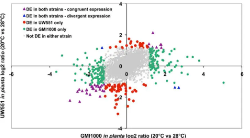
Temperature did not affect the expression of most genes in the two strains (Fig 1). Scatter plots showing mean signal intensities of all genes in UW551 and GMI1000 at 20°C and 28°Cin plantahad very high correlation coefficients (0.93 for UW551 and 0.91 for GMI1000) (S1 Fig). The in culture transcriptomes of both strains were also highly correlated at the two tempera-tures (data not shown). We used an EB-LNN critical threshold of 0.01 and a two-fold differ-ence in expression to define differentially expressed (DE) genes. The differential expression of selected genes was confirmed with qPCR analysis of the same RNA samples used for the micro-array analysis (S1 Table).In planta, tropical strain GMI1000 had more genes DE by tempera-ture than temperate strain UW551. Overall, 4.2% of UW551 genes (181 of 4318) were DE by temperaturein planta(S2 Table), and 3.2% (137 genes) were DE in culture (S3 Table). In strain GMI1000, 7.9% of genes (398 of 5061) were DE by temperaturein planta(S4 Table), and 1.8% (89 genes) were DE in culture (S5 Table). There was little overlap between the GMI1000 and UW551 genesets that were DE by temperaturein plantaand in culture (S2 Fig), indicating that the tropical and temperate strains had distinct transcriptional responses to low temperature under the two experimental conditions. A COG analysis did not identify any noteworthy pat-terns, with the majority of DE genes in the‘unassigned’or‘unknown function’categories in all four conditions. Since responses in the host plant environment are most relevant toR. solana-cearumbiology, the following analysis focuses on thein plantadata, unless otherwise noted.
complex Core Genome, as defined by analyses of 18 strains from all four phylotypes [27]. Genes encoding known bacterial wilt virulence factors like EPS, pectinases, and swimming and twitching motility were not DE by temperature in either strain, although several genes involved in Type 3 secretion were slightly upregulated at 20°C in both strains. Among the strain-specific genes, 66 (8.1%) were differentially expressed in UW551 and 135 (9.8%) were differentially expressed in GMI1000. In both strains, strain-specific genes were more often differentially expressed than orthologs in response to cool temperature, suggesting that UW551 and GMI1000 have fundamentally different responses to temperate conditions. This divergence could reflect distinct, independently evolved responses to chilling, and may offer useful clues to understanding the cool virulence trait of R3bv2 strains.
Several stress and defense-related pathways were up-regulated at cool
temperatures in tropical strain GMI1000
TheR.solanacearum naggene cluster, which putatively encodes degradation of the potent plant defense signal molecule salicylic acid, was also up-regulated in GMI1000 at 20°C (S6 Table). In addition, at the lower temperature GMI1000 had 3- to 6-fold higher expression of the genes encoding degradation of plant phenolic hydroxycinnamic acids (S3 Fig). Finally, sev-eral genes putatively or known to be involved in toxin efflux were more highly expressed in tropical strain GMI1000 during pathogenesis at 20°C than at 28°C (S7 Table). Collectively, this expression profile suggests that infecting a plant at cool temperatures is stressful for this tropi-cal pathogen.
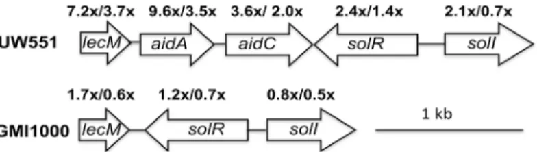
R3bv2 genes induced by cool temperature
Genes encoding the SolI/R quorum sensing system were differentially up-regulated at 20°C rel-ative to 28°C in temperate strain UW551 in culture, indicating that their expression is respon-sive to low temperature (Fig 2). An adjacent gene cluster, which was upregulated at 20°C both in culture andin planta, encodes LecM, a mannose-fucose binding lectin, and AidA and AidC, two proteins of unknown function. The GMI1000 genome does not contain eitheraidAor aidC, and GMI1000’slecMhomolog was not differentially expressed by temperature. We con-firmed the microarray expression results forlecM,aidA, andaidCby measuring their
Fig 1. Scatter plot of log2 expression ratio for orthologous genes at 20°C compared to 28°Cin planta.
The orthologs shared between UW551 and GMI1000 were divided into five groups based on expression pattern. Gray dots in the middle represent orthologs not differentially expressed by temperature in either strain; symbols in colors represent orthologs differentially expressed in at least one strain. Each symbol represents one ortholog.
expression in culture at both temperatures using quantitative real-time PCR (qPCR). For each gene, the expression trend was consistent between microarray and qPCR, although qPCR indi-cated a higher fold-induction (S1 Table), probably due to the higher sensitivity of the method. These results suggested that this region could be involved in cool virulence and thus warranted further study.
Mutation of
lecM
,
aidA
, or
aidC
differentially reduced UW551 virulence at
20°C
To test the hypothesis that genes in the lectin region contribute to the cool virulence of UW551, we generated deletion mutant strains UW551ΔlecM, UW551ΔaidA, and UW551ΔaidC, as well as a UW551ΔsolI mutant. qPCR analysis of cDNA from the mutants confirmed that there was no detectable expression of the deleted gene in each mutant. All the mutants grew indistinguish-ably from wild type in rich broth culture and in tomato leaves at both 20°C and 28°C, indicating that their phenotypes were not the result of a growth defect in culture orin planta. We measured the virulence of mutants on tomato plants using a biologically representative soil-soak inocula-tion that requires bacteria to actively find and invade unwounded host plant roots from the soil. At 28°C, UW551ΔlecM was almost as virulent as its wild type parent, while UW551ΔaidA and UW551ΔaidC had slight virulence defects. In contrast, at 20°C, all three mutants were signifi-cantly reduced in virulence relative to wild type (Fig 3). We quantified this differential virulence by comparing the area under the disease progress curve (AUDPC) for each strain at both tem-peratures to that of wild type. Strains lackinglecM,aidA, oraidCcaused significantly less disease under temperate conditions (P<0.01). For example, at 28°C the AUDPC of UW551ΔlecM was 96% of wild type, but it was only 72% of wild type at 20°C (Fig 3E). All three mutants were also significantly reduced in virulence when they were inoculated directly into the tomato xylem via a cut leaf petiole (Fig 3F,P<0.01, repeated measures ANOVA), indicating that R3bv2 strain UW551 requireslecM,aidA, andaidCfor mid-stage bacterial wilt disease virulence, and not only for early infection. Together, these results demonstrate thatlecM,aidA, andaidCencode functions necessary for cool virulence of UW551.
Lectin-specific sugars inhibited
R
.
solanacearum
attachment to tomato
roots
Lectins, which are carbohydrate-binding proteins, play important roles in cell–cell interactions, including microbial pathogenesis [30]. To test the hypothesis that LecM contributes toR. sola-nacearumadhesion to plants, we compared the ability of UW551 and the UW551ΔlecM mutant to attach to tomato seedling roots at 20°C, both separately and in a 1:1 competition
Fig 2. A cluster of genes adjacent to those encoding the SolI/R quorum sensing system were up-regulated inR.solanacearumstrain UW551 at 20°C, in culture.Some were also upregulatedin planta. Arrows represent open reading frames. The numbers above the arrows indicate the expression fold-change for each gene at 20°C compared to 28°C, in culture/in planta, determined by whole-genome microarray analysis as described in the text. The arrangement and expression levels of the corresponding genes in strain GMI1000 are also shown.
assay. ThelecM-deficient strain had no measurable defect in attachment in either condition. This could be the result of functional redundancy between LecM and LecX, the other man-nose-fucose binding lectin encoded by the UW551 genome, especially sincelecXwas also strongly up-regulatedin plantacompared to its expression in CPG culture at the same temper-ature (Table 1).
LecM is 70% identical to LecB of the opportunistic human pathogenPseudomonas aerugi-nosa; LecB contributes toP.aeruginosavirulence via involvement in biofilm formation, adhe-sion, host recognition, and inhibition of host defenses [31] [32–34]. Patients infected byP. aeruginosahave been successfully treated with a solution of sugars specifically bound by its two lectins, LecA and LecB [35,36]. The sugars compete for and block the lectin sugar-binding sites, thus inhibiting bacterial attachment to host cells.
To determine if lectins have a similar function inR.solanacearum, we incubated strain UW551 in a solution of sugars that bind to its two lectins (LecM and LecX) to see if this would
Fig 3. Mutation ofR.solanacearumstrain UW551lecM,aidA, oraidCbut notsolIdifferentially reduced bacterial virulence at 20°C.Virulence was measured on wilt-susceptible tomato plants at 20°C and 28°C via soil soak inoculation (A-E) or at 20°C via cut petiole inoculation (F). Each point represents the mean of three biological replicates, each containing 16 plants per strain per temperature. The area under disease progress curve (AUDPC) was measured for each strain in A-C and each mutant’s AUDPC relative to wild type is shown (E). Asterisks indicate that virulence of wild type and mutant strains were significantly different (*P<0.05,**P<0.01, ANOVA). Each mutant was also significantly reduced in virulence (P<0.01, repeated measures ANOVA) compared to the wild type following cut petiole inoculation (F).
affect its ability to attach to plant roots. UW551 cells pre-incubated with 10 mM D-mannose and 10 mM L-fucose were significantly reduced in attachment to tomato roots compared with the untreated bacteria (1.5x10⁷cfu/g root as compared to 2.2x10⁶cfu/g root, P<0.01, t-test). Exposure to the sugar solution did not affect bacterial viability and a control treatment with 10 mM glucose had no effect on bacterial attachment.
LecM was required for biofilm formation
in vitro
R.solanacearumforms biofilms on PVC plastic plates and biofilm-like aggregations on tomato seedling roots, although the biological significance of biofilms in this species is not known [37,38].P.aeruginosaLecB specifically binds to cells in biofilms and contributes to biofilm for-mation. Exopolysaccharides are key components of microbial biofilms, and mannose, which is preferentially bound by both LecB and LecM, is a primary constituent of EPS from bothP. aeruginosaandR.solanacearum[39–41]. We therefore tested the hypothesis that LecM is involved in biofilm formation. On PVC microtiter plates, UW551ΔlecM formed 9.15-fold less biofilm than its wild type parent (P<0.01). Together, these results suggest that LecM may func-tion both in attaching bacteria to host plant cells (adhesion), and in bacterial cell–cell binding in biofilms (cohesion).
LecM was required for normal host colonization
To measure the effect of LecM on bacterial success inside host plants, after the adhesion and entry stages, we quantifiedR.solanacearumpopulation growth in stems of tomato inoculated directly into the xylem via a cut petiole with either UW551 or UW551ΔlecM and incubated at 20°C. Two days after inoculation, plants inoculated with wild type and UW551ΔlecM con-tained similar numbers of bacteria, with mean population sizes just over 1×10⁶cfu g−1. How-ever, eight days after inoculation, the mean population size of UW551 in tomato stem was 2.83×10¹¹cfu g−1, more than 50-fold larger than that of UW551ΔlecM, which was 5x10⁹cfu g−1(P<0.01). This result indicated thatlecMcontributes to xylem colonization by UW551. The colonization defect at day 8 corresponds to the difference in time of disease onset between wild type and UW551ΔlecM in the cut-petiole virulence assay (Fig 3F).
UW551
Δ
lecM and UW551
Δ
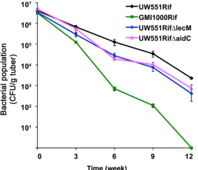
aidC survived poorly in potato tubers at 4°C
We previously found that R3bv2 strain UW551 survived much longer than non-R3bv2 strains in tubers of potato, a socioeconomically important natural host, at the common seed tuber storage temperature of 4°C [9]. We inoculated potato tubers withlecMandaidCmutants and
Table 1. Expression of lectin genes inR.solanacearumstrains GMI1000 and UW551 under various conditions.
Gene name GMI1000 gene expression UW551 gene expression Locus tag
20°C CPG 28°C CPG 20°C IP 28°C IP 20°C CPG 28°C CPG 20°C IP 28°C IP GMI1000 UW551
lecF 16.7a 16.6 16.7 16.6 npb np np np RSc2107 np
lecM 14.5 13.8 15.9 16.5 13.3 10.5 14.6 12.6 RSc3288 RRSL_02788
lecX 16.7 16.6 16.8 16.6 14.9 14.7 16.2 16.1 RS03909 RRSL_03943
aValues shown are scaled log2 signal intensities from strain-speci
fic whole genome microarrays hybridized to labeled cDNA extracted fromR. solanacearumcells grown in rich culture medium (CPG) orin planta(IP). These absolute expression values were used to allow comparisons across strains.
bnp: gene not present in this strain.
measured their population sizes over time. As previously observed, tropical strain GMI1000 survived poorly in potato tubers at 4°C compared to UW551. At 6, 9 and 12 weeks after inocu-lation, the population sizes of UW551ΔlecM and UW551ΔaidC in tubers were around 5-fold lower than those of the wild type parent strain (Fig 4,P<0.05), indicating that LecM and AidC contribute to cold survival of UW551 as well as to temperate virulence.
Adding the UW551 lectin region to GMI1000 did not confer cool
virulence on GMI1000
SinceaidAandaidCare not present in GMI1000 and there is some sequence variation between the two strains in this region, we tested the hypothesis that inserting the UW551lecMcluster into a neutral site in the GMI1000 chromosome would increase its virulence in temperate con-ditions. However, neither a fragment containing the UW551lecM,aidA, andaidCORFs (plus 753 bp of upstream sequence) nor a larger fragment containing the UW551lecM,aidA,aidC, soIR, andsolIORFs increased the virulence of GMI1000 at 20°C. This negative result suggests that either the necessary UW551-typical regulation of these genes did not occur in GMI1000, or that additional elements are required to confer cool virulence on non-R3bv2 strains.
LecM, AidA, and AidC are required for cool virulence in other R3bv2
strains
We attempted to complement each of these three mutations, firstin transon a low-copy num-ber plasmid and then by insertion of single copies into the selectively neutral chromosomalatt site. In all cases the merodiploid strains had pleiotropic phenotypes: they were hypermotile and several overproduced an acylhomoserine lactone quorum sensing signal molecule. We there-fore used an alternative method to confirm that the insertions in the target genes were
Fig 4. UW551ΔlecM and UW551ΔaidC had reduced survival in potato tubers at 4°C.Potato tubers were injected with differentR.solanacearumstrains, and bacterial cell numbers were counted by grinding and dilution plating tubers at different times after inoculation. The experiment was repeated three times, with three tubers per strain per time point. At 6, 9 and 12 weeks after inoculation, the population sizes of UW551ΔlecM and UW551ΔaidC in tubers were significantly lower than those of the wild type parent strain (P<0.05, ANOVA).
responsible for the cool virulence phenotypes we observed, and to demonstrate that this result was not unique to UW551. We independently recreated thelecM,aidA, andaidCmutations in two additional R3bv2 strains ofR.solanacearum, UW553 and UW560. The resulting mutants did not have the pleiotropic phenotypes observed in the merodiploid“complemented”strains, nor did they have any growth defect compared to the wild type strains. We tested the virulence of these mutants at 20°C via soil-soak inoculation. Consistent with the findings in UW551, strain UW553 and UW560 mutants lackinglecM,aidA, oraidChad similarly reduced viru-lence at 20°C compared to their respective wild type parents (S4 Fig,P<0.01, repeated mea-sures ANOVA). These results confirmed that each of these genes is important for the temperate virulence of R3bv2 strains.
The l
ecM
region is regulated by the SolI/R quorum sensing system
Since thelecMgene cluster is adjacent to thesolI/Racyl-homoserine lactone (AHL) quorum sensing (QS) system, andaidAis controlled bysolI/R[28], we used qRT-PCR to determine if other genes in this region were also regulated by SolI/R. WhensolIwas deleted, transcription in culture oflecM,aidA, andaidCwere all lower than in a wild-type background, although to dif-fering degrees. Expression oflecMwas 54.7-fold lower in UW551ΔsolI, whileaidAexpression was 31-fold lower andaidCwas 2.2-fold lower. These results demonstrate that, at least in cul-ture,lecM,aidA, andaidCare all positively regulated by the SolI/R QS system.
Discussion
We compared the transcriptional profiles of two ecologically distinct strains of a high-impact plant pathogen at different temperatures in culture and during disease in a natural host. There was only minimal overlap between the differentially expressed gene sets in culture andin plantafor both temperate R3bv2 strain UW551 and tropical strain GMI1000, confirming our previous finding thatR.solanacearumregulates gene expression very differentlyin vitroandin planta[29]. These results offer further evidence that data fromin vitrostudies do not reliably reflect pathogen behaviorin vivoand therefore should be used and interpreted with caution [42].
Thisin plantacomparative analysis yielded insights into ways that temperature has shaped two related pathogens. Changing the temperature of pathogenesis from a tropical 28°C to a cool temperate 20°C altered expression of relatively few genes in bothR.solanacearumstrains. Nonetheless, almost twice as many genes were differentially expressed at the lower temperature in GMI1000 than in UW551, and the number of genes up-regulated at 20°C in GMI1000 was almost three times greater than the number up-regulated in UW551. This was not unexpected, since UW551 is a successful pathogen at both temperate and tropical temperatures, and is thus well adapted to broad environmental parameters. When UW551 causes disease at 20°C, it alters expression of less than 5% of its genome relative to when it wilts tomato plants at 28°C. In contrast, tropical strain GMI1000 had a more significant transcriptional response to cool temperatures, including up-regulating diverse stress-related functions, possibly because it is not adapted to this condition.
because our experiment did not expose bacteria to a sudden change in temperature. It is also likely that 20°C was not low enough to induce cold shock.
R.solanacearumstrain GMI1000 is only weakly virulent on tomato at 20°C [9]. At this cool temperature, the tropical strain increased expression of diverse self-protective functions that were not up-regulated in temperate strain UW551 under the same condition. Like most bacte-ria,R.solanacearumstrains have multiple toxin efflux pumps that confer tolerance of deleteri-ous compounds and are often up-regulated as part of a generalized stress response [47,48]. We previously found that AcrAB, an RND family efflux pump, contributes to bacterial wilt viru-lence by protectingR.solanacearumfrom host antimicrobial compounds [49]. Genes encoding six toxin efflux pumps, including AcrAB, were differentially up-regulated during GMI1000 pathogenesis of tomato at 20°C, suggesting that this tropicalR.solanacearumstrain confronts toxic plant defenses in temperate conditions.
Thenaggenes, which encode degradation of the plant signaling molecule salicylic acid (SA) were also differentially up-regulated at 20°C in GMI1000. The SA defense pathway is activated during tomato resistance toR.solanacearum[50], and SA itself can directly reduce bacterial fit-ness and virulence factor production [51]. Elevatednagcluster expression may reflect a bacte-rial response to high SA levels in tomato plants infected by GMI1000 at 20°C. We previously found that at 28°C, GMI1000-infected tomato plants expressed SA defense genes faster and to a greater degree than plants infected with UW551 [52]. At 28°C, both GMI1000 and UW551 can successfully overcome SA-dependent defenses in tomato, and UW551 is still able to do this at 20°C. GMI1000 may increase expression of its SA degradation pathway at 20°C in an unsuc-cessful attempt to overcome SA-triggered plant defenses.
Finally, during pathogenesis at 20°C, GMI1000 differentially upregulated genes encoding degradation of hydroxycinnamic acids, which are major plant cell wall-reinforcing phenolic compounds that also have direct antimicrobial properties [53–58].R.solanacearumelicits pro-duction of cell-wall-bound phenolic compounds in tomato, and increased phenolic levels cor-related with lowerR.solanacearumpopulation sizes [59,60]. Furthermore, we recently found that the ability to degrade hydroxycinnamic acids is required for full virulence of GMI1000 at 28°C [61]. The increased expression of the hydroxycinnamic acid degradation pathway at 20°C suggests that GMI1000 may confront higher levels of phenolics at cool temperatures, where it struggles to cause disease. Consistent with this idea, we observed that tomato plants infected by GMI1000 at 20°C exhibit extensive vascular browning, which is indicative of plant phenolic production. We speculate that, like resistant plants, tomatoes infected with GMI1000 at 20°C produce more phenolic compounds. This may help explain the strain’s weak virulence at this temperature.
Taken together, this expression profile suggests that GMI1000 confronts significant stress in tomato plants at 20°C, and it may up-regulate toxin efflux pumps and enzymes that degrade SA and phenolic compounds in an ineffectual attempt to overcome host defenses. Alterna-tively, GMI1000 and UW551 may experience similar levels of host defense under these condi-tions, but UW551 is better able to tolerate them.
extracted from bacteria co-cultured with tomato seedlings in static liquid MS medium, whereas the transcriptomic study measured mRNA from bacteria extracted from the xylem vessels of diseased whole tomato plants. Thus, both theR.solanacearumstrains and their host plants experienced significantly different physiological conditions in these two studies.
A separate study found that cool virulent strains exhibited reduced twitching motility, which is required for bacterial wilt virulence [63]. However, no known twitching motility genes were DE by temperature in either the cool virulent R3bv2 strain or tropical strain GMI1000. Again, the experimental conditions were not comparable; cool virulent strains may use twitch-ing motility on solid surfaces like agar plates (or plant roots) but not in shaktwitch-ing broth culture or in tomato xylem, which were the two conditions we assayed here.
Three genes in a cluster that was up-regulated at 20°C in R3bv2 strain UW551 contributed significantly to cool virulence on tomato plants and to survival in potato tubers at 4°C. To our knowledgelecM,aidA, andaidCare the first genes known to be involved in temperature adap-tation in plant pathogenic bacteria. However, their exact mechanisms of action remain to be determined. Our data suggest that the mannose/fucose binding lectin LecM increases pathogen attachment to other bacteria during biofilm formation. Pre-treatment of the bacterium with a solution of mannose and fucose reduced root attachment, which is indirect evidence that that lectins may also helpR.solanacearumattach to host surfaces.
Binding affinities and crystal structures have been determined for three lectins fromR. sola-nacearum, originally named RSL, RS-IIL, and RS20L, but their biological functions remain unknown. LecF (RSL), which is not present in UW551, preferably binds L-fucose [64–66]. LecX (RS20L) binds L-fucose, D-mannose and D-xylose [67]. LecM (RS-IIL), which was signif-icantly induced in UW551 at 20°C, binds D-mannose, L-fructose, L-fucose and D-arabinose [65]. These sugars are common constituents of plant cell wall polysaccharides [68,69].
Interestingly, all lectin genes from both strains were highly expressed under all conditions tested, although with differing expression patterns. Specifically,lecFandlecXwere consistently highly expressed in GMI1000; indeed, their expression levels were so high that the chip may have been saturated, preventing detection of expression differences across conditions. In UW551,lecXwas up-regulatedin planta, but was not differentially expressed by temperature, andlecMwas up-regulated in response to both growthin plantaand low temperature.lecM was also inducedin plantain GMI1000, though not by low temperature. The high expression levels of all lectin genes suggest these proteins are important forR.solanacearumfitnessin planta. It would be interesting to determine the virulence of a triple mutant lacking all three lectin structural genes.
Plant lectins play important diverse roles in plant-microbe interactions [70–73], but to our knowledge this is the first indication that bacterial lectins may contribute to pathogen attach-ment to plants. Our finding that lectin-specific sugars could inhibit attachattach-ment of UW551 cells to tomato roots is consistent with a novel role forR.solanacearumlectins in bacterial adher-ence to plant cells. Because the UW551ΔlecM mutant was significantly reduced in virulence even when it was introduced directly into the tomato xylem through a cut petiole, this lectin apparently also functions inside the plant, not only during initial root attachment.
AidA and AidC were previously wholly cryptic, but this work demonstrated that they both play significant roles in bacterial wilt virulence, contributing disproportionately at low temper-atures. They are not, however, unique to cool-temperate R3bv2 strains ofR.solanacearum. AlthoughaidAandaidCare absent from African (phylotype III) and Asian (phylotype I) strains, they are present in tropical Indonesian phylotype IV strains and in tropical and warm-temperate phylotype II strains from North and South America, suggesting that their function extends beyond cool virulence [26]. ThelecM-aidA-aidCgene order is retained inBurkholderia cenocepaciaandPseudomonas entomophila, although the gene sequences are not well con-served. TheaidAgene was originally identified inR.solanacearumstrain AW1 because it is regulated by the SolIR quorum sensing system [28]. An AidA homolog inBurkholderia cenoce-paciacontributes to slow killing of nematodes [77]. However,R.solanacearumAidA is not necessary for slow killing of nematodes and its function remains unknown [78]. AidC is about 24% identical to AidA at the amino acid level and its function is also unknown;aidCis located directly downstream ofaidAin allR.solanacearumgenomes that containaidA.
The SolI/R quorum sensing system regulated expression oflecM,aidA, andaidCat 20°C in strain UW551. Consistent with a previous study of asolImutant inR.solanacearumstrain AW1 [28], we found that the UW551ΔsolImutant had no virulence defect at either 20°C or 28°C. This might seem counterintuitive, since SolI/R positively regulates expression oflecM, aidA, andaidC, which were all required for virulence, especially at 20°C. Our microarrays and qPCR assays measured the transcription ofR.solanacearumcells at densities higher than 1x10⁸ cfu g−1, which is above the threshold for activation of the SolI/R regulatory system. However, expression of these genes was induced not only by quorum sensing but also by low temperature and by a condition or signal present in plants, so it is possible thatlecM,aidA, andaidCare expressedin plantaeven in the absence ofsolIR. We and others previously showed that viru-lence traits controlled by another quorum sensing regulator, PhcA, are regulated very differ-ently in culture andin planta[29,79]. It is also possible that inactivation ofsolIRdid not affect virulence because of redundancy between the virulence-regulating quorum sensing systems in R.solanacearum. The AHL-responsive SolI/R autoinduction system inR.solanacearumis part of a more complex autoregulatory hierarchy. Expression ofsolRandsolIrequires PhcA, which is itself controlled by a second quorum sensing system that responds to 3-OH-PAME [28]. As mentioned above,solIRexpression is additionally dependent on the alternate sigma factor RpoS [75].
Overall, these studies reveal that cool virulence is a complex multigenic trait inR. solana-cearum. Unexpectedly, we found that a bacterial lectin contributes differentially to bacterial wilt virulence at lower temperatures. Our results generated diverse testable hypotheses for fur-ther laboratory and field studies and also identified biochemical pathways that plant breeders may be able to manipulate in tomato varieties to restrict growth of vascular pathogens and gen-erate disease-resistant crops.
Materials and Methods
Bacterial strains and culture conditions
Laboratories (Detroit, MI) and other chemicals were purchased from Sigma-Aldrich (St. Louis, MO).
RNA extraction
For the in culture samples, RNA was extracted from 30 ml cultures of strains UW551 and GMI1000 grown in rich CPG broth to log phase (~6x10⁸cfu ml−1), using a modified hot-phe-nol extraction followed by four DNase treatments [82]. For thein plantasamples, wilt-suscep-tible Bonny Best tomatoes were soil-soak inoculated at 28°C and 20°C with strain UW551 or strain GMI1000 as described for the virulence assay below. Before inoculation, roots of plants inoculated with GMI1000 at 20°C were disturbed by gently lifting the plant stem a few mm to increase numbers of symptomatic plants at this non-conducive temperature. Bacterial RNA was harvested from plants showing early wilting symptoms (disease index = 1, as described below). Since xylem vessels are the primary habitat ofR.solanacearumduring pathogenesis, we harvested bacteria from infected plants by centrifuging 7 cm sections of infected tomato stems at 13,500gfor 5 min in 15 ml conical tubes containing 3 ml transcriptional stop solution (95% ethanol, 5% phenol). The resulting bacterial pellet was stored at -80°C until the bacterial popu-lation size in the stem was confirmed by dilution plating a ground stem section. Pellets from stems colonized with 1x10⁸cfu g−1to 1x10⁹cfu g−1were pooled for RNA extraction by the hot phenol method. Absence of genomic DNA contamination in RNA was confirmed by PCR with R.solanacearumuniversal primers 759/760 [10]. Quality of RNA samples was assessed using Nanodrop and the Agilent Bioanalyzer 2100 nanochip system (Agilent Technologies). RNA samples with A260/A280>2.0, A260/A230>2.0, and RNA Integrity Number (RIN)>8 were used for microarray and qPCR analysis. We did four biological replicates for each condition of the microarray experiment.
Transcriptome analysis with microarrays
Quantitative RT-PCR (qPCR)
qPCR was performed as described [52] using an ABI PRISM 7300 Real-Time PCR System (Applied Biosystems, Foster, CA) with SYBR-Green chemistry. Based on observed stable expression in the microarrays under all conditions, threeR.solanacearumgenes,oxyR,rplM, andserC, were selected to normalize expression values for all other genes. The qPCR primer sequences are listed inS9 Table. Reaction parameters for qPCR were: 10 min polymerase acti-vation, followed by 40 cycles of 95°C for 15 sec and 57°C for 1 min.
Comparative genomics
Orthologous (homologous) proteins between genomes were identified using BLAST-based methods. Orthologs were assigned to genes whose similarity scores and percent identity fell within cutoff values of>70% for pair-wise BLASTP searches. Whole genome alignments using Mauve v2.1 [87] allowed us to use gene context as an arbiter in cases where multiple paralogs were possible and to designate orthologs with improved specificity and precision. When true orthology was critical, downstream analyses were limited to high confidence datasets.
DNA manipulations
Cloning, restriction digestion, sequencing, and PCR were performed using standard methods [88].R.solanacearumandE.coliwere transformed by electroporation as previously described [89]. DNA sequencing and oligonucleotide synthesis were performed at the University of Wis-consin-Madison Biotechnology Center. DNA sequence was analyzed using the DNASTAR software package (DNASTAR, Inc., Madison, WI, U.S.A.). Unless otherwise noted, molecular biology reagents and kits were purchased from Promega (Madison, WI).
Mutant strain construction
Splicing by overlap extension PCR (SOE-PCR) was used to create in-frame deletion constructs using primers listed inS9 Table[90]. Deletion constructs were introduced into the chromo-some of wild-typeR.solanacearumstrain UW551 by double homologous recombination as previously described [38] to create UW551ΔlecM, UW551ΔaidA, UW551ΔaidC and UW551Δ -solI, respectively. The correct allelic replacement in each mutant was confirmed by PCR and sequencing analyses. To generate GMI1000 strains carrying the putative cool virulence region from UW551, the 2,534-bp DNA fragment containing thelecM-aidA-aidCregion plus 753bp of upstream oflecMor a 4,149-bp fragment containing thelecM-aidA-aidC-solR-solIregion were amplified from UW551 genomic DNA using the PCR primer pairs Up lecM_ F/Com aidC _R and ComWhole _F/ComWhole _R, respectively, and cloned into theSmaI site of pUC18-mini-Tn7T-Gm [91]. The resulting products were introduced into strain GMI1000 at the selectively neutralattTn7 site 25bp downstream of theglmSgene [92] as described [93] and the correct integration was confirmed with PCR.
Natural transformation of
R
.
solanacearum
strains
Virulence assays
To compare the virulence ofR.solanacearumstrains at different temperatures, we used a natu-ralistic soil soak inoculation and a more direct petiole inoculation of the wilt-susceptible tomato cv. Bonny Best, as previously described [95], except that half the plants were moved from 28°C to 20°C growth chambers one day after transplanting. For the soil soak assay, a bac-terial suspension was poured onto the soil of pots that each contained an unwounded 16-day-old tomato plant to a final density of approximately 1x10⁸cfu g−1soil. For the petiole inocula-tion, 2x10³bacteria in a 2μL volume were placed onto the freshly cut petiole of the first true leaf of a 21-day-old tomato plant. Plants were monitored daily for disease progress by a rater blind to treatment identity, and symptoms were scored on a 0-to–4 disease index, where 0 indi-cates no disease, 1 indiindi-cates 1 to 25% of leaves wilted, 2 indiindi-cates 26 to 50% of leaves wilted, 3 indicates 51 to 75% of leaves wilted, and 4 indicates 76 to 100% of leaves wilted. Each experi-ment contained a minimum of 16 plants per strain at each temperature, and experiexperi-ments were repeated at least three times. The disease index for each day is the average of 48 plants from three experiments.
Multiplication of
R
.
solanacearum
strains in tomato leaves
To ensure that mutant strains had not lost the ability to multiply in the host, we measured growth of strains uniformly infused into tomato leaves.R.solanacearumcells grown overnight in CPG were collected by centrifugation, resuspended in water, adjusted to OD600= 0.01, and
infiltrated into fully expanded tomato leaves with a syringe. Plants were incubated at either 28°C or 20°C. Every 12 hours for three days, leaves were sampled with a #5 cork borer, three leaf discs were pooled, ground, and dilution plated on TZC medium to enumerate colony form-ing units cm−2leaf tissue. The experiment was repeated three times, with at least six plants per strain per temperature.
Colonization of tomato stems by
R
.
solanacearum
strains
To measure bacterial colonization of tomato stems at 20°C, plants were inoculated with either the wild-type strain or the mutant using the cut petiole method as described above. Population sizes of each strain in tomato plants were quantified as described [38]. Briefly, at different time points after inoculation, five plants inoculated with each strain were randomly chosen and a 1 cm stem segment spanning the inoculation site was collected, weighed, and ground in 1 ml sterile deionized water. The resulting homogenate was dilution plated on TZC plates supple-mented with 100 mg l−1cycloheximide and appropriate antibiotics.R.solanacearum popula-tion sizes were determined as cfu g−1of plant tissue. All experiments were repeated three times.
Measuring bacterial attachment to tomato roots
Attachment ofR.solanacearumcells to tomato seedling roots was quantified as described [80]. Briefly, susceptible tomato cv.‘Bonny Best’seedlings were grown axenically on Murashige and Skoog medium with vitamins (Caisson Laboratories, North Logan, UT) for 2 weeks, when sec-ondary roots were well developed. These were incubated withR.solanacearumcell suspensions in water (OD600= 0.01) at 20°C for 1h. For competition studies, wild type and mutant cell
were prepared as described above, except they were resuspended either in sterile water or water containing 0.01M D-mannose and 0.01M L-fucose, or 0.01M glucose as a control, and incu-bated at room temperature for half an hour. The bacterial suspensions were then incuincu-bated with sterile tomato seedlings at 20°C for an hour as described above.
Biofilm assays
Production of biofilms byR.solanacearumstrains was measuredin vitrousing a minor modifi-cation of the polyvinylchloride (PVC) microtiter plate assay [96]. Briefly, 5μL overnight cul-tures ofR.solanacearumadjusted to OD600= 0.1 were used to inoculate 95μL of CPG broth in
wells of a PVC microtiter plate and incubated without shaking for 24h at 28°C. Biofilms were quantified by absorbance at 600nm following crystal violet staining.
Survival in potato tubers
We measured the survival ofR.solanacearumstrains in susceptible 15 to 17 mm potato mini-tubers (cv.‘Russet Norkotah’, Sklarczyk Seed Farm, Johannesburg, MI) at the typical seed potato storage temperature of 4°C as previously described [9]. Briefly, both ends of each potato tuber were injected with 2μL of 1x10⁹cfu ml−1bacterial suspensions. Tubers were stored in the dark at 4°C and sampled at intervals. Tissue was weighed, ground, and dilution plated to determine cfu g−1. Survival rates were calculated based on triplicate experiments using two to three individual tubers per sampling time point. The detection limit was 1 log cfu g−1potato tissue and a value of 1 log cfu g−1was also used to report samples below the detection limit. Experiments were performed with rifampicin-resistantR.solanacearumstrains to facilitate pathogen detection in the natural microbial background.
Supporting Information
S1 Fig. Scatter plots of expression levels of allR.solanacearumUW551 and GMI1000 at 20°C and 28°C during tomato pathogenesis.Within each strain, gene expression was highly correlated at temperate and tropical temperatures. Scatter plots showingin plantamean signal intensities of genes in the genomes of UW551 (A) and GMI1000 (B) at 20°C and 28°C, as determined by whole-genome microarray analysis. Each dot represents a gene, and the log2 signal intensity for each gene shown is the average of four biological replicates.
(PDF)
S2 Fig. Venn diagrams ofR.solanacearumUW551 and GMI1000 genes differentially expressed by temperature in culture and during tomato pathogenesis.A small number of genes were differentially expressed (DE) at 20°C compared to 28°C, with minimal overlap between those DE in culture andin planta. The numbers in the circles indicate the number of genes DE under different conditions (>2-fold difference in expression by EBArray analysis with the false discovery rate set at 0.01).
(PDF)
S3 Fig. TheR.solanacearumhydroxycinnamic acid degradation pathway and effect of cool temperature on its expression.The hydroxycinnamic acid degradation pathway was up-regu-lated inR.solanacearumstrain GMI1000 during tomato pathogenesis at 20°C. Above each arrow is shown the gene(s) encoding each enzyme, together with their expression level fold-changes at 20°C compared to 28°Cin plantaare shown above the arrows.
S4 Fig. Virulence effects of mutatinglecM,aidA, andaidCin two additional R3bv2R. sola-nacearumstrains.Mutation oflecM,aidA, oraidAinR.solanacearumR3bv2 strains UW553 (A) and UW560 (B) resulted in significantly lower bacterial wilt virulence than the correspond-ing wild-type parent strain at 20°C (P<0.01 by repeated measures ANOVA). Virulence was measured on wilt-susceptible tomato plants at 20°C via soil soak inoculation. The experiment was repeated twice, each replicate containing 16 plants per treatment per strain. Results from a representative experiment are shown.
(PDF)
S1 Table. qPCR validation of expression levels for someR.solanacearumstrain UW551 genes differentially expressed in the microarray analysis.
(PDF)
S2 Table.R.solanacearumstrain UW551 genes differentially expressedin plantaat 20°C compared to 28°C.
(PDF)
S3 Table.R.solanacearumstrain UW551 genes differentially expressed in rich culture medium (CPG) at 20°C compared to 28°C.
(PDF)
S4 Table.R.solanacearumstrain GMI1000 genes differentially expressedin plantaat 20°C compared to 28°C.
(PDF)
S5 Table.R.solanacearumstrain GMI1000 genes differentially expressed in rich culture medium (CPG) at 20°C compared to 28°C.
(PDF)
S6 Table. Genes involved in the salicylic acid degradation pathway were selectively up-regu-lated inR.solanacearumstrain GMI1000 during tomato pathogenesis at 20°C.
(PDF)
S7 Table. Genes involved in multidrug efflux were up-regulated inR.solanacearumstrain GMI1000 during tomato pathogenesis at 20°C.
(PDF)
S8 Table. Bacterial strains used in this study. (PDF)
S9 Table. Sequences of PCR primers used in this study. (PDF)
Acknowledgments
Author Contributions
Conceived and designed the experiments: FM LB JMJ CA. Performed the experiments: FM JMJ. Analyzed the data: FM LB JMJ CA. Wrote the paper: FM LB JMJ CA.
References
1. Hayward AC (1991) Biology and epidemiology of bacterial wilt caused byPseudomonas solanacearum. Annu Rev Phytopathol 29: 65–87. PMID:18479193
2. Allen C, Prior P, Hayward AC, editors (2005) Bacterial Wilt Disease and the Ralstonia solanacearum species complex. St Paul: APS Press. 510 p.
3. Genin S, Denny TP (2012) Pathogenomics of theRalstonia solanacearumspecies complex. Annu Rev Phytopathol 50: 67–89. doi:10.1146/annurev-phyto-081211-173000PMID:22559068
4. Schell MA (2000) Control of virulence and pathogenicity genes ofRalstonia solanacearumby an elabo-rate sensory network. Annu Rev Phytopathol 38: 263–292. PMID:11701844
5. Champoiseau P, Jones J, Allen C (2009)Ralstonia solanacearumRace 3 biovar 2 causes tropical losses and temperate anxieties. Plant Health Progress.
6. Elphinstone JG (2005) The current bacterial wilt situation: A global overview. In: Allen C, Prior P, Hay-ward AC, editors. Bacterial Wilt: The Disease and the Ralstonia solanacearum Species Complex. St Paul: American Phytopathological Society Press. pp. 9–28.
7. Ciampi L, Sequeira L (1980) Influence of temperature on virulence of Race 3 strains ofPseudomonas solanacearum. Am Potato J 57: 307–317.
8. Tung PX, Rasco ET, VanderZaag P, Schmiediche P (1990) Resistance toPseudomonas solana-cearumin the potato: II. Aspects of host-pathogen-environment interaction. Euphytica 45: 211–215.
9. Milling A, Meng F, Denny TP, Allen C (2009) Interactions with hosts at cool temperatures, not cold toler-ance, explain the unique epidemiology ofRalstonia solanacearumRace 3 biovar 2. Phytopathology 99: 1127–1134. doi:10.1094/PHYTO-99-10-1127PMID:19740025
10. Fegan M, Prior P (2005) How complex is the "Ralstonia solanacearumspecies complex"? In: Allen C, Prior P, Hayward AC, editors. Bacterial Wilt Disease and theRalstonia solanacearumspecies complex. St. Paul: APS Press. pp. 449–461.
11. Poussier S, Trigalet-Demery D, Vandewalle P, Goffinet B, Luisetti J, Trigalet A (2000) Genetic diversity ofRalstonia solanacearumas assessed by PCR-RFLP of thehrpgene region, AFLP and 16S rRNA sequence analysis, and identification of an African subdivision. Microbiology 146: 1679–1692. PMID:
10878132
12. Janse JD, van den Beld HE, Elphinstone J, Simpkins S, Tjou-Tam-Sin NAA, van Vaerenbergh J (2004) Introduction to Europe ofRalstonia solanacearumbiovar 2, race 3 inPelargonium zonalecuttings. J Plant Pathol 86: 147–155.
13. Kim SH, Olson TN, Schaad NW, Moorman GW (2003)Ralstonia solanacearumRace 3, biovar 2, the causual agent of brown rot of potato, identified in geranium in Pennsylvania, Delaware, and Connecti-cut. Plant Dis 87: 450.
14. Swanson J, Montes L, Mejia L, Allen C (2007) Detection of latent infections ofRalstonia solanacearum Race 3 biovar 2 in geranium. Plant Disease 91: 828–834.
15. Williamson L, Kazuhiro N, Hudelson B, Allen C (2002)Ralstonia solanacearumRace 3, Biovar 2 strains isolated from geranium are pathogenic on potato. Plant Disease 86: 987–991.
16. Elphinstone J (1996) Survival and possibilities for extinction of Pseudomonas solanacearum (Smith) in cool climates. Potato Res 39: 403–410.
17. Elphinstone J, Stanford HM, Stead D (1998) Detection ofRalstonia solanacearumin potato tubers, Solanum dulcamara, and associated irrigation water. In: Prior P, Allen C, Elphinstone J, editors. Bacte-rial wilt disease: Molecular and ecological aspects. Berlin, Germany: Springer. pp. 133–139.
18. van Elsas JD, Kastelein P, van Bekkum P, van der Wolf JM, de Vries PM, van Overbeek L S (2000) Sur-vival ofRalstonia solanacearumbiovar 2, the causative agent of potato brown rot, in field and micro-cosm soils in temperate climates. Phytopathology 90: 1358–1366. doi:10.1094/PHYTO.2000.90.12. 1358PMID:18943377
19. van Elsas JD, Kastelein P, de Vries PM, van Overbeek LS (2001) Effects of ecological factors on the survival and physiology ofRalstonia solanacearumbv. 2 in irrigation water. Can J Microbiol 47: 842–
854. PMID:11683466
21. Agrios GN (2005) Plant Pathology. Amsterdam: Elsevier Academic Press.
22. Large EC (1940) The Advance of the Fungi. New York: Henry Holt and Co.
23. Schumann G, D'Arcy C (2012) Hungry Plant: Stories of Plant Diseases. St. Paul: APS Press.
24. Salanoubat M, Genin S, Artiguenave F, Gouzy J, Mangenot S, Arlat M, et al. (2002) Genome sequence of the plant pathogenRalstonia solanacearum. Nature 415: 497–502. PMID:11823852
25. Gabriel DW, Allen C, Schell M, Denny TP, Greenberg JT, Duan YP, et al. (2006) Identification of open reading frames unique to a select agent:Ralstonia solanacearumRace 3 Biovar 2. Mol Plant-Microbe Interact 19: 69–79. PMID:16404955
26. Remenant B, Cambiaire J-CD, Cellier G, Barbe V, Medigue C, Jacobs JM, et al. (2011) Phylotype IV strains of Ralstonia solanacearum, R. syzygii and the Blood Disease Bacterium form a single genomic species despite their divergent life-styles. PLoS One 6: e24356.
27. Guidot A, Prior P, Schoenfeld J, Carrere S, Genin S, Boucher C (2007) Genomic structure and phylog-eny of the plant pathogenRalstonia solanacearuminferred from gene distribution analysis. J Bacteriol 189: 377–387. PMID:17085551
28. Flavier AB, Ganova-Raeva LM, Schell MA, Denny TP (1997) Hierarchical autoinduction inRalstonia solancearum: control of acyl-homoserine lactone production by a novel autoregulatory system respon-sive to 3-hydroxypalmitic acid methyl ester. Journal of Bacteriology 179: 7089–7097. PMID:9371457 29. Jacobs JM, Babujee L, Meng F, Milling A, Allen C (2012) Thein plantatranscriptome ofRalstonia
sola-nacearum: conserved physiological and virulence strategies during bacterial wilt of tomato. mBio 3: e00114–00112. doi:10.1128/mBio.00114-12PMID:22807564
30. Gabius HJ, Andre S, Kaltner H, Siebert HC (2002) The sugar code: functional lectinomics. Biochim Bio-phys Acta 1572: 165–177. PMID:12223267
31. Tielker D, Hacker S, Loris R, Strathmann M, Wingender J, Wilhelm S, et al. (2005)Pseudomonas aeru-ginosalectin LecB is located in the outer membrane and is involved in biofilm formation. Microbiology 151: 1313–1323. PMID:15870442
32. Gilboa-Garber N, Sudakevitz D, Sheffi M, Sela R, Levene C (1994) PA-I and PA-II lectin interactions with the ABO(H) and P blood group glycosphingolipid antigens may contribute to the broad spectrum adherence ofPseudomonas aeruginosato human tissues in secondary infections. Glycoconj J 11: 414–417. PMID:7696845
33. Imberty A, wimmerova M, Mitchell EP, Gilboa-Garber N (2004) Structures of the lectins from Pseudo-monas aeruginosa: insight into the molecular basis for host glycan recognition. Microbes Infect 6: 221–
228. PMID:15049333
34. Adam EC, Mitchell BS, Schumacher DU, Grant G, Schumacher U (1997)Pseudomonas aeruginosaII lectin stops human ciliary beating: therapeutic implications of fucose. Am J Respir Crit Care Med 155: 2102–2104. PMID:9196121
35. von Bismarck P, Schneppenheim R, Schumacher U (2001) Successful treatment ofPseudomonas aer-uginosarespiratory tract infection with a sugar solution–-a case report on a lectin based therapeutic principle. Klin Padiatr 213: 285–287. PMID:11582527
36. Hauber HP, Schulz M, Pforte A, Mack D, Zabel P, Schumacher U (2008) Inhalation with fucose and galactose for treatment ofPseudomonas aeruginosain cystic fibrosis patients. Int J Med Sci 5: 371–
376. PMID:19043609
37. Kang Y, Liu H, Genin S, Schell MA, Denny TP (2002)Ralstonia solanacearumrequires type–4 pili to adhere to multiple surfaces and for natural transformation and virulence. Mol Microbiol 46: 427–437. PMID:12406219
38. Yao J, Allen C (2006) Chemotaxis is required for virulence and competitive fitness of the bacterial wilt pathogenRalstonia solanacearum. J Bacteriol 188: 3697–3708. PMID:16672623
39. Ma L, Lu H, Sprinkle A, Parsek MR, Wozniak DJ (2007)Pseudomonas aeruginosaPsl is a galactose-and mannose-rich exopolysaccharide. J Bacteriol 189: 8353–8356. PMID:17631634
40. Orgambide G, Montrozier H, Servin P, Roussel J, Trigalet-Demery D, Trigalet A (1991) High heteroge-neity of the exopolysaccharides ofPseuodomonas solanacearumstrain GMI1000 and the complete structure of major polysaccharide. J Biol Chem 266: 8312–8321. PMID:2022648
41. Wozniak DJ, Wyckoff TJ, Starkey M, Keyser R, Azadi P, O'Toole GA, et al. (2003) Alginate is not a sig-nificant component of the extracellular polysaccharide matrix of PA14 and PAO1Pseudomonas aerugi-nosabiofilms. Proc Natl Acad Sci U S A 100: 7907–7912. PMID:12810959
42. Mobley HLT (2015) Measuring bacterial gene expression and metabolism during infections provides a more comprehensive view of virulence in action. Microbe 10:239–246.
44. Goldstein J, Pollitt NS, Inouye M (1990) Major cold shock protein ofEscherichia coli. Proc Natl Acad Sci U S A 87: 283–287. PMID:2404279
45. Phadtare S (2004) Recent developments in bacterial cold-shock response. Curr Issues Mol Biol 6: 125–136. PMID:15119823
46. Phadtare S, Alsina J, Inouye M (1999) Cold-shock response and cold-shock proteins. Curr Opin Micro-biol 2: 175–180. PMID:10322168
47. Martinez JL, Sanchez MB, Martınez-Solano L, Hernandez A, Garmendia L, Fajardo A, et al. (2008)
Functional role of bacterial multidrug efflux pumps in microbial natural ecosystems. FEMS Microbiol Rev 33: 430–449.
48. Ma D, Alberti M, Lynch C, Nikaido H, Hearst J. E. (1996) The local repressor AcrR plays a modulating role in the regulation ofacrABgenes ofEscherichia coliby global stress signals. Mol Microbiol 19: 101–112. PMID:8821940
49. Brown DG, Swanson JK, Allen C (2007) Two host-inducedRalstonia solanacearumgenes,acrAand dinF, encode multidrug efflux pumps and contribute to bacterial wilt virulence. Appl Environ Microbiol 73: 2777–2786. PMID:17337552
50. Chen YY, Lin YM, Chao TC, Wang J-F, Liu AC, Ho FI, et al. (2009) Virus-induced gene silencing reveals the involvement of ethylene-, salicylic acid- and mitogen-activated protein kinase-related defense pathways in the resistance of tomato to bacterial wilt. Physiol Plantarum 136: 324–335.
51. Prithiviraj B, Bais HP, Weir T, Suresh B, Najarro EH, Dayaker BV, et al. (2005) Down regulation of viru-lence factors ofPseudomonas aeruginosaby salicylic acid attenuates its virulence onArabidopsis thali-anaandCaenorhabditis elegans. Infect Immun 73: 5319–5328. PMID:16113247
52. Milling AS, Babujee L, Allen C (2011)Ralstonia solanacearumextracellular polysaccharide is a specific elicitor of defense responses in wilt-resistant tomato plants. PLoS One 6: e15853. doi:10.1371/journal. pone.0015853PMID:21253019
53. Fry SC (1982) Phenolic components of the primary cell wall. Feruloylated disaccharides of D-galactose and L-arabinose from spinach polysaccharide. Biochem J 203: 493–504. PMID:7115300
54. Carnachan SM, Harris PJ (2000) Ferulic acid is bound to the primary cell walls of all gymnosperm fami-lies. Biochem Syst Ecol 28: 865–879. PMID:10913848
55. Mathew S, Abraham TE (2004) Ferulic acid: an antioxidant found naturally in plant cell walls and feru-loyl esterases involved in its release and their applications. Crit Rev Biotechnol 24: 59–83. PMID:
15493526
56. Smith BG, Harris PJ (2001) Ferulic acid is esterified to glucuronoarabinoxylans in pineapple cell walls. Phytochemistry 56: 513–519. PMID:11261585
57. Harris V, Jiranek V, Ford CM, Grbin PR Inhibitory effect of hydroxycinnamic acids onDekkeraspp. Appl Microbiol Biotechnol 86: 721–729. doi:10.1007/s00253-009-2352-6PMID:19957080
58. Nicholson RL H R (1992) Phenolic compounds and their role in disease resistance. Annual Review of Phytopathology 30: 369–389.
59. Mandal S, Mitra A, Mallick N (2009) Time course study on accumulation of cell wall-bound phenolics and activities of defense enzymes in tomato roots in relation toFusariumwilt. World J Microbiol Biotech 25: 795–802.
60. Zhu HH, Yao Q (2004) Localized and systemic increase of phenols in tomato roots induced byGlomus versiformeinhibitsRalstonia solancearum. J Phytopathol 152: 537–542.
61. Lowe TM, Ailloud F, Allen C (2015) Hydroxycinnamic acid degradation, a broadly conserved trait, pro-tectsRalstonia solanacearumfrom chemical plant defenses and contributes to root colonization and vir-ulence. Mol Plant-Microbe Interact 28: 286–297. doi:10.1094/MPMI-09-14-0292-FIPMID:25423265 62. Bocsanczy AM, Achenbach UCM, Mangravita-Novo A, Chow M, Norman DJ (2014) Proteomic
compar-ison ofRalstonia solanacearumstrains reveals temperature dependent virulence factors. BMC Geno-mics 15: 280. doi:10.1186/1471-2164-15-280PMID:24725348
63. Bocsanczy AM, Achenbach UCM, Mangravita-Novo A, Yuen J, Norman DJ (2012) Comparative effect of low temperature on virulence and twitching motility ofRalstonia solanacearumstrains present in Flor-ida. Phytopathology 102: 185–194. doi:10.1094/PHYTO-05-11-0145PMID:21936660
64. Sudakevitz D, Imberty A, Gilboa-Garber N (2002) Production, properties and specificity of a new bacte-rial L-fucose- and D-arabinose-binding lectin of the plant aggressive pathogenRalstonia solanacearum, and its comparison to related plant and microbial lectins. J Biochem 132: 353–358. PMID:12153735 65. Sudakevitz D, Kostlanova N, Blatman-Jan G, Mitchell EP, Lerrer B, Wimmerová M, et al. (2004) A
newRalstonia solanacearumhigh-affinity mannose-binding lectin RS-IIL structurally resembling thePseudomonas aeruginosafucose-specific lectin PA-IIL. Mol Microbiol 52: 691–700. PMID:
66. Arnaud J, Claudinon J, Tröndle K, Trovaslet M, Larson G, Thomas A, et al. (2013) Reduction of lectin valency drastically changes glycolipid dynamics in membranes but not surface avidity. ACS Chemical Biology 8: 1918–1924. doi:10.1021/cb400254bPMID:23855446
67. Šulák O, Kostlánová N., Adam J., Mitchell E., Imberty A., and Wimmerová M (2007) Bacterial lectin
RS20L fromRalstonia solanacearum: corelation between structure and function. XI Setkání biochemiků a molekulrních biologů. Brno: Masarykova Univerzita. pp. 37–38
68. Carpita NC, Gibeaut DM (1993) Structural models of primary cell walls in flowering plants: consistency of molecular structure with the physical properties of the walls during growth. Plant J 3: 1–30. PMID:
8401598
69. Somerville C, Bauer S, Brininstool G, Facette M, Hamann T, Milne J, et al. (2004) Toward a systems approach to understanding plant cell walls. Science 306: 2206–2211. PMID:15618507
70. Schmidt EL (1979) Initiation of plant root-microbe interactions. Annu Rev Microbiol 33: 355–376. PMID:386926
71. Halverson LJ, Stacey G (1986) Signal exchange in plant-microbe interactions. Microbiol Rev 50: 193–
225. PMID:3523189
72. Hirsch AM (1999) Role of lectins (and rhizobial exopolysaccharides) in legume nodulation. Curr Opin Plant Biol 2: 320–326. PMID:10458994
73. Peumans WJ, van Damme EJ (1995) The role of lectins in plant defence. Histochem J 27: 253–271. PMID:7635758
74. Winzer K, Falconer C, Garber NC, Diggle SP, Camara M, Williams P (2000) ThePseudomonas aerugi-nosalectins PA-IL and PA-IIL are controlled by quorum sensing and by RpoS. J Bacteriol 182: 6401–
6411. PMID:11053384
75. Flavier AB, Schell MA, Denny TP (1998) An RpoS (sigmaS) homologue regulates acylhomoserine lac-tone-dependent autoinduction inRalstonia solanacearum. Mol Microbiol 28: 475–486. PMID:9632252 76. Valls M, Genin S, Boucher C (2006) Integrated regulation of the type III secretion system and other
viru-lence determinants inRalstonia solanacearum. PLoS Pathog 2: 798–807.
77. Huber B, Feldmann F, Kothe M, Vandamme P, Wopperer J, Reidel K, et al. (2004) Identification of a novel virulence factor inBurkholderia cenocepaciaH111 required for efficient slow killing of Caenor-habditis elegans. Infect Immun 72: 7220–7230. PMID:15557647
78. Lynch N, Denny T. P. (2009)Ralstonia solanacearumPhc confinement-sensing system is required for slow-killing of the nematodeCaenorhabditis elegans. Phytopathology 99: S78
79. Monteiro F, Genin S, vanDijk I, Valls M (2012) A luminescent reporter evidences active expression of Ralstonia solanacearumtype III secretion system genes throughout plant infection. Microbiology 158: 2107–2116. doi:10.1099/mic.0.058610-0PMID:22609750
80. Meng F, Yao J, Allen C (2011) A MotN mutant ofRalstonia solanacearumis hypermotile and has reduced virulence. J Bacteriol 193: 2477–2486. doi:10.1128/JB.01360-10PMID:21421761
81. Miller JH (1992) A short course in bacterial genetics: a laboratory manual and handbook for Escherichia coli and related bacteria. Cold Spring Harbor, NY: Cold Spring Harbor Laboratory Press.
82. Jahn CE, Charkowski AO, Willis DK (2008) Evaluation of isolation methods and RNA integrity for bacte-rial RNA quantification. J Microbiol Meth 75: 318–324.
83. Irizarry RA, Hobbs B, Collin F, Beazer-Barclay YD, Antonellis KJ, et al. (2003) Exploration, normaliza-tion, and summaries of high density oligonucleotide array probe level data. Biostatistics 4: 249–264. PMID:12925520
84. Bolstad BM, Irizarry RA, Astrand M, Speed TP (2003) A Comparison of normalization methods for high density oligonucleotide array data based on bias and variance. Bioinformatics 19: 185–193. PMID:
12538238
85. Efron B, Tibshirani R (2002) Empirical Bayes methods and false discovery rates for microarrays. Genet Epidemiol 23: 70–86. PMID:12112249
86. Reimers M, Carey VJ (2006) Bioconductor: an open source framework for bioinformatics and computa-tional biology. Methods Enzymol 411: 119–134. PMID:16939789
87. Darling AC, Mau B, Blattner FR, Perna NT (2004) Mauve: multiple alignment of conserved genomic sequence with rearrangements. Genome Res 14: 1394–1403. PMID:15231754
88. Ausubel F, Brent R, Kingston R, Moore D, Seidman J, et al. (1995) Short Protocols in Molecular Biol-ogy. New York: John Wiley and Sons.
89. Allen C, Huang Y, Sequeira L (1991) Cloning of genes affecting polygalacturonase production in Pseu-domonas solanacearum. Mol Plant-Microbe Interact 4: 147–154.
91. Choi KH, Schweizer HP (2006) Mini-Tn7 insertion in bacteria with singleattTn7 sites: the example of Pseudomonas aeruginosa. Nat Protoc 1: 153–161. PMID:17406227
92. Choi K-H, Gaynor JB, White KG, Lopez C, Bosio CM, Karkhoff-Schweizer RR et al. (2005) A Tn7-based broad-range bacterial cloning and expression system. Nat Methods 2: 443–448. PMID:
15908923
93. Yao J, Allen C (2007) The plant pathogenRalstonia solanacearumneeds aerotaxis for normal biofilm formation and interactions with its tomato host. J Bacteriol 189: 6415–6424. PMID:17601784 94. Bertolla F, Van Gijsegem F, Nesme X, Simonet P (1997) Conditions for natural transformation of
Ral-stonia solanacearum. J Bacteriol 180: 27–34.
95. Tans-Kersten J, Huang H., and Allen C. (2001)Ralstonia solancearumneeds motility for invasive viru-lence on tomato. J Bacteriol 183: 3597–3605. PMID:11371523