115 Radiol Bras. 2009 Mar/Abr;42(2):115–120
Educational software as a tool for teaching & learning
of digital mammography*
Uso de software como ferramenta pedagógica no processo de ensino-aprendizagem da mamografia digital
Simone Elias1, Silvio Ricardo Pires2, Ana Claudia Patrocinio1, Regina Bitelli Medeiros3
OBJECTIVE: The present study was aimed at evaluating the performance of residents trained in the reading of conventional and digital mammography images with a specific computational tool. MATERIALS AND METHODS: The training was accomplished in the Laboratory of Medical Images Qualification (QualIM – Laboratório de Qualificação de Imagens Médicas). Residents in radiology performed approximately 1,000 readings of a set of 60 images acquired from a statistical phantom (Alvim®) presenting microcalcifications and fibers with different sizes. The analysis of the residents’ performance in the detection of these structures was based on statistical parameters. RESULTS: Values for detection probability were respectively 0.789 and 0.818 for conventional and digital systems. False-positive rates were 8% and 6%, and true-positive rates, 66% and 70% respectively. The total kappa value was 0.553 for readings on the negatoscope (hard-copy readings), and 0.615 on the monitor (soft-copy readings). The area under the ROC curve was 0.716 for hard-copy readings and 0.810 for soft-copy readings. CONCLUSION: The training has showed to be effective, with a positive impact on the residents’ performance, representing an interesting educational tool. The results of the present study suggest that this method of training based on the reading of images from phantoms can improve the practitioners’ performance in the interpretation of mammographic images.
Keywords: Digital image; Teaching & learning; Mammography; Educational tool; Soft-copy reading; Hard-copy reading.
OBJETIVO: Avaliar o impacto sobre o treinamento de residentes utilizando uma ferramenta computacional dedicada à avaliação do desempenho da leitura de imagens radiológicas convencionais e digitais. MATERIAIS E MÉTODOS: O treinamento foi realizado no Laboratório de Qualificação de Imagens Médicas (QualIM). Os residentes de radiologia efetuaram cerca de 1.000 leituras de um total de 60 imagens obtidas de um simu-lador estatístico (Alvim®) que apresenta fibras e microcalcificações de dimensões variadas. O desempenho dos residentes na detecção dessas estruturas foi avaliado por meio de parâmetros estatísticos. RESULTA-DOS: Os resultados da probabilidade de detectabilidade foram de 0,789 e 0,818 para os sistemas conven-cional e digital, respectivamente. As taxas de falso-positivos foram de 8% e 6% e os valores de verdadeiro--positivos, de 66% e 70%, respectivamente. O valor de kappa total foi 0,553 para as leituras em negatos-cópio e 0,615 em monitor. A área sob a curva ROC foi de 0,716 para leitura em filme e 0,810 para monitor. CONCLUSÃO: O treinamento proposto mostrou ser efetivo e apresentou impacto positivo sobre o desempe-nho dos residentes, constituindo-se em interessante ferramenta pedagógica. Os resultados sugerem que o método de treinamento baseado na leitura de simuladores pode produzir um melhor desempenho dos profis-sionais na interpretação das imagens mamográficas.
Unitermos: Imagem digital; Ensino-aprendizagem; Mamografia; Ferramenta pedagógica; Leitura em filme; Leitura em monitor.
Abstract
Resumo
* Study developed in the Department of Imaging Diagnosis at Universidade Federal de São Paulo/Escola Paulista de Medicina (Unifesp/EPM), São Paulo, SP, Brazil.
1. PhDs, Fellows PhD degree, Department of Imaging Diag-nosis, Universidade Federal de São Paulo/Escola Paulista de Medicina (Unifesp/EPM), São Paulo, SP, Brazil.
2. PhD, Collaborator, Departamento of Imaging Diagnosis, Universidade Federal de São Paulo/Escola Paulista de Medicina (Unifesp/EPM), São Paulo, SP, Brazil.
3. Post-doctorate, Associate Professor, Department of Imaging Diagnosis, Universidade Federal de São Paulo/Escola Paulista de Medicina (Unifesp/EPM), São Paulo, SP, Brazil.
Mailing address: Dra. Simone Elias. Rua Mirassol, 313, Vila Clementino. São Paulo, SP, Brazil, 04044-010. E-mail: simone. elias@cfhr.epm.br
Information technology has contributed in this process, promoting professional capacitation, thus representing an alterna-tive means to transform the pedagogical practice into an accessible, flexible and dynamic structure.
Mammography is one of the most de-manding imaging methods in terms of pro-fessional knowledge and experience in the interpretation of lesions. However, it is not a certainty that the benefits from this
expe-Elias S, Pires SR, Patrocinio AC, Medeiros RB. Educational software as a tool for teaching & learning of digital mammography. Radiol Bras. 2009;42(2):115–120.
INTRODUCTION
Professionals in the medical area are constantly required to acquire new skills in order to get updated with the technologi-cal progress within their specialities. Edu-cation is also affected by these changes, requiring new pedagogical models.
rience can be extended to digital mammog-raphy, a new version of a well established diagnostic modality (1).
Breast images interpretation, in particu-lar, is a topic of such a relevance that the Society of Breast Imaging runs a teaching & learning program dedicated to this spe-ciality. This program is designed with three objectives: providing guidance for teach-ers and program leadteach-ers; evaluating and refining the training of the specialist in breast diagnosis; guiding specialized prac-titioners to keep themselves updated(2).
The Food and Drug Administration (FDA) requires a minimum eight-hour training in digital mammography for a practitioner to be able to interpret this new image standard(3). Some authors, however,
highlight the necessity of further, wider studies for determining if such training workload is sufficient to assure the control over the technology, maintaining the diag-nostic accuracy(4).
Individual preferences and time of ex-perience have a direct influence on the radiologist’s option for softcopy or hard-copy reading. Generally, radiologists with more than 30 years of experience prefer hardcopy reading. In terms of sensitivity, there is no significant difference between the two modalities, but it seems there be a difference in the specificity and positive predictive value for three in every four practitioners who prefer softcopy reading. However, it is still to be established if the specialist presents better performance and less subjectivity with this reading modal-ity(5).
However, differences in the monitor’s luminance, utilization of softwares as tools for images manipulation and other charac-teristics involving the display of the image on the monitor screen can directly influ-ence the practitioner’s performance(6,7).
It is assumed that softcopy reading can qualify a professional to take advantage of the digital system, considering that per-sonal preferences and subjectivity in the images interpretation seem to affect the specificity and sensitivity for lesions detec-tion(5).
The adaptation of the professional plays a significant role in this period of transition from the conventional to the digital technology, where the recognition
of the new image standard (particularly in terms of brightness and contrast resolu-tion) and the accurate differentiation be-tween actual structures and artifacts have been gradually integrated in order to main-tain the potential benefits of the popula-tional screening, namely, early detection and fast reading(5).
A method supported by a dedicated soft-ware is proposed to foster this transition. The present study is aimed at evaluating the impact of the utilization of this computa-tional tool for training residents in the read-ing of digital images.
MATERIALS AND METHODS
In 2006, a pilot project approved by the Committee for Ethics in Research was de-veloped with residents of the Department of Imaging Diagnosis at Universidade Fed-eral de São Paulo/Escola Paulista de Medi-cina (Unifesp/EPM), São Paulo, SP, Bra-zil. The residents were given a training developed for digital technology applied to mammography, besides the official content of the medical residency program. The training consists of theoretical activities (classes about digital systems, digital im-aging and pre-processing; monitors; qual-ity-control; BI-RADS® classification) and
practical activities (on negatoscopes and monitors) developed in a dedicated labora-tory within the institution.
The laboratory infrastructure encom-passes software and hardware for training, including high-resolution Clinton® 3
Mpixel LCD monitors (Clinton Electron-ics Corporation; Rockford, USA) and LCD
Barco® 5 Mpixel monitors (BarcoView;
Kortrijk, Belgium), specific Planilux®
nega-toscope (Planilux; Warstein, Germany) and environment with controlled luminance, in compliance with the FDA recommenda-tions(3). Each resident performance in
terms of detectability is measured by means of images reading on films (hard-copy reading), and digitized images read-ing on monitors (softcopy readread-ing). These images were acquired from a statistical mammographic phantom, and stored in a data/image bank.
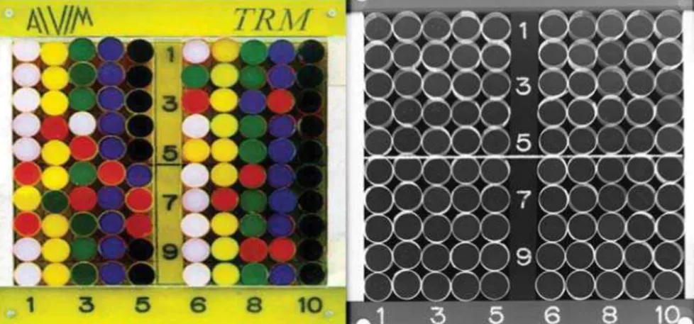
The mammographic phantom (Figure 1) model Alvim Statistical Phantom 18-209 (R&D Ltd.; Jerusalem, Israel) is composed of a main acrylic 1.5 cm-thick plate and other three, secondary 1.0 cm-thick plates which, as a whole, simulate a typically compressed breast (4.5 cm). Within the internal structure of the main plate, there are 100 cylinders which may be randomly distributed, 25 of them including objects simulating microcalcifications and fibers of different sizes(8).Generally, this type of
phantom is utilized only for research pur-poses, because its utilization as a routine in quality control programs implies a method of difficult application.
In the first phase of the program, the residents perform the reading of phantom images on the dedicated negatoscope and classify their findings into five confidence levels, according to the following scoring system: 100 (certainty about the presence of the object), 75 (probable presence of the object), 50 (uncertainty about the presence of the object), 25 (improbable presence of the object), 0 (definitely absent object). In
the second phase, the training is developed with high-resolution monitors. The soft-ware GQM® version 2(9), developed by the
laboratory dedicated to the training pro-cess, allows the images manipulation by means of digital tools for brightness and contrast settings, magnification and Gray-scale inversion (Figure 2).
Additionally, this program allows the automatized analysis of the residents’ per-formance in the detection of signs present on the images. The statistical analysis de-fines the observer’s profile, calculating the following evaluation indices: detection probability (Pdet), kappa (κ) values(10),
true-positive results, false-negative results, and area under the ROC curve(11) for
microcalcifications and fibers of different sizes.
Other capabilities include comparison between softcopy and hardcopy readings, evaluation of intra- and interobserver agreement in the different training phases, and monitoring of the time spent for each digital image reading.
Once these phases are completed, the professionals are given access to an image bank with mammograms acquired in differ-ent equipmdiffer-ent of several radiological units and classified in accordance with different levels of complexity. A golden-standard data set (stored in the data bank after double-reading by specialists) registers the diagnostic impression based on these im-ages. This images bank is continuously updated for utilization in the third phase of the training. In this phase, the professional undergoing the training gathers the infor-mation required for preparing his/her own report that is stored in a specific interface. A golden-standard report is available for self-training and comparison of the diag-nostic impressions. A tutorial is also avail-able for instruction in this phase.
The results of the analysis of 2,000 phantom images interpreted by experi-enced specialists with a minimum eight-hour training in digital systems, were adopted as a parameter for evaluating the residents’ performance(9), besides the
val-ues established by regulation of the Brazil-ian Ministry of Health (12). These
param-eters are shown on Tables 1 and 2. The performance indices utilized for evaluating the residents were: κ value,
Figure 2. Interface for training in phantom images interpretation. The software provides digital tools similar to the ones of a digital mammography workstation. On the right upper corner, there is a window to be fulfilled with the user’s rate of detection probability, and at right, the visualization of the image histo-gram.
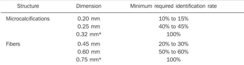
Table 1 Reference values for the reading of about 2,000 phantom images interpreted by specialists(9)
and respective parameters of the Brazilian Ministry of Health(12).
Structure
Microcalcifications
Fibers
Dimension
0.20 mm 0.25 mm 0.32 mm*
0.45 mm 0.60 mm 0.75 mm*
Minimum required identification rate
10% to 15% 40% to 45%
100%
20% to 30% 50% to 60%
100%
* Values established by technical regulation of the Brazilian Ministry of Health, Order (Portaria) No. 453, dated June 2, 1998.
Table 2 Reference values for reading of about 2,000 phantom images interpreted by specialists(9).
Index
Kappa
Area under the ROC curve
True-positive
False-positive
Reference values
> 0,7
> 0,8
> 85%
Up to 8%
Pdet, area under the ROC curve, besides true-positive and false-positive results.
The κ value measures the degree of agreement between answers (presence or absence of structures); Pdet indicates the chance of structures identification with a determined degree of certainty either with true-positive or false-positive results. The data evaluation based on a ROC curve(11)
allows a graphic and fast analysis of the relation between sensitivity and specificity both individually and in group.
RESULTS
A total of 1,003 Alvim®-type phantom
image readings were performed by the
resi-dents as follows: 588 readings on nega-toscope, and 415 readings on high-resolu-tion monitors.
Figure 3 presents the mean values of performance indices among hardcopy and softcopy readings.
Figure 4 shows the histograms with the distribution of κ values with different read-ing methods.
Figure 5 shows ROC curves for conven-tional and digital readings.
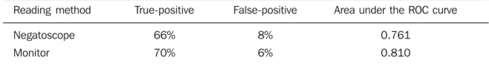
Table 3 presents the true-positive and false-positive rates, besides the area under the ROC curve in both reading methods.
The mean time for reading an image on the monitor at the beginning of the train-ing was 7.5 minutes, and, at the end, 4.0 minutes.
DISCUSSION
Users can benefit from the utilization of the digital technology in the interpretation of images; however, the professional must be trained for adaptation to the new visual patterns.
Figure 5. Area under the ROC curve in conventional (AZ = 0.761) and
digital systems (AZ = 0.810).
Figure 4. Histograms of kappa val-ues distribution in different reading systems.
Advantages can be clearly observed when the practitioners are appropriately trained and qualified(13).
The present study is intended to aid in the training of professionals during the period of introduction of the digital tech-nology and contributing to the reduction of variations or disagreements in the images interpretation, minimizing false-positive and false-negative results.
Images interpretation on monitor screens (softcopy reading) have been widely studied to evaluate the practitioners’ performance as compared with the conven-tional system (hardcopy reading)(14,15). The
majority of these studies have concluded that the images interpretation on monitor screens is widely accepted, the adaptation of the specialist is rapid, and the accuracy and time spent in the interpretation com-pare to the conventional system, provided the professional is duly trained(16,17).
An overall analysis demonstrated that the Pdet by the residents during the learn-ing process was higher in the digital sys-tem. However, considering the character-istic of this indicator, false-positive results should be evaluated for a correct interpre-tation. As far as the reference values are considered, a false-positive results rate of up to 8% is acceptable.
The higher Pdet (= 0.818), associated with the lower rate of false-positive results (6%) confirms a better identification of structures on the monitor screen (softcopy reading). These results indicate that the digital reading presents both higher sensi-tivity and specificity.
Also, κ results corroborate a higher de-tectability of structures at digital reading for both microcalcifications and fibers. But, it was in the detection of fibers that a clear improvement could be observed, sug-gesting that the manipulation of images brightness and contrast allowed by the digi-tal system may have influenced this result.
Based on the results of κ values, a his-togram with the distribution of these data could be constructed, allowing the evalua-tion of the interobserver reading variabil-ity (Figure 4). On this Figure, a narrower base can be observed with a symmetrical distribution (similar to a typical distribution curve), demonstrating a lower rate of sub-jectivity in the softcopy readings.
The evaluation of the time spent in the digital reading has shown that, with the progression of the training, this time de-creased without, however, affecting the residents’ performance, which demon-strates the effectiveness of the training.
The overall analysis demonstrated that, for a less experienced professional, false-positive results have a negative influence on the final accuracy of the reading(13). This
lower accuracy is primarily a result of the lack of perception and experience during the training in mammography, which re-stricts the resident’s capability to recognize “objects”, complicating the recognition of real lesions and artifacts. The solution pro-posed is a systematic training tutorial asso-ciating the image perception with the feed-back on reasons that aid in the decision making process(18).
Reading velocity and improved perfor-mance of experienced observers in the terpretation of mammographic images in-volve a shift in the mechanism of image perception from a “search-to-find” pro-cess(18) to a relatively faster holistic
mode(19). This shift depends, among others,
on the observer’s expertise in the method and the presence of an actual or virtual tu-torial.
Even though the results of the present study indicate a better performance in this reading mode (digital phantom images al-lowing manipulation), some inherent as-pects may have contributed to this conclu-sion. The training with films (hardcopy) preceded the training with the digital
sys-tem; therefore the Professional, although short, have acquired some experience in the reading of phantom images. The personal motivation may also have influenced the observers’ concentration at the moment o f the softcopy reading. Additionally, the initial contact with the technological inno-vation may have positively influenced the professionals’ interest in this phase of the training.
It is important to observe that the results of the present study were based on phan-tom images reading, with the objective of quantifying data and evaluating the influ-ence of the training on the overall profes-sionals’ performance. Additionally, it should be noted that, in the reading of mammographic images, where the fibro-glandular tissue and lesion present with different patterns, the detection process becomes even more complex, requiring more experience of the professional.
CONCLUSION
The method of training proposed in the present study has shown to be effective, with a positive impact on the residents’ performance, constituting an interesting pedagogical tool.
The results suggest that the method of training based on phantom reading can pro-duce a better performance of the practitio-ners in the interpretation of mammographic images.
It is the authors’ opinion that this train-ing method with a dedicated software plays a significant role as an ancillary tool in the complex, multifactorial process of learning in mammography.
Acknowledgements
To Fundação de Amparo à Pesquisa do Estado de São Paulo (Fapesp), for the fi-nancial support.
REFERENCES
1. Elmore JG, Wells CK, Howard DH. Does diagnos-tic accuracy in mammography depend on radi-ologists’ experience? J Womens Health. 1998;7: 443–9.
2. The Society of Breast Imaging – SBI (2005-2008). ACR – Breast imaging training. [cited 2008 Sep 25]. Available from: http://sbi-online.org
3. Radiological Health. MQSA: Mammographic Quality Standards Act – final regulation program.
Table 3 Values of the area under the ROC curve, true-positive and false-positive results on the different reading methods. Reading method Negatoscope Monitor True-positive 66% 70% False-positive 8% 6%
Area under the ROC curve
0.761
0.810
[cited 2008 Sep 25]. Available from: http://www. fda.gov/cdrh/mammography/digital.html 4. Monsees B. The Mammography Quality
Stan-dards Act. An overview of the regulations and guid-ance. Radiol Clin North Am. 2000;38:759–72. 5. Obenauer S, Hermann KP, Marten K, et al. Soft
copy versus hard copy reading in digital mam-mography. J Digit Imaging. 2003;16:341–4.
6. Pisano ED, Chandramouli J, Hemminger BM, et al. Does intensity windowing improve the detec-tion of simulated calcificadetec-tions in dense mammo-grams? J Digit Imaging. 1997;10:79–84. 7. Krupinski E, Roehrig H, Furukawa T. Influence
of film and monitor display luminance on ob-server performance and visual search. Acad Radiol. 1999;6:411–8.
8. Gurvich VA. Statistical approach for image qual-ity evaluation in daily medical practice. Med Phys. 2000;27:94–100.
9. Pires SR. “Software” gerenciador de uma base de
dados e de imagens mamográficas classificadas segundo um índice de qualidade [tese de mes-trado]. São Paulo: Universidade Federal de São Paulo; 2003.
10. Altman DG. Practical statistics for medical re-search. 1st ed. London: Chapman & Hall; 1991. 11. Metz CE. Receiver operating characteristic analy-sis: a tool for the quantitative evaluation of ob-server performance and imaging systems. J Am Coll Radiol. 2006;3:413–22.
12. Brasil. Ministério da Saúde. Secretaria de Vigi-lância Sanitária. Diretrizes de proteção radioló-gica em radiodiagnóstico médico e odontológico. Portaria nº 453, de 2/6/1998. Brasília: Diário Ofi-cial da União; 1998.
13. Nodine CF, Kundel HL, Mello-Thoms C, et al. How experience and training influence mammog-raphy expertise. Acad Radiol. 1999;6:575–85.
14. Pisano ED, Gatsonis C, Hendrick E, et al. Diag-nostic performance of digital versus film
mam-mography for breast-cancer screening. N Engl J Med. 2005;353:1773–83.
15. Krug KB, Stützer H, Girnus R, et al. Image qual-ity of digital direct flat-panel mammography ver-sus an analog screen-film technique using a phan-tom model. AJR Am J Roentgenol. 2007;188: 399–407.
16. Kundel HL, Polansky M. Measurement of ob-server agreement. Radiology. 2003;228:303–8.
17. Hemminger BM. Soft copy display requirements for digital mammography. J Digit Imaging. 2003; 16:292–305.
18. Kundel HL, Nodine CF, Conant EF, et al. Holis-tic componentofimageperceptionin mammo-gram interpretation: gaze-tracking study. Radiol-ogy.2007;242:396–402.