Trypanosoma cruzi
Metacyclic Trypomastigotes Enables
Resistant Strains to Invade Eukaryotic Cells
Igor Cestari, Marcel I. Ramirez*
Laborato´rio de Biologia Molecular de Parasitas e Vetores, Instituto Oswaldo Cruz - Fiocruz, Rio de Janeiro, Brazil
Abstract
The complement system is the main arm of the vertebrate innate immune system against pathogen infection. For the protozoanTrypanosoma cruzi, the causative agent of Chagas disease, subverting the complement system and invading the host cells is crucial to succeed in infection. However, little attention has focused on whether the complement system can effectively controlT. cruziinfection. To address this question, we decided to analyse: 1) which complement pathways are activated byT. cruziusing strains isolated from different hosts, 2) the capacity of these strains to resist the complement-mediated killing at nearly physiological conditions, and 3) whether the complement system could limit or controlT. cruzi invasion of eukaryotic cells. The complement activating molecules C1q, C3, mannan-binding lectin and ficolins bound to all strains analysed; however, C3b and C4b deposition assays revealed thatT. cruziactivates mainly the lectin and alternative complement pathways in non-immune human serum. Strikingly, we detected that metacyclic trypomastigotes of someT. cruzi strains were highly susceptible to complement-mediated killing in non-immune serum, while other strains were resistant. Furthermore, the rate of parasite invasion in eukaryotic cells was decreased by non-immune serum. Altogether, these results establish that the complement system recognizesT. cruzimetacyclic trypomastigotes, resulting in killing of susceptible strains. The complement system, therefore, acts as a physiological barrier which resistant strains have to evade for successful host infection.
Citation:Cestari I, Ramirez MI (2010) Inefficient Complement System Clearance ofTrypanosoma cruziMetacyclic Trypomastigotes Enables Resistant Strains to Invade Eukaryotic Cells. PLoS ONE 5(3): e9721. doi:10.1371/journal.pone.0009721
Editor:Anne Charlotte Gruner, Agency for Science, Technology and Research (A*STAR), Singapore
ReceivedOctober 14, 2009;AcceptedFebruary 23, 2010;PublishedMarch 16, 2010
Copyright:ß2010 Cestari, Ramirez. This is an open-access article distributed under the terms of the Creative Commons Attribution License, which permits unrestricted use, distribution, and reproduction in any medium, provided the original author and source are credited.
Funding:This work was supported by The National Council for Scientific and Technological Development (CNPq) (476737/2004-4); I.S. Cestari was a recipient of CNPq (141757/2006-0) and Coordenac¸a˜o de Aperfeic¸oamento de Pessoal de Nı´vel Superior (CAPES) (PDEE4261/06-2) scholarships. The funders had no role in study design, data collection and analysis, decision to publish, or preparation of the manuscript.
Competing Interests:The authors have declared that no competing interests exist.
* E-mail: [email protected]
Introduction
Chagas disease is an illness that affects 18 million people in Latin America [1]. It is caused by the protozoan parasite
Trypanosoma cruziand is mainly transmitted by triatomine insects, although congenital, oral and blood transfusion transmission has also been reported [2,3,4,5,6,7]. In the insect vector,T. cruziare present as replicative non-infective epimastigotes, which differen-tiate to non-replicative infective metacyclic trypomastigotes. During the infection, metacyclic trypomastigotes have to evade the host innate immune system and infect cells to progress in the infection. Parasites that succeed in infecting host cells differentiate to amastigotes, an intracellular replicative stage, which after several divisions differentiate to bloodstream trypomastigotes that disrupt cells, infecting new cells, or are taken by insect vectors restarting the life cycle [8].
The first barrier against a microbe’s infection of vertebrates is the innate immune system, encompassing the complement system and phagocytic and natural killer cells. The complement system is one of the most effective innate mechanisms of host defense against a pathogen infection. It is composed of several proteins activated in a cascade that culminate with membrane attack complex formation on the pathogen surface, causing killing by cell lysis [9]. The complement system can be activated by three
pathways: classical, activated when IgG or IgM bind to the pathogen surface and associate with the C1 complex (C1qr2s2),
which cleaves C2 and C4 to form C3 convertase (C4b2a); lectin, activated when mannan-binding lectins (MBLs), L-ficolins or H-ficolins bind to the pathogen surface and associate with MBL-associated serine protease-2 (MASP2) to cleave C2 and C4, generating C3 convertase (as in the classical pathway); and alternative, activated when C3b binds to the pathogen surface and associates with factor B to form C3 convertase (C3bBb) [9].
interact with the surface deposited C3b [18]. On the other hand, a rapid binding of MBLs, L-ficolins and H-ficolins on the surface of metacyclic trypomastigotes has been shown, providing evidence that the lectin pathway is activated in non-immune serum byT. cruzi [19]. Furthermore, bloodstream trypomastigotes sensitized with antibodies from acutely infected animals were killed by the complement system in human serum [20,21], and anti-a -galactosyl antibodies recognizing O-linked oligosaccharides on
T. cruzi mucin-like GPI-anchored proteins were identified in chronic phase Chagas disease patients [22,23]. Altogether, these data support the idea that the complement system can recognize and kill the infectious stages ofT. cruzi.
The expression of complement system inhibitors, such as complement C2 receptor inhibitor trispanning, complement regulatory protein, calreticulin and decay accelerating factor, has been demonstrated in T. cruzi; these molecules help T. cruzi to avoid complement killing [24,25,26,27]. However, the levels of expression and functionality of these molecules could be different amongT. cruzistrains, since resistance to complement killing has been demonstrated to vary among them [20,24]. Despite the mechanisms of resistance to the complement system, the differences ofT. cruzistrains and the high parasite load required for infection suggest that not all metacyclic trypomastigotes succeed in infecting the host [12,28,29,30,31].
Aiming to understand howT. cruzimetacyclic trypomastigotes interact with the human complement system, we analysed the effect of the complement system on the lysis and eukaryotic cell invasion by severalT. cruzistrains. Firstly, through C3b and C4b deposition assays we found thatT. cruzimainly activate the lectin and alternative pathways in non-immune serum. In vitro assays revealed that metacyclic trypomastigotes of several strains of T. cruzidiffer in their capacity to resist the complement system, and some of them were killed after incubation in non-immune human serum. Moreover, the rate ofT. cruziinvasion of eukaryotic cells was significantly reduced in the presence of non-immune human serum, showing that the complement system acts as a physiological barrier forT. cruzi infection and strains resistant to complement system have a higher chance to succeed in the infection.
Results
1- Complement activating factors bind to the surface of T. cruzistrains
To determine whether different strains ofT. cruziwould activate the complement system in human serum, the binding of complement activating factors to these strains was analyzed.T. cruzi epimastigotes were used because they are sensitive to complement killing; therefore, they activate the complement system. Strains from congenitally transmitted Chagas disease patients, in addition to recently isolated strains from primate, opossum and rodent were studied (Table 1). The strains Y, Silvio X10/6, and CL Brener are used by several laboratories and were also included in some of the experiments.
Binding of C1q, involved in classical pathway activation; MBLs, L-ficolins and H-ficolins, for the lectin pathway, and C3 for the alternative pathway were analyzed by ELISA. All these molecules were detected bound to the parasites after incubation with non-immune human serum (Figure 1A to E). Although a slight difference in the binding was detected among the strains, the results show that all T. cruzi strains are recognized by the complement activating molecules.
2-T. cruzistrains activate mainly the lectin and alternative complement pathways
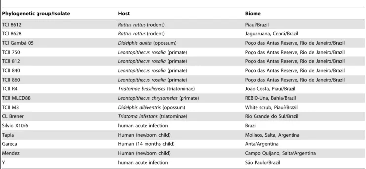
To determine the contribution of each pathway to the complement activation by T. cruzi strains, kinetic assays were performed to detect C3b and C4b deposition on the parasite surface. To analyze the classical and lectin pathway contribution, C4b deposition assays were performed with normal human serum (classical and lectin pathway activation), with serum depleted of MBLs and L-ficolins (mainly classical pathway activation) and with serum depleted of C1q (lectin pathway activation). C4b deposition in normal human serum was rapid and similar between all strains, reaching 50% (absorbance = 1.0) in less than 5 minutes (Figure 2A). However, C4b deposition in MBLs/ficolins-depleted serum was slower than in normal human serum (Figure 2B). C4b
Table 1.Trypanosoma cruzistrains, hosts and biome.
Phylogenetic group/Isolate Host Biome
TCI 8612 Rattus rattus(rodent) Piauı´/Brazil
TCI 8628 Rattus rattus(rodent) Jaguaruana, Ceara´/Brazil
TCI Gamba´ 05 Didelphis aurita(opossum) Poc¸o das Antas Reserve, Rio de Janeiro/Brazil
TCII 750 Leontopithecus rosalia(primate) Poc¸o das Antas Reserve, Rio de Janeiro/Brazil
TCII 812 Leontopithecus rosalia(primate) Poc¸o das Antas Reserve, Rio de Janeiro/Brazil
TCII 840 Leontopithecus rosalia(primate) Poc¸o das Antas Reserve, Rio de Janeiro/Brazil
TCII 860 Leontopithecus rosalia(primate) Poc¸o das Antas Reserve, Rio de Janeiro/Brazil
TCII R4 Triatomae brasilienses(triatominae) Joa˜o Costa, Piauı´/Brazil
TCII MLCD88 Leontopithecus chrysomelas(primate) REBIO-Una, Bahia/Brazil
TCII M3 Didelphis albiventris(opossum) White scrub, Piauı´/Brazil
CL Brener Triatoma infestans(triatominae) Rio Grande do Sul/Brazil
Silvio X10/6 human acute infection Brazil
Tapia Human (newborn child) Molinos, Salta, Argentina
Gareca Human (14 months child) Anta/Argentina
Mendez Human (newborn child) Campo Quijano, Salta/Argentina
Y human acute infection Sa˜o Paulo/Brazil
deposition in MBLs/ficolins-depleted serum varied among the strains. Between 2 to 4 times less deposited C4b was detected in assays with MBLs/ficolins-depleted serum than with normal human serum, even after 1 hour of incubation. To examine the ability of the lectin pathway to recognizeT. cruziand activate the complement, the C4b deposition was performed in serum depleted of C1q. The velocity of C4b deposition in this serum was slightly slower than in normal human serum, but higher than in serum depleted of MBLs/ficolins (Figure 2C). Substantial depletion of MBLs/L-ficolins and C1q from sera was confirmed by ELISA (Figure S1). These results indicate that T. cruzi activate both classical and lectin pathways; although, as observed before [19],
the lectin pathway seems to be more effective than the classical pathway in recognizingT. cruziin non-immune serum. To verify if these strains activate the alternative pathway, C3b deposition was analyzed in serum treated with EGTA to chelate Ca2+. Importantly, the classical and lectin pathways are Ca2+- and Mg2+-dependent, whereas the alternative pathway is only Mg2+ -dependent [9]. C3b deposition increased gradually through time for all strains (Figure 2D). It is noteworthy that strains 8612, Y and Gamba 05 activated the alternative pathway less than the other strains analyzed. Together, these results indicate that upon contact with human serum,T. cruzistrains activate mainly the lectin and alternative pathways of the complement system.
Figure 1. Complement activating factors bind toT. cruzisurface.A) ELISA for detection of C1q bound toT. cruziepimastigotes of several strains. Parasites adsorbed in ELISA plates were incubated with 5% NHS for 1 h at 37uC. Anti-C1q antiserum was used for detection and reactions were developed with ABTS-peroxidase solution measured at 405 nm. B) Detection of C3b bound toT. cruzi. C) Detection of MBL bound toT. cruzi. D) Detection of L-ficolins bound toT. cruzi. E) Detection of H-ficolins bound toT. cruzi. Experiments in B, D and E were performed as in A, except that for detection were used antiserum against C3, L-ficolins and H-ficolins, respectively. Experiments in C were performed as in A, except that 20% NHS and anti-MBL antiserum were used.
doi:10.1371/journal.pone.0009721.g001
Figure 2.T. cruziactivate mainly the lectin and alternative complement pathways.A) C4b deposition onT. cruziepimastigotes of several strains. Parasites adsorbed in ELISA plates were incubated in 5% NHS from 5 to 60 minutes at 37uC, followed by anti-C4 antiserum incubation. Reactions were developed with ABTS-peroxidase solution measured at 405 nm. B) C4b deposition in epimastigotes ofT. cruzi. Experiments were performed as in A, except that MBLs/L-ficolins-depleted sera were used. C) C4b deposition in epimastigotes ofT. cruzi. Experiments were performed as in A, except that C1q-depleted sera were used. D) C3b deposition inT. cruziepimastigotes. Experiments were performed as in A; except that sera treated with 10 mM EGTA and 7 mM MgCl2were used. Detection was made with anti-C3 antiserum.
3-T. cruzimetacyclic trypomastigotes are killed by the complement system
We have seen previously that the majority of epimastigotes ofT. cruzi strains are susceptible to complement killing in normal human serum at nearly physiological conditions (50% of serum at 37uC) after incubations ranging from 5 to 30 minutes (not shown). We decided to analyze whether the metacyclic trypomastigotes of
T. cruzistrains would be resistant to complement killing. Strikingly, someT. cruzistrains were killed by human complement at nearly physiological serum concentration, 50% (Figure 3 A and B). Parasites from the strains Y, R4 and 8628 were completely killed between 30 to 60 minutes of incubation, whereas the survival rate of the strains Silvio X10/6, 840, 750, M3 and 812 were no more than 30% after a 60 minute incubation. The survival rate of the strains CL Brener, 860 and 8612 was about 50%; and higher survivals were detected for strains MLCD88 and Gamba 05, about 75%. We also did not find any correlation between the complement killing susceptibility of the strains and their respective phylogenetic groups. It is noteworthy that the purity of metacyclic trypomastigotes used in these assays was always above 95%, as confirmed by morphological parameters, and they also expressed the antigen-specific gp82, detected by Western blotting (exempli-fied in Figure S2).T. cruziis refractory to the African trypanosome lytic factor apolipoprotein-I [32,33,34], excluding the possibility that this factor mediates the lysis. Furthermore, inhibition of the complement cascade by chelating the Ca2+
and Mg2+
from the serum with EDTA, treatment of the serum with protease inhibitors or the use of a complement consumed serum failed to lyseT. cruzi
metacyclic trypomastigotes (Figure S3). To obtain further evidence that metacyclic trypomastigotes are killed by activation of the complement system, we decided to compare the deposition of C3b and C4b between epimastigotes and metacyclic trypomastigotes. There was a higher deposition of C3b and C4b on epimastigotes than on metacyclic trypomastigotes at 10 minutes incubation with non-immune serum (Figure 3C and D). However, by 30 minutes no significant difference for C4b deposition on epimastigotes and metacyclic trypomastigotes was detected (Figure 3D), confirming that metacyclic trypomastigotes activate the complement system.
Using a live/dead cells fluorescence-based assay we confirmed that metacyclic trypomastigotes were being killed after human serum incubation. For this assay, metacyclic trypomastigotes of the strain 812 were incubated with or without non-immune human serum at concentration of 50% for 30 minutes at 37uC, followed by labeling with calcein-AM (green, to detect live cells) and ethidium homodimer-1 (red, for dead cells). Serum incubation resulted in almost complete parasite killing (red), while non-treated parasites were alive (green) and motile. There were few parasites survivors after serum treatment, as can be observed in Figure 3E (arrows). Together, these results show that T. cruzi metacyclic trypomastigotes activate the complement system, and there are strains sensitive and resistant to complement-mediated killing by human serum.
4-T. cruziinvasion of eukaryotic cells is limited by the complement system
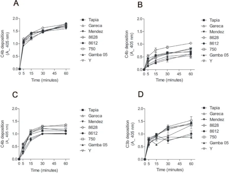
After showing that differentT. cruzistrains vary in their ability to resist the complement system, and that some strains are susceptible to complement killing, we decide to investigate how efficiently the complement system can control metacyclic trypo-mastigotes invasion of eukaryotic cells in an in vitro assay that simulates physiological conditions.
Firstly, kinetics of invasion with CL Brener, Silvio X10/6, Gamba 05, 860, 812 and R4 strains in Vero cells were determined
using a ratio of 5 parasites to each cell. After 30 minutes incubation we detected invasion with all strains analysed (Figure 4A). The highest invasion rate was detected for the strain Cl Brener, 26 parasites per 100 cells after a 120 minute incubation, whereas the strains Silvio X10/6, R4 and Gamba 05 had similar rates of invasion, 12, 14 and 15 parasites per 100 cells, respectively (Figure 4A). Lower rates were detected for the strains 860 and 812, 5 and 7 parasites per 100 cells after 120 minutes, respectively (Figure 4A). To determine whether the complement system would affect the parasite invasion, assays were performed by incubating parasites with cells in the presence of 50% non-immune human serum for 60 minutes at 37uC, and the intracellular parasites were quantified. The amount of intracellular parasites decreased in the presence of normal human serum when compared to invasion in the absence of serum for the complement-sensitive strains (Figure 4B), and a slight difference in invasion rates was detected between them. However, the Gamba 05 strain (complement-resistant strain) maintained similar invasion rate in the presence of serum (Figure 4B), indicating that the decrease in cell invasion was caused by complement-mediated lysis. Furthermore, the invasion rates increased for all strains when the ratios of parasites per cell were increased from 5 to 10 parasites to each cell (Figure 4B); suggesting that the parasite load may affect the success of invasion in the presence of serum. To certify that the complement was limiting the parasite invasion, the assays were performed incubating the parasites and cells for a long time period (3 hours). The longer incubation led to an increase in cell invasion in the absence of serum (Figure 4C); however, in the presence of serum, the rates of invasion of the complement-sensitive strains did no increase significantly. In contrast, the rate of invasion of the strains Gamba 05 (complement-resistant strain) remained similar to the rate of invasion in the absence of serum (Figure 4C). These results indicate that the complement system limit the invasion of sensitive strains likely through the complement-mediated lysis. Importantly, intracellular amastigotes and cell culture-derived trypomastigotes were detected (in the supernatant) after 72 hours of infection in the presence of serum, indicating that parasites that succeeded in infecting the cells were able to progress in their life cycle (not shown).
Discussion
The host innate immune system is the major barrier for pathogens to succeed in infection, and the complement system is one of the main arms of host defense [9]. Several molecules have been reported to control the complement activation and killing in
T. cruzi [24,25,26,27]. However, little work has focused on the interactions between insect-transmitted metacyclic trypomastigotes and the complement system in non-immune serum; in addition, whether the complement system can effectively control T. cruzi
infection.
Kinetic studies of complement activation by metacyclic trypomastigotes have not been reported, and we decided to study several strains from different origins to understand this mecha-nism. Here, we show evidence that the complement system can kill
T. cruzimetacyclic trypomastigotes limiting the parasite to succeed in invading the host cells.
alternative pathway to recognize and control pathogen infection, including trypanosomatids, has been extensively explored [17,18,35,36]. However, the lectin pathway has only recently emerged as an important host defense mechanism due to pattern associated recognition molecules, such as MBLs and ficolins, which can recognize a broad range of carbohydrates on invading pathogens [37]. Also, mutations in lectin pathway components have been shown to increase the susceptibility to infection by several microbes [38,39,40,41,42]. We have recently shown that
T. cruzi activate the complement lectin pathway [19], and the current results confirm thatT. cruzistrains isolated from different hosts also activate this pathway. Other pathogens, such as
Leishmania brasiliensis, Salmonella typhimuriumand Nisseria gonorrheae, have been shown to activate the lectin pathway [43,44], strengthening the importance of studying the mechanisms that these pathogens evolved to inhibit this activation and complement killing. On the other hand, as shown here and elsewhere [17,18,19,45] the classical pathway has a limited involvement in Figure 3.T. cruzimetacyclic trypomastigotes are killed by the complement system.A and B) Kinetics of complement-mediated killing assays with metacyclic trypomastigotes of severalT. cruzistrains. Parasites were incubated with 50% NHS at 37uC for 5 to 120 minutes and survivors were counted. C) C3b deposition in metacyclic trypomastigotes of severalT. cruzistrains. Parasites adsorbed on ELISA plates were incubated in 5% sera treated with 10 mM EGTA and 7 mM MgCl2for 10 and 30 minutes at 37uC, followed by anti-C3 antiserum incubation. Reactions were developed
with ABTS-peroxidase solution measured at 405 nm. D) C4b deposition in metacyclic trypomastigotes of severalT. cruzistrains. Experiments were performed as in C, except that 5% NHS anti-C4 antiserum was used. E) Live/dead viability fluorescence assay. Metacyclic trypomastigotes (812 strain) were incubated in 50% non-immune human serum for 30 minutes at 37uC (plus NHS), or in PBS as control (buffer). After that, parasites were incubated with 2mM of calcein-AM and 4mM of ethidium homodimer-1 for 30 minutes at RT, and slides were analysed by fluorescence microscopy.
Calcein-AM dye is retained by live cells and produces green fluorescence after activation by cytoplasmic esterase, whereas ethidium homodimer-1 is excluded by the plasma membrane of live cells, enters cells with damaged membranes, and produces red fluorescence upon binding to nucleic acids in dead cells. *p,0.05; **p,0.01.
doi:10.1371/journal.pone.0009721.g003
Figure 4.T. cruziinvasion in eukaryotic cells is limited by the complement system.A) Kinetics invasion of Vero cells byT. cruzimetacyclic trypomastigotes. Parasites (56105) were incubated with Vero cells (1.06105, ratio 5:1, parasites:cells) for 15 to 120 minutes at 37uC, and intracellular
parasites in at least 500 cells were quantified. B) Invasion assays of Vero cells by metacyclic trypomastigotes ofT. cruzistrains with or without NHS. Parasites (56105or 1.06106, ratios 5:1 and 10:1, parasites:cells) were incubated with Vero cells (1.06105) in 50% NHS for 60 minutes at 37uC. As
controls, parasites and cells (ratio 5:1) were incubated without NHS. Intracellular parasites in at least 500 cells were quantified. C) Invasion assays of Vero cells by metacyclic trypomastigotes ofT. cruzistrains with or without NHS. Parasites (56105, ratio 5:1, parasites:cells) were incubated with Vero
cells (1.06105) in 50% NHS for 3 hours at 37uC. As controls, parasites and cells (ratio 5:1) were incubated without NHS. Intracellular parasites in at least
the complement activation by T. cruzi in non-immune serum, suggesting that natural antibodies would have a limited capacity to recognizeT. cruziand activate the complement system.
Assays of complement-mediated lysis, and deposition of C3b and C4b, demonstrated that T. cruzimetacyclic trypomastigotes activate the complement system. Furthermore, we have found that someT. cruzistrains are highly susceptible to lysis in non-immune serum, whereas other strains differ in their capacities to resist complement killing. Other trypanosomatids, such asLeishmania sp.
and Chritidia sp., have been shown to be highly susceptible to complement-mediated killing in human serum [36,46,47], indi-cating that the complement system can act as a physiological barrier for trypanosome infection. Since they must have evolved a strategy to evade the complement attack; it appears that, for some pathogens, this strategy actually uses host/vector factors. For example, phagocytosis by macrophages and neutrophils during this period may help them to escape the complement-mediated killing [12,36,48]. Furthermore, it has been reported that the saliva and intestinal content of the insect vectors contain complement inhibitors [49], which could also help the parasites to escape the lysis. Although complement regulators could be contributing to complement evasion, it is possible that other unknown evasion strategies are displayedin vivo; or perhaps the complement sensitive strains are being transmitted by other routes of infection, such as orally. It is important to consider that the mechanisms employed byT. cruzistrains to escape the killing could have evolved differently according to the hosts. Complement system activity is different among humans, opossums, rodents and chickens [50,51,52,53], and their effectiveness in eliminating the parasites could be different. On the other hand, the expression and functionality of complement regulators onT. cruzisurface may be different in the strains, resulting in their different abilities to resist complement killing. The expression analysis of complement regulators in resistant and susceptibleT. cruzi strains could open the possibility to understand this difference.
We have also investigated how the complement system affects the invasion of eukaryotic cells byT. cruzi. Experimental invasion
in vitro in the presence of non-immune human serum at nearly physiological conditions showed that the complement system can limit, but not avoid parasite invasion. Some strains invaded the host cells as soon as 15 minutes, and the invasion increased according with time. Furthermore, in the presence of human serum the levels of invasion of the complement-sensitive strains were dramatically decreased. However, the human serum did not affect the complement-resistant strains to invade the cells. These results suggest that, for the complement system to control the infection, complete clearance during the first minutes of infection is necessary to stop parasites from invading host cells; otherwise, resistant strains can invade the cells and progress in their life cycle.
T. cruzi metacyclic trypomastigotes differ in their capacity to invade the host cells. There are high and low invasive strains [31,54], which has been related to the presence of different parasite surface molecules [31]. It is possible that a long parasite exposure to the complement system, before the cell invasion, can affect the success of the infection according to the capacity of the strain to resist the lysis.
Our data support the idea that resistance to complement-mediated killing is not a strict characteristic of the metacyclic trypomastigote stage ofT. cruzi; rather, there are strains sensitive and resistant to complement killing by human serum. The features that make some strains resistant and others sensitive are still unknown. However, the understanding that T. cruzi metacyclic trypomastigotes can be lysed by the complement system or the parasites could express transiently complement regulators
facili-tating them to resist the lytic effect of the complement and produce infection; open new possibilities to investigate parasite extrinsic mechanisms involved in immune evasion.
A detailed study of the mechanisms of complement system activation and host cell invasion on a molecular level is essential to understand the disease and the development of new therapies. Drug screening initiatives have been focused mainly on parasite metabolism targets rather than on immune evasion strategies [55]. Nevertheless, recent discoveries of complement receptors and immune evasion strategies may reveal promising targets for therapeutic intervention [18,19,24,26,56], which could be used to control Chagas disease.
In summary, this study demonstrates that the human comple-ment system can kill metacyclic trypomastigotes ofT. cruzi, and the ability to resist the complement system varies between the strains. Indeed, the complement system acts as a physiological barrier during the infection which parasites have to evade to successfully infect the host.
Methods
Parasites and cells
T. cruziepimastigotes were cultivated in liver infusion tryptose medium (bovine liver infusion at 5 g/l, tryptose at 5 g/l, NaCl at 4 g/l, KCl at 0.4 g/l, Na2HPO4 at 8 g/l, glucose at 2 g/l;
pH 7.2), supplemented with haemin at 10 mg/l and fetal bovine serum 10%, at 27uC. Metacyclic trypomastigote forms were obtained as previously described [57]. Vero cells were grown in RPMI 1640 supplemented with 10% FBS at 37uC and 5% CO2.
Complement-mediated killing assay
Normal human serum was obtained from healthy voluntary donors and pooled. Aliquots were stored at280uC until use. For complement assays,T. cruzimetacyclic trypomastigotes (5.06105) in 100ml of veronal buffer (VB, 10 mM barbital/NaCl 145 mM/
CaCl2 0.15 mM/MgCl2 0.5 mM, pH 7.4) were incubated with
100ml of normal human serum (NHS) for 5, 10, 30, 60 and 120 minutes at 37uC. Reactions were stopped by the addition of 800ml of ice-cold VB. Parasite survivors were quantified in a Neubauer chamber by light microscopy.
Depletion of MBLs and L-ficolins from NHS
Depletion of MBL and L-ficolin from human serum was performed as previously described [19]. Firstly, to deplete NHS of MBL, mannan-agarose (SIGMA-Aldrich) resin equilibrated in HEPES buffer (10 mM HEPES/140 mM NaCl buffer containing 0.15 mM CaCl2 and 0.5 mM MgCl2, pH 7.4) were incubated
with NHS for 2 hour at 4uC on ice, the serum was dialysed against 2 litres of HEPES buffer. Afterwards, the same serum (MBL-depleted) was used for L-ficolins depletion. The serum was incubated for 2 hours on ice with N-acetyl-glucosamine-Sepharose resins (SIGMA-Aldrich, prepared according to manufacture’s instructions) equilibrated in 10 mM HEPES/640 mM NaCl buffer, pH 7.4 and 5 mM ethylene diamine tetracetic acid (EDTA). The resin was collected by centrifugation and depleted sera (and non-treated serum) were dialyzed against 10 mM HEPES/140 mM NaCl buffer, pH 7.4. After dialysis, MgCl2
and CaCl2 were added to final concentrations of 0.5 mM and
dialyzed against 10 mM HEPES/140 mM NaCl buffer, pH 7.4. After dialysis, MgCl2 and CaCl2 were added to final
concentra-tions of 0.5 mM and 0.15 mM, respectively. Serum depletion was confirmed by ELISA.
ELISA for complement assays
T. cruziepimastigote or metacyclic trypomastigote forms were fixed in 4% paraformaldehyde, and 100ml (1.06106) of parasites were incubated in each well of a 96 well ELISA MaxiSorp plate (Nalge Nunc International) overnight at 4uC with coating buffer (0.1M Na2CO3/0.1M NaHCO3, pH 9.5). The wells were washed
three times with phosphate buffered saline (PBS, 137 mM NaCl, 2.7 mM KCl, 4.3 mM Na2HPO4, 1.47 mM KH2PO4), then
blocked with PBS 3% bovine serum albumin (BSA) for 2 hours at RT. For binding of C1q, C3, L- and H-ficolins, 100ml of 5% NHS
(diluted in VB) were added to each well, followed by a 60 minute incubation at 37uC. For MBL binding, 20% NHS was added to each well and incubated for 60 minutes at 37uC. Wells were washed with PBS, then affinity purified polyclonal goat anti-MBL antiserum (1:100) (Santa Cruz), polyclonal rabbit anti-L-ficolin antiserum (1:500), anti-H-ficolin antiserum (1:500), anti-C1q antiserum (1:500) and anti-C3 antiserum (1:500, diluted in PBS 3% BSA) (MRC Immunochemistry Unit, Oxford, England) were added. After a 2 hours incubation at RT, the plate was washed as above, and either rabbit anti-goat IgG-HRP conjugate (1:2000, diluted in PBS 3% BSA) (Kirkegaard and Perry Laboratories) for anti-MBL or goat anti-rabbit IgG-HRP conjugate (Bio-Rad) (1:2000, diluted in PBS 3% BSA) for others were added and incubated in the wells for 2 hours at RT. The wells were washed as above, and 100ml of ABTS peroxidase solution (Kirkegaard and Perry Laboratories) was added. Reactions were incubated at RT for 5 to 10 minutes and stopped with the addition of 50ml of 1% SDS. Absorbances (at 405 nm) were obtained through spectrophotometric measurement. For kinetics of C3 and C4 deposition, wells previously adsorbed with parasites were incubat-ed with either 5% NHS, or 5% MBLs/L-ficolins-depletincubat-ed serum, or 5% NHS treated with 10 mM EGTA and 7 mM MgCl2for 5
to 60 minutes at 37uC. Afterwards, the wells were washed with PBS, incubated with rabbit polyclonal anti-C3 antiserum (1:500) (MRC Immunochemistry Unit) or goat polyclonal anti-C4 antibodies (1:500, diluted in PBS 3% BSA) (Calbiochem) for 2 hours at RT. After that, wells were washed, incubated with goat anti-rabbit IgG-HRP conjugate (1:2000, diluted in PBS 3% BSA) (Bio-Rad) and rabbit anti-goat IgG-HRP conjugate (Kirkegaard and Perry Laboratories) (1:2000, diluted in PBS 3% BSA), respectively and incubated for 2 hours at RT. For development, 100ml of ABTS peroxidase solution (Kirkegaard and Perry Laboratories) were added, incubated at RT for 5 to 10 minutes and stopped with 50ml of 1% SDS in H2O. Absorbances
(at 405 nm) were obtained through spectrophotometric
measurement.
Live/dead viability fluorescence assay
T. cruzimetacyclic trypomastigotes of the strain 812 (5.06106in 250ml of PBS) were incubated with 250ml of NHS or 250ml of PBS (as control) for 30 minutes at 37uC. After that, cells were washed three times in PBS, resuspended in 1 ml of PBS containing 2mM of calcein-AM and 4mM of ethidium homodimer-1 (Live/ Dead Viability/Cytotoxity kit, Molecular Probes) and incubated for 30 minutes at RT. Afterwards, cells were centrifuged for 5 minutes at 5,000 rpm, resuspended in 10ml of PBS, and mounted on slides with Vectashield mounting medium (Vector Laborato-ries). For fluorescence microscopy an Olympus IX81 inverted
microscope, equipped with a U-CMAD3 camera, was used (Olympus).
Invasion assays
Vero cells from logarithmically grown culture were treated with trypsin, washed once with PBS and seeded on 13 mm cover slips in 24 well plates (1.06105cells per well). After 24 h, cells were washed with serum-free RPMI 1640 and incubated with 5.06105
T. cruzimetacyclic trypomastigote forms for 15, 30, 60 and 120 minutes at 37uC with 5% CO2. Cells were then washed with PBS,
fixed with absolute methanol (MERCK) for 5 minutes, washed with H2O and stained with Giemsa for 1 h at RT. Then, they
were washed with H2O and slides were mounted with aqueous
mounting medium (Biomeda). Intracellular parasites were quan-tified by light microscopy, counting at least 500 cells per slide. For invasion in the presence of NHS, cells were seeded as described above and metacyclic trypomastigotes 5.06105 (ratio 5:1, para-sites:cells) and 1.06106(ratio 10:1, parasites:cells) were added in the presence of 50% NHS (diluted in serum-free RPMI 1640, in a final volume of 500ml), followed by 60 or 180 minutes incubation
at 37uC with 5% CO2. As controls, 5.06105parasites were added to wells containing Vero cells without NHS. Staining and quantification were performed as described above.
Data presentation and statistical analysis
All data shown are the average of two or three experiments performed at least in triplicate. All data are shown as mean+/2 SD. Comparisons among groups were made by the unpaired t-test for repeated measures using GraphPad Prism version 5.00 for Windows, GraphPad Software, San Diego California USA. Values of p,0.05 with confidence interval of 90% were considered statistically significant unless otherwise specified.
Supporting Information
Figure S1 ELISA to analyse human serum depleted of MBL/L-ficolins and C1q. A) 100ml of 5% normal human serum, 5% C1q-depleted serum and 5% MBL/L-ficolins-C1q-depleted serum were incubated with T. cruzi epimastigotes of the strain X10/6 (previously adsorbed in ELISA plates) for 1 hour at 37uC. Polyclonal antibodies anti-C1q was used for C1q detection. Reactions were developed with ABTS peroxidase solution and absorbances obtained at 405 nm. NHS (normal human serum), MBL/L-fic-dep (MBL and L-ficolins-depleted serum), C1q-dep (C1q-depleted serum). B) Similar to A, except that polyclonal antibodies anti-MBL was used for detection. C) Similar to A, except that polyclonal antibodies anti-L-ficolins was used for detection. D) Similar to A, except that polyclonal antibodies anti-C3 was used for detection. Significance is shown on each graph. Data from depleted serum was compared to normal human serum.
Found at: doi:10.1371/journal.pone.0009721.s001 (2.26 MB TIF)
mono-clonal antibodies anti-3F6 (which recognizes gp82). D) T. cruzi
strain Gamba 05 stained with Giensa. 1 and 2, epimastigotes (4 days of culture); 3 and 4, metacyclic trypomastigotes. Metacyclic trypomastigotes were obtained as described in A. The efficiency of the purification was 99%. C) Western blotting to detect the expression of the metacyclic trypomastigotes stage-specific protein gp82 in the strain Gamba 05. The procedure was as described in B, except that 2.06107 parasites/well was used. E, epimastigotes; M, metacyclic trypomastigotes.
Found at: doi:10.1371/journal.pone.0009721.s002 (5.99 MB TIF)
Figure S3 Serum lysis ofT. cruzimetacyclic trypomastigotes is dependent on the complement system. Lysis ofT. cruzimetacyclic trypomastigotes with human serum is inhibited by serum treatment with ethylenediamine tetraacetic acid (EDTA), protease inhibitors or using complement consumed serum. A) Metacyclic trypomastigotes (5.06105) of the strain Silvio X10/6 were incubated for 1 hour at 37uC with normal human serum (NHS), normal human serum treated with 10 mM EDTA (NHS+EDTA), normal human serum treated with protease inhibitors (NHS+P Inhibitors; 1 mM phenylmethylsulfonyl fluoride, 1mM aprotinin and 50 pM soybean trypsin inhibitor, added just before the
complement lysis assay) or complement consumed normal human serum (NHS Consumed; consumption of complement were obtained by incubating the serum at 37uC for 2 hours with protein extract of 5.06106 T. cruzi epimastigotes). Parasite survivors were quantified. B) Similar to A, but with the strain 860. Significance is shown on each graph. Data from NHS treatment were compared to the values from EDTA, NHS-PI and NHS Cons separately using unpaired t-test.
Found at: doi:10.1371/journal.pone.0009721.s003 (0.38 MB TIF)
Acknowledgments
We are very grateful to Prof RB Sim for antiserum against L-Ficolins, H-ficolins, C1q and C3; Dr AM Jansen, Dr MA Basombrio, Dr MC Mora, Dr O Sanchez and Dr F Sanchez forT. cruzistrains; and to Dr Lindsay Carpp for valuable suggestions to the manuscript.
Author Contributions
Conceived and designed the experiments: IC MIR. Performed the experiments: IC. Analyzed the data: IC MIR. Contributed reagents/ materials/analysis tools: MIR. Wrote the paper: IC MIR.
References
1. Teixeira AR, Nitz N, Guimaro MC, Gomes C, Santos-Buch CA (2006) Chagas disease. Postgrad Med J 82: 788–798.
2. Gurtler RE, Ceballos LA, Ordonez-Krasnowski P, Lanati LA, Stariolo R, et al. (2009) Strong Host-Feeding Preferences of the Vector Triatoma infestans Modified by Vector Density: Implications for the Epidemiology of Chagas Disease. PLoS Negl Trop Dis 3: e447.
3. Munoz J, Coll O, Juncosa T, Verges M, del Pino M, et al. (2009) Prevalence and vertical transmission of Trypanosoma cruzi infection among pregnant Latin American women attending 2 maternity clinics in Barcelona, Spain. Clin Infect Dis 48: 1736–1740.
4. Piron M, Verges M, Munoz J, Casamitjana N, Sanz S, et al. (2008) Seroprevalence of Trypanosoma cruzi infection in at-risk blood donors in Catalonia (Spain). Transfusion 48: 1862–1868.
5. Sanchez Negrette O, Mora MC, Basombrio MA (2005) High prevalence of congenital Trypanosoma cruzi infection and family clustering in Salta, Argentina. Pediatrics 115: e668–672.
6. Valente SA, da Costa Valente V, das Neves Pinto AY, de Jesus Barbosa Cesar M, dos Santos MP, et al. (2009) Analysis of an acute Chagas disease outbreak in the Brazilian Amazon: human cases, triatomines, reservoir mammals and parasites. Trans R Soc Trop Med Hyg 103: 291–297.
7. Yoshida N (2008) Trypanosoma cruzi infection by oral route: how the interplay between parasite and host components modulates infectivity. Parasitol Int 57: 105–109.
8. Buscaglia CA, Campo VA, Frasch AC, Di Noia JM (2006) Trypanosoma cruzi surface mucins: host-dependent coat diversity. Nat Rev Microbiol 4: 229–236. 9. Lambris JD, Ricklin D, Geisbrecht BV (2008) Complement evasion by human
pathogens. Nat Rev Microbiol 6: 132–142.
10. Kollien AH, Schaub GA (1998) Development of Trypanosoma cruzi after starvation and feeding of the vector - a review. Tokai J Exp Clin Med 23: 335–340.
11. Kollien AH, Schaub GA (2000) The development of Trypanosoma cruzi in triatominae. Parasitol Today 16: 381–387.
12. Bijovsky AT, Milder RV, Abrahamsohn IA, Sinhorini IL, Mariano M (1984) The influence of lymphatic drainage in experimental Trypanosoma cruzi infection. Acta Trop 41: 207–214.
13. Schuster JP, Schaub GA (2000) Trypanosoma cruzi: skin-penetration kinetics of vector-derived metacyclic trypomastigotes. Int J Parasitol 30: 1475–1479. 14. Leon-Perez F, Gomez-Garcia L, Alejandre-Aguilar R, Lopez R, Monteon VM
(2007) Mexican Trypanosoma cruzi isolates: in vitro susceptibility of epimas-tigotes to anti-trypanosoma cruzi drugs and metacyclic forms to complement-mediated lysis. Vector Borne Zoonotic Dis 7: 330–336.
15. Yoshida N, Araguth MF (1987) Trypanolytic activity and antibodies to metacyclic trypomastigotes of Trypanosoma cruzi in non-Chagasic human sera. Parasite Immunol 9: 389–393.
16. Joiner K, Hieny S, Kirchhoff LV, Sher A (1985) gp72, the 72 kilodalton glycoprotein, is the membrane acceptor site for C3 on Trypanosoma cruzi epimastigotes. J Exp Med 161: 1196–1212.
17. Krettli AU, Pontes de Carvalho LC (1985) Binding of C3 fragments to the Trypanosoma cruzi surface in the absence of specific antibodies and without activation of the complement cascade. Clin Exp Immunol 62: 270–277.
18. Joiner K, Sher A, Gaither T, Hammer C (1986) Evasion of alternative complement pathway by Trypanosoma cruzi results from inefficient binding of factor B. Proc Natl Acad Sci U S A 83: 6593–6597.
19. Cestari I, Krarup A, Sim RB, Inal JM, Ramirez MI (2009) Role of early lectin pathway activation in the complement-mediated killing of Trypanosoma cruzi. Mol Immunol 47: 426–437.
20. Krettli AU, Weisz-Carrington P, Nussenzweig RS (1979) Membrane-bound antibodies to bloodstream Trypanosoma cruzi in mice: strain differences in susceptibility to complement-mediated lysis. Clin Exp Immunol 37: 416–423. 21. Kipnis TL, Tambourgi DV, Alves MJ, Silva WD (1992) Comparison of the
C-mediating killing activity and C-activating properties of mouse monoclonal and polyclonal antibodies against Trypanosoma cruzi. Mediators Inflamm 1: 309–312.
22. Almeida IC, Milani SR, Gorin PA, Travassos LR (1991) Complement-mediated lysis of Trypanosoma cruzi trypomastigotes by human anti-alpha-galactosyl antibodies. J Immunol 146: 2394–2400.
23. Almeida IC, Ferguson MA, Schenkman S, Travassos LR (1994) Lytic anti-alpha-galactosyl antibodies from patients with chronic Chagas’ disease recognize novel O-linked oligosaccharides on mucin-like glycosyl-phosphatidylinositol-anchored glycoproteins of Trypanosoma cruzi. Biochem J 304 (Pt3): 793–802. 24. Cestari I, Evans-Osses I, Freitas JC, Inal JM, Ramirez MI (2008) Complement
C2 receptor inhibitor trispanning confers an increased ability to resist complement-mediated lysis in Trypanosoma cruzi. J Infect Dis 198: 1276–1283. 25. Atayde VD, Neira I, Cortez M, Ferreira D, Freymuller E, et al. (2004) Molecular basis of non-virulence of Trypanosoma cruzi clone CL-14. Int J Parasitol 34: 851–860.
26. Norris KA (1998) Stable transfection of Trypanosoma cruzi epimastigotes with the trypomastigote-specific complement regulatory protein cDNA confers complement resistance. Infect Immun 66: 2460–2465.
27. Tambourgi DV, Kipnis TL, da Silva WD, Joiner KA, Sher A, et al. (1993) A partial cDNA clone of trypomastigote decay-accelerating factor (T-DAF), a developmentally regulated complement inhibitor of Trypanosoma cruzi, has genetic and functional similarities to the human complement inhibitor DAF. Infect Immun 61: 3656–3663.
28. Roellig DM, Ellis AE, Yabsley MJ (2009) Oral transmission of Trypanosoma cruzi with opposing evidence for the theory of carnivory. J Parasitol 95: 360–364.
29. Bahia MT, Tafuri WL, Caliari MV, Veloso VM, Carneiro CM, et al. (2002) Comparison of Trypanosoma cruzi infection in dogs inoculated with blood or metacyclic trypomastigotes of Berenice-62 and Berenice-78 strains via intraperitoneal and conjunctival routes. Rev Soc Bras Med Trop 35: 339–345. 30. Roellig DM, Ellis AE, Yabsley MJ (2009) Genetically different isolates of Trypanosoma cruzi elicit different infection dynamics in raccoons (Procyon lotor) and Virginia opossums (Didelphis virginiana). Int J Parasitol 39: 1603–1610.
31. Yoshida N (1983) Surface antigens of metacyclic trypomastigotes of Trypano-soma cruzi. Infect Immun 40: 836–839.
33. Perez-Morga D, Vanhollebeke B, Paturiaux-Hanocq F, Nolan DP, Lins L, et al. (2005) Apolipoprotein L-I promotes trypanosome lysis by forming pores in lysosomal membranes. Science 309: 469–472.
34. Pays E, Vanhollebeke B, Vanhamme L, Paturiaux-Hanocq F, Nolan DP, et al. (2006) The trypanolytic factor of human serum. Nat Rev Microbiol 4: 477–486. 35. Kipnis TL, Krettli AU, Dias da Silva W (1985) Transformation of trypomastigote forms of Trypanosoma cruzi into activators of alternative complement pathway by immune IgG fragments. Scand J Immunol 22: 217–226.
36. Dominguez M, Moreno I, Lopez-Trascasa M, Torano A (2002) Complement interaction with trypanosomatid promastigotes in normal human serum. J Exp Med 195: 451–459.
37. Garlatti V, Belloy N, Martin L, Lacroix M, Matsushita M, et al. (2007) Structural insights into the innate immune recognition specificities of L- and H-ficolins. Embo J 26: 623–633.
38. Turner MW, Hamvas RM (2000) Mannose-binding lectin: structure, function, genetics and disease associations. Rev Immunogenet 2: 305–322.
39. Thiel S, Frederiksen PD, Jensenius JC (2006) Clinical manifestations of mannan-binding lectin deficiency. Mol Immunol 43: 86–96.
40. Stengaard-Pedersen K, Thiel S, Gadjeva M, Moller-Kristensen M, Sorensen R, et al. (2003) Inherited deficiency of mannan-binding lectin-associated serine protease 2. N Engl J Med 349: 554–560.
41. Rantala A, Lajunen T, Juvonen R, Bloigu A, Silvennoinen-Kassinen S, et al. (2008) Mannose-binding lectin concentrations, MBL2 polymorphisms, and susceptibility to respiratory tract infections in young men. J Infect Dis 198: 1247–1253.
42. Ivanova M, Ruiqing J, Matsushita M, Ogawa T, Kawai S, et al. (2008) MBL2 single nucleotide polymorphism diversity among four ethnic groups as revealed by a bead-based liquid array profiling. Hum Immunol 69: 877–884. 43. Devyatyarova-Johnson M, Rees IH, Robertson BD, Turner MW, Klein NJ,
et al. (2000) The lipopolysaccharide structures of Salmonella enterica serovar Typhimurium and Neisseria gonorrhoeae determine the attachment of human mannose-binding lectin to intact organisms. Infect Immun 68: 3894–3899. 44. Ambrosio AR, De Messias-Reason IJ (2005) Leishmania (Viannia) braziliensis:
interaction of mannose-binding lectin with surface glycoconjugates and complement activation. An antibody-independent defence mechanism. Parasite Immunol 27: 333–340.
45. Barrett VJ, Leiby DA, Odom JL, Otani MM, Rowe JD, et al. (1997) Negligible prevalence of antibodies against Trypanosoma cruzi among blood donors in the southeastern United States. Am J Clin Pathol 108: 499–503.
46. Moreno I, Molina R, Torano A, Laurin E, Garcia E, et al. (2007) Comparative real-time kinetic analysis of human complement killing of Leishmania infantum promastigotes derived from axenic culture or from Phlebotomus perniciosus. Microbes Infect 9: 1574–1580.
47. Pearson RD, Steigbigel RT (1980) Mechanism of lethal effect of human serum upon Leishmania donovani. J Immunol 125: 2195–2201.
48. Peters NC, Sacks DL (2009) The impact of vector-mediated neutrophil recruitment on cutaneous leishmaniasis. Cell Microbiol 11: 1290–1296. 49. Barros VC, Assumpc¸a˜o JG, Cadete AM, Santos VC, Cavalcante RR, et al.
(2009) The role of salivary and intestinal complement system inhibitors in the midgut protection of triatomines and mosquitoes. PLoS ONE 4: 1–10. 50. Wirtz GH, Westfall SA (1967) Immune complement of the opossum.
Immunochemistry 4: 61–63.
51. Parmentier HK, Baelmans R, Savelkoul HF, Dorny P, Demey F, et al. (2004) Serum haemolytic complement activities in 11 different MHC (B) typed chicken lines. Vet Immunol Immunopathol 100: 25–32.
52. Koene RA, McKenzie IF (1973) A comparison of the cytolytic action of guinea pig and rabbit complement on sensitized nucleated mouse cells. J Immunol 111: 1894–1901.
53. Ish C, Ong GL, Desai N, Mattes MJ (1993) The specificity of alternative complement pathway-mediated lysis of erythrocytes: a survey of complement and target cells from 25 species. Scand J Immunol 38: 113–122.
54. Yoshida N (2006) Molecular basis of mammalian cell invasion by Trypanosoma cruzi. An Acad Bras Cienc 78: 87–111.
55. Decastro SL (1993) The challenge of Chagas’ disease chemotherapy: an update of drugs assayed against Trypanosoma cruzi (a review). Acta Trop 53: 83–98. 56. Ferreira V, Valck C, Sanchez G, Gingras A, Tzima S, et al. (2004) The classical
activation pathway of the human complement system is specifically inhibited by calreticulin from Trypanosoma cruzi. J Immunol 172: 3042–3050.