Jemds.com
Original article
J. Evolution Med. Dent. Sci./eISSN- 2278-4802, pISSN- 2278-4748/ Vol. 05/ Issue 34/ Apr. 28, 2016 Page 1950
PROSPECTIVE STUDY OF MANAGEMENT OF FRACTURE SHAFT OF HUMERUS WITH LOCKING
COMPRESSION PLATING
Mohammed Abdul Wahed1, P. N. Prasad2, Nishith Reddy 3
1Associate Professor, Department of Orthopaedics, Shadan Institute of Medical Sciences, Hyderabad, Telangana State. 2Professor, Department of Orthopaedics, Shadan Institute of Medical Sciences, Hyderabad, Telangana State.
3Resident, Department of Orthopaedics, Shadan Institute of Medical Sciences, Hyderabad, Telangana State.
ABSTRACT OBJECTIVES
To study the concept of Management of fracture shaft of humerous with locking compression plating.
MATERIAL AND METHODS
During the period of one and half years between Nov. 2013 to April 2015,30 cases have been studied for the effect of Management of fracture shaft of Humerous with locking compression plating at shadan institute of medical sciences, Hyderabad, T. S. Maximum follow period was 2 years.
RESULTS
The study concludes that locking compression plating is establishing its position as the implant of choice in the management of fracture shaft of humerus.
KEYWORDS:
Fracture Shaft of Humerous, Internal Fixation, Locking Compression Plating, Porosis.
HOW TO CITE THIS ARTICLE:Wahed MA, Prasad PN, Reddy N. Prospective study of management of fracture shaft of humerus
with locking compression plating. J. Evolution Med. Dent. Sci. 2016;5(34):1950-1953, DOI: 10.14260/jemds/2016/460
INTRODUCTION
Fractures of humerus account for nearly 3% of all fractures. Although most of the humeral shaft fractures can be managed conservatively with good results, the matter of consideration is of maintaining their alignment, length, rotations and early mobilization of the neighbouring joints. The A.O. Group has devised locking compression plate, in which the screws are locked into the threads provided in the screw hole of the plate making the plate and screw become single assembly. The advantage is that there would not be any backing out of the screw resulting in loosening of the plate and failure of fixation and implant failure, especially in case of osteoporotic bone, metaphyseal fixation, poor quality of bone, etc., it offers numerous fixation possibilities and has proven its worth in complex fracture situations and in revision after failure of other implants.
AIMS AND OBJECTIVES
This study is undertaken to understand the use of locking compression plate system in the treatment of fresh fractures of humerus. Advantages of the technique over the prevailing techniques along with the attendant complications have been studied.
Financial or Other, Competing Interest: None. Submission 09-03-2016, Peer Review 04-04-2016, Acceptance 11-04-2016, Published 28-04-2016. Corresponding Author:
Dr. Mohammed Abdul Wahed,
#12-2-823/A/86, FL. No. 301, Double Tree Apts. Santosh Nagar Colony,
Mehdipatnam, Hyderabad-500028. E-mail: [email protected] DOI: 10.14260/jemds/2016/460
Review of Literature
Internal fixation of fractures of long bones of extremity with plate and screws as a mode of treatment has evolved progressively since 19th century when Hugh Owen Thomas (1831-1891) stressed the importance of un-interrupted and prolonged immobilization in fracture treatment. In 1948 Eggers and Associates studied the effect of compression on healing of experimental fractures in animals and concluded that compression forces applied to healing bone fragments could influence the rate of healing. The results of the first general study of various locking compression plates were published in 2003 by Sommer C et al. They concluded that the LCP was a technically mature and proven its worth in complex fracture situations and in revision operations after the failure of other implants.1
In 2006, Niemeyer P et al, described that locking compression plate is represented by combination of two completely different anchorage techniques and two opposed principles of osteosynthesis in one implant. It combines the principles of conventional plate synthesis for direct anatomical reduction with those of bridging plate osteosynthesis.2 A biomechanical
study on LCP conducted in 2006 by Ahmed M et al, opined that if an LCP is being used then it is desirable to place the plate at or less than 2 mm from the bone as it maintains the periosteal blood supply to the bone beneath the plate and also allows a mechanically stable environment at the fracture site to allow fracture healing to continue undisturbed.3
In 2013, a study conducted by Soumya Ghosh et al, they have compared locking plate with intramedullary nailing in 60 humeral shaft fractures through which they suggested that LCP shows early union and excellent-to-good functional outcome in 73% than intramedullary interlocking nail in 60%.4 In another study by Neuhaus V et al, in 2012 they
Jemds.com
Original article
J. Evolution Med. Dent. Sci./eISSN- 2278-4802, pISSN- 2278-4748/ Vol. 05/ Issue 34/ Apr. 28, 2016 Page 1951
extremity. There is a clear trend to choose operative treatment for these fractures, because the angular stability allows stable fixation and early functional mobilization.5
The Evolution of Locking Compression Plate
In conventional plating since the stability is achieved by creating friction between the plate and the bone, the newly developed internal fixators (Pc Fix, LISS) consists of plate and screw systems where the screws are locked in the plate, hence minimizing the compressive forces exerted by the plate on the bone and also reducing the contact area between bone and plate. The advantage of reduced contact area between bone and plate and of fixed angle anchorage of the screws in the plate was demonstrated for the PC-fix in laboratory testing and clinical application.
Not only the angular stability was guaranteed, but also the axial stability was proven. This was achieved technically by matching a conical thread in both screw head and plate hole. This method of plate fixation means that the plate need not touch the bone at all. The most promising idea to compensate for this disadvantage was to merge a DCU hole geometry of the DCP with the conical thread hole of PC-FIX and LISS, the result being combi hole. Thus, the locking compression which is a symbiosis of various techniques of plate osteosynthesis and a result of experience gained in research and clinic.
General Principles for using LCP.6
Conventional compression plating needs good bone quality and precise anatomical reduction. In multi-fragmentary shaft fractures, precise anatomical reduction is often not possible without a great risk of an iatrogenic soft tissue trauma. The primary and the secondary loss of reduction leading to malalignment and instability. Conventional plating leads to compression of the periosteum, which causes a disturbance of the blood supply. Plate and screw systems where the screw can be locked in the plate form one stable system and the stability of the fracture depends on the stiffness of the construct.
No compression of the plate on the bone is required to suppress the risk of primary loss of reduction and preserve the blood supply. Locking the screws in the plate ensures both angular as well as axial stability and eliminates the possibility of the screw to toggle, slide or be dislodged.
The LCP with combination holes allows the surgeons to use it as a conventional plate as well as an internal fixator with locking head screws. There are different indications to use LCP for different techniques and biomechanical principles.
The Benefits of Locking Compression Plate
1. The plate and screws form one stable system and the stability of the fracture depends on the stiffness of the construct. Locking the screw into the plate to ensure angular as well as axial stability eliminates the possibility for the screw to toggle, slide or dislodged and thus strongly reduces the risk of post-operative loss of reduction.
2. Multiple angle stable screw fixation in the epiphyseal and metaphyseal region allows for fixation of many fractures that are not treatable with standard devices.
3. Improved stability in multi-fragmentary, complex fractures, which have loss of medial/lateral buttress or have bone loss in which double plating is avoided. 4. The fixed angle stability avoids subsidence of fixation in
metaphyseal areas. This allows for less precise contouring of the plate
5. Improved biology for healing achieved by avoiding compressive forces and also by elastic fixation in bridging techniques.
6. Better fixation in osteoporotic bone, especially in epiphyseal and metaphyseal regions. Divergent locked screws improve the pull out resistance of the entire construct.
7. No or less need for primary bone grafting as most fractures fixed with bridging technique with elastic fixation and also because of angle stable constructs.
MATERIAL AND METHODS
The study consists of fresh humeral shaft fractures of traumatic aetiology, meeting the inclusion criteria between Nov. 2013 and April 2015.
Inclusion Criteria
1. Age >16 years.
2. All patients with closed displaced fracture shaft of humerus.
3. Polytrauma patients.
Exclusion Criteria
1. Gustilo Anderson open type IIIB, IIIC fractures. 2. Pathological fractures.
3. Non-union.
(In the present study, Muller et al of AO/ASIF group classification of humeral shaft fractures was used). The size and length of plate determined as per the level, line of the fracture, number of fragments.
Operative Technique
1. Anaesthesia: General/Brachial block as per feasibility. 2. Post-operative management:
a. Limb is elevated, patient was encouraged to move the fingers.
3. The results were assessed based on pain, deformity, range of movements both of shoulder and elbow. 4. Fracture union clinically and radiologically.
5. Functional outcome as per UCLA.7,8 and MEPI.9 scale.
6. Complications like non-union, infection and radial nerve injury.
RESULTS
In the present study, 30 cases of fracture shaft of humerus treated with locking compression plating between Nov. 2013 and April 2015.
THE FOLLOWING OBSERVATIONS WERE MADE IN THE PRESENT STUDY
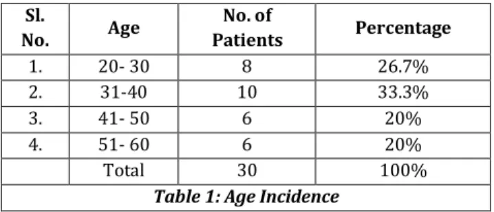
Sl.
No. Age
No. of
Patients Percentage
1. 20- 30 8 26.7%
2. 31-40 10 33.3%
3. 41- 50 6 20%
4. 51- 60 6 20%
Total 30 100%
Jemds.com
Original article
J. Evolution Med. Dent. Sci./eISSN- 2278-4802, pISSN- 2278-4748/ Vol. 05/ Issue 34/ Apr. 28, 2016 Page 1952
Sl. No.
Mode of Injury
No. of
Patients Percentage
1. RTA 25 83.4%
2. Self-Fall 2 6.6%
3. Assault 2 6.6%
4. Domestic Injury 1 3.4%
Total 30 100%
Table 2: Mode of Injury
Sl. No.
Fracture Union in Weeks
No. of
Patients Percentage
1. 13 – 16 15 50%
2. 17 – 20 9 30%
3. 21 -24 2 6.6%
4. >/= to 25 2 6.7% 5. Non-union 2 6.7%
Total 30 100%
Table 3: Fracture Union in Weeks
Sl. No.
Hospital Stay (Weeks)
No. of
Patients Percentage
1. 1 6 20%
2. 1.5 1 3.3%
3. 2 15 50%
4. 2.5 3 10%
5. 3 5 16.7%
Total 30 100%
Table 4: Total Hospital Stay Duration
Sl.
No. Pain
No. of
Patients Percentage
1. No Pain 22 73.4%
2. Mild 7 23.3%
3. Moderate 1 3.3%
4. Severe 0 0
Total 30 100
Table 5: Functional Evaluation
Sl. No.
Functional
Outcome UCLA % MEPI %
1. Excellent 18 60% 18 60% 2. Good 16 20% 7 23.3% 3. Fair 3 10% 2 6.7%
4. Poor 3 10% 3 10%
Total 30 100% 30 100%
Table 6: Functional Outcome as per UCLA, MEPI Score
Sl.
No. Complications
No. of
Patients Percentage
1. Radial Nerve Injury 1 3.3% 2. Delayed Union 1 3.3%
3. Non-Union 2 6.7%
4. Infection 0 0%
5. None 26 86.7%
Total 30 100%
Table 7: Complications
Sl.
No. Result Number Percentage
1. Excellent 16 53.3%
2. Good 7 23.4%
3. Fair 4 13.3%
4. Poor 3 10%
Total 30 100%
Table 8: Results
Fracture Union
Excellent (12–16 wks.), Good (17–20 wks.), Fair (21–24 wks.), Poor (>25 wks.).
Sl.
No. Series Years
Average Age (Years)
1. Sam G. Hunter 1982 38 2. M. J. Bell 1985 31.5
3. Robert Vander
Griend 1986 36 4. Augusto Sarmiento 1990 28 5. Tzu-Liang Hsu 2005 46.2 6. Present study 2015 37.8
Table 9: Comparative Studies
The results of Robert Vander Griend et al and Sam G. Hunter et al showed similar results.
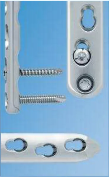
Fig. 1: LCP Combi Hole
Jemds.com
Original article
J. Evolution Med. Dent. Sci./eISSN- 2278-4802, pISSN- 2278-4748/ Vol. 05/ Issue 34/ Apr. 28, 2016 Page 1953
Fig. 3: LCP and Two Types of Screws Pg-10
Fig. 4: Locking Compression Plate
Fig. 5: LCP with Screws
Fig. 6: Schematic Diagram of LCP
CONCLUSION
In the present study, 30 patients with diaphyseal fracture of humerus were surgically managed with locking compression plating and the results conclude that locking compression plating is the superior method of surgical management of diaphyseal fractures of humerus.
REFERENCES
1. Sommer C, Gautier E, Muller M, et al. First clinical results of locking compression plate (LCP). Injury 2003;34(Suppl 2):B43-54.
2. Niemeyer P, Sudkamp NP. Principles and clinical application of the locking compression plate (LCP). Acta Chirurgiae Orthopaedicae Et Traumatology Cechosl 2006;73:221-8.
3. Ahmed M, Nanda R, Bajwa AS, et al. Biomechanical testing of the locking compression plate: when does the distance between bone and implant significancy reduce construct stability? Injury 2007;38(3):358-64.
4. Ghosh S, Halder TC, Chaudhari A, et al. Comparative study by locking plate vs intramedulary interlocking nail. Medical journal of Dr. D.Y. Patil University 2013;6(4):390-4.
5. Neuhaus V, King JD, Jupiter JB. Fixation of osteoporosis of fractures with LCP. Acta Chirurgie Orthopaedicae Et Traumatology Cechosl 2012;79(5):404-10.
6. Wagner M. General principle for the clinical use of LCP. Injury 2003;34(Suppl 2):B31–42.
7. Ellman H, Hanker G, Bayer M. Repair of of the rotator cuff. End-result study of factors influencing reconstruction. JBJS Am 1986;68(8):1136-44.
8. Ellman H. Arthroscopic subacromial nail decompression: analysis of one-to three-year result. Arthroscopy 1987;3(3):173-81.