Original Article
Higher Body Mass Index and Increased Prevalence
of Paranasal Sinus Disease
Yusuke Kabeya
1,2, Kiyoe Kato
2, Masuomi Tomita
2, Takeshi Katsuki
2,
Yoichi Oikawa
2, and Akira Shimada
21Division of General Internal Medicine, Department of Internal Medicine, Tokai University Hachioji Hospital, Hachioji, Tokyo, Japan 2Department of Internal Medicine, Tokyo Saiseikai Central Hospital, Tokyo, Japan
Received May 11, 2015; accepted August 21, 2015; released online January 30, 2016
Copyright © 2016 Yusuke Kabeya et al. This is an open access article distributed under the terms of Creative Commons Attribution License, which permits unrestricted use, distribution, and reproduction in any medium, provided the original author and source are credited.
ABSTRACT
Background:We hypothesized that higher body mass index (BMI) was associated with increased prevalence of paranasal sinus disease and examined the hypothesis in Japanese adults.
Methods:This was a cross-sectional study including 1350 Japanese adults aged 40 years or more who participated in a health check-up program focusing on brain diseases and metabolic syndrome. Participants were divided into quartiles of BMI levels. Paranasal sinus disease was confirmed by a head MRI scan. The association between BMI and paranasal sinus disease was examined using logistic regression analysis, which was adjusted for age, sex, waist:hip ratio, hemoglobin A1c, systolic blood pressure, smoking status, alcohol intake, and white blood cell count. Results:Of the 1350 participants, 151 (11.2%) had paranasal sinus disease. In relation to those in the lowest quartile of BMI, the odds ratios of having the disease among those in the 2nd, 3rd, and 4th quartiles of BMI were 1.89 (95% confidence interval [CI], 1.03–3.48), 2.26 (95% CI, 1.20–4.23) and 2.26 (95% CI, 1.14–4.51), respectively. When BMI was analysed as a continuous variable, an increase of one unit in BMI was significantly associated with increased odds of having the disease, with an OR of 1.08 (95% CI, 1.01–1.16).
Conclusions:The present study suggests that patients with higher BMI are more likely to have paranasal sinus disease.
Key words:obesity; body mass index; paranasal sinus disease
INTRODUCTION
The epidemic of obesity prevails worldwide, along with changes in lifestyles and dietary patterns. The numbers of overweight and obese adults in 2005 were estimated to be 937 million and 396 million worldwide, which accounted for 23% and 10% of the world’s adult population, respectively.1 The estimated numbers of overweight and obese adults are projected to be 2.16 billion and 1.12 billion, respectively, by 2030. The increase would impose serious health burdens. The underlying pathophysiology of these conditions is characterized by chronic inflammation with excess adipose tissue,2,3which could lead to the development
of obesity-related diseases in various organ systems, including the respiratory system. Recently, several epidemiological studies have reported that higher BMI was associated with increased risk of asthma.4–6 Since asthma is considered to
be a state of airway inflammation, it has been proposed that both conditions could have a common inflammatory pathway.7
Likewise, paranasal sinus disease, which is closely related to asthma according to the concept of unified airway,8,9
develops as a result of inflammation in the sinus cavities. Few studies have examined the association of obesity with paranasal sinus disease, especially among adults, with only one study10 examining large-scale data on the Medical
Expenditure Panel Survey in the U.S. reporting an epidemiological link between obesity and paranasal sinus disease. The study concluded that obesity was significantly associated with increased risk of having chronic rhinosinusitis, with an odds ratio of 1.31.10 Although the study examined
a large sample, the diagnosis of paranasal sinus disease was based on diagnosis codes. Difficulties in confirming a diagnosis of paranasal sinus disease might pose an obstacle
to facilitating clinical research on the epidemiological link between paranasal sinus disease and obesity.
Previously, we performed a series of analyses11,12 investigating associations of glycemic status with lung functions and paranasal sinus disease in a sample of Japanese adults who underwent a health check-up program. The program included a head MRI scan, which enabled us to assess paranasal sinus disease in an objective way and provided the opportunity to examine the association between obesity and paranasal sinus disease. In the present study, we hypothesized that higher BMI was associated with increased prevalence of paranasal sinus disease and examined the hypothesis in a sample of Japanese adults.
METHODS
Study design and study population
This cross-sectional study was performed as part of analyses designed to investigate associations of brain diseases and paranasal sinus disease with other chronic diseases in a Japanese health check-up population. The study included 1351 Japanese adults aged 40 years or older who participated in a health check-up program focusing on metabolic syndrome and brain diseases from January 2007 to December 2011. Participants were apparently healthy and visited the hospital not for symptomatic diseases but for the purpose of a health check-up. One participant was excluded because of missing data. As a result, 1350 participants were included in the analysis. The study protocol was reviewed and approved by the ethics committee of Saiseikai Central Hospital.
Anthropometric and laboratory measurements Since the present study was performed as part of a larger set of analyses, the detailed methods of collecting anthropometric and laboratory data have been reported elsewhere.11,12 In
brief, information on age, sex, weight, height, body mass index (BMI), waist:hip ratio, blood pressure, white blood cell (WBC) count, fasting plasma glucose levels, and hemoglobin A1c (HbA1c) levels was collected. Information on smoking status and alcohol intake was obtained using a self-administered questionnaire. As for smoking status, participants were sorted into three groups (non-smoker, past smoker, or current smoker). With regard to alcohol intake, they were categorized into three groups according to frequency of alcohol intake (abstainer, 1–4 days per week, or 5 days or more per week).
Paranasal sinus disease
In the present study, paranasal sinus disease was diagnosed if a participant had any abnormalfindings (mucosal thickening, polyps, or sinus opacification) in at least one of the paranasal sinuses. The criteria were established by referencing the Lund-Mackay system,13 which is a widely accepted method for
grading paranasal sinus abnormalities.14In the Lund-Mackay
system,13 paranasal sinus abnormality was categorized as normal, partially opacified (includingfluid retention, polyps, and mucosal thickening), or totally opacified. The findings were detected via head MRI scan with a 1.5-Tesla scanner (SIGNA HDxt 1.5T; GE Healthcare Japan, Tokyo, Japan). The images were assessed by trained neurologists who did not know the purpose of the study.
Statistical analysis
The participants were divided into quartiles of BMI levels according to the study population distribution. Characteristics of the participants were compared across the BMI categories. One-way analysis of variance tests were used for comparing continuous variables, whileχ2tests were used for categorical variables. Regarding WBC count, the log-transformed values were compared because of the skewed distribution of WBC count. Then, the association between BMI and the prevalence of paranasal sinus disease was graphically and statistically examined. The observed prevalence of paranasal sinus disease for each 0.4 unit of BMI band was plotted, and the expected prevalences of paranasal sinus disease and their 95% confidence intervals (CIs) in each BMI level were plotted in the same graph using a restricted cubic spline model. The model confirmed that the log-odds of having paranasal sinus disease were a linear function of BMI level. Finally, the odds ratios (ORs) and 95% CIs of having paranasal sinus disease were estimated for quartiles of BMI levels using a multiple logistic regression model. The ORs and the 95% CIs were adjusted for age, sex, waist:hip ratio, HbA1c, systolic blood pressure, smoking status, alcohol intake, and WBC count. To test for linear trend, the BMI data were scored from 1 (lowest) to 4 (highest) quartile. Then, trend tests were performed by modelling these quartile-based scores as a continuous variable. In the multiple logistic regression, we did not stratify the analysis by sex, since no significant effect modification of the association by sex was observed. In this process, models with and without the interaction term of sex and BMI were constructed and compared using a chi-squared log-likelihood ratio test (χ2= 0.47, P= 0.49), which found no significant effect modification.
Statistical analyses were performed using STATA software version 11 (StataCorp, College Station, TX, USA). All statistical tests were two-sided, and p-values less than 0.05 were considered statistically significant.
RESULTS
pressure, diastolic blood pressure, fasting plasma glucose, HbA1c, and WBC count were also significantly different across the quartiles. The differences in age and alcohol intake were significant but less pronounced.
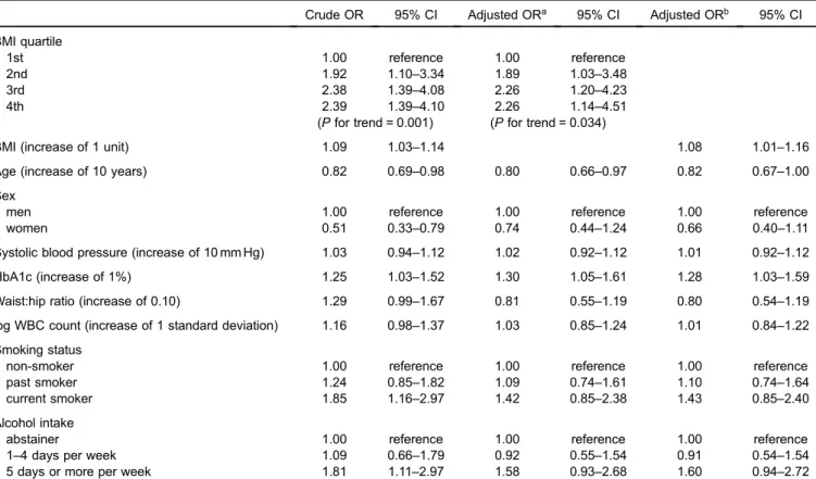
The prevalence of paranasal sinus disease was further investigated using a restricted cubic spline model, which showed a positive association between BMI levels and the presence of paranasal sinus disease, although the association appeared attenuated in those with high BMI levels (Figure).
Table 2 shows the result of the logistic regression analysis. Although the variables listed in Table 2 produced significance or borderline significance in the crude analysis, the significant associations remained in the multivariate analysis only for age, BMI, and HbA1c levels. In relation to those in the lowest quartile of BMI, the odds of having paranasal sinus disease were significantly higher among those in the 2nd, 3rd, and 4th quartiles of BMI, with ORs of 1.89 (95% confidence interval [CI], 1.03–3.48), 2.26 (95% CI, 1.20–4.23) and 2.26 (95% CI,
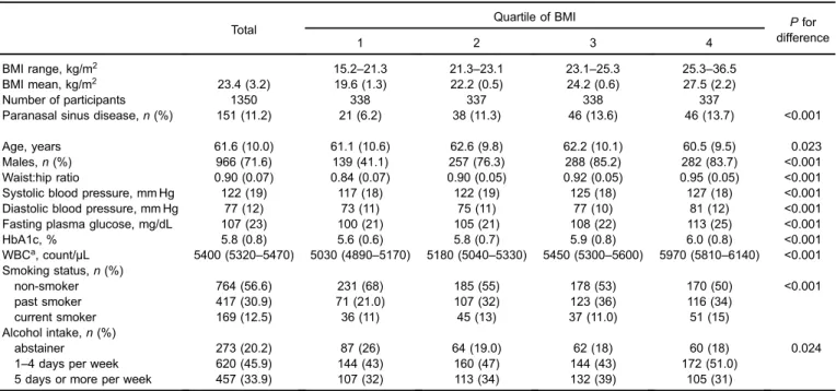
Table 1. Characteristics of study participants
Total Quartile of BMI Pfor
difference
1 2 3 4
BMI range, kg/m2 15.2–21.3 21.3–23.1 23.1–25.3 25.3–36.5
BMI mean, kg/m2 23.4 (3.2) 19.6 (1.3) 22.2 (0.5) 24.2 (0.6) 27.5 (2.2)
Number of participants 1350 338 337 338 337
Paranasal sinus disease,n(%) 151 (11.2) 21 (6.2) 38 (11.3) 46 (13.6) 46 (13.7) <0.001
Age, years 61.6 (10.0) 61.1 (10.6) 62.6 (9.8) 62.2 (10.1) 60.5 (9.5) 0.023
Males,n(%) 966 (71.6) 139 (41.1) 257 (76.3) 288 (85.2) 282 (83.7) <0.001
Waist:hip ratio 0.90 (0.07) 0.84 (0.07) 0.90 (0.05) 0.92 (0.05) 0.95 (0.05) <0.001 Systolic blood pressure, mm Hg 122 (19) 117 (18) 122 (19) 125 (18) 127 (18) <0.001 Diastolic blood pressure, mm Hg 77 (12) 73 (11) 75 (11) 77 (10) 81 (12) <0.001 Fasting plasma glucose, mg/dL 107 (23) 100 (21) 105 (21) 108 (22) 113 (25) <0.001
HbA1c, % 5.8 (0.8) 5.6 (0.6) 5.8 (0.7) 5.9 (0.8) 6.0 (0.8) <0.001
WBCa, count/µL 5400 (5320–5470) 5030 (4890–5170) 5180 (5040–5330) 5450 (5300–5600) 5970 (5810–6140) <0.001 Smoking status,n(%)
non-smoker 764 (56.6) 231 (68) 185 (55) 178 (53) 170 (50) <0.001
past smoker 417 (30.9) 71 (21.0) 107 (32) 123 (36) 116 (34)
current smoker 169 (12.5) 36 (11) 45 (13) 37 (11.0) 51 (15)
Alcohol intake,n(%)
abstainer 273 (20.2) 87 (26) 64 (19.0) 62 (18) 60 (18) 0.024
1–4 days per week 620 (45.9) 144 (43) 160 (47) 144 (43) 172 (51.0)
5 days or more per week 457 (33.9) 107 (32) 113 (34) 132 (39) 105 (31)
BMI, body mass index; HbA1c, hemoglobin A1c; WBC, white blood cell.
Data are presented as mean (standard deviation) for continuous variables and count (%) for categorical variables except WBC. aWBC count is presented as geometric mean (95% confidence interval) because of the skewed distribution.
Body mass
index (kg/m
2)
15
20
25
30
Prevalence (%)
0
20
40
60
Observed prevalence Expected prevalence 95% confidence interval
1.14–4.51), respectively. A significant linear trend was observed (P for trend = 0.034). When BMI was analysed as a continuous variable, an increase of one unit of BMI was significantly associated with increased odds of having paranasal sinus disease, with an OR of 1.08 (95% CI, 1.01–1.16).
DISCUSSION
The present study examined the association between BMI levels and the prevalence of paranasal sinus disease in a Japanese health check-up population. Overall, we found a positive association between BMI levels and the prevalence of paranasal sinus disease. In addition, we found that younger age and higher HbA1c levels were significantly associated with increased prevalence of paranasal sinus disease.
The multivariate analysis demonstrated that the association of BMI with paranasal sinus disease remained significant after adjustment for HbA1c levels. Since BMI and glycemic status are closely related to each other, the apparent association of higher BMI with paranasal sinus disease may be confounded by glycemic status. However, the results showed that both HbA1c and BMI were significantly and independently associated with paranasal sinus disease. Although there may
be some overlap in the mechanisms of the association, the results imply that higher glycemic status and higher BMI might affect paranasal sinus disease in different manners.
The multivariate analysis also showed that the association of BMI with paranasal sinus disease was independent of waist:hip ratio, which is a marker of central obesity.15 According to a past report,16 body fat distribution, which
reflects systemic inflammation, may be a more accurate measure of obesity than BMI. We expected that the association of BMI with paranasal sinus disease became attenuated after adjustment for the waist:hip ratio in the present study. However, the association did not change substantially after such adjustment. This finding suggests that the link between higher BMI and paranasal sinus disease was not necessarily mediated through inflammation. Other mechanisms, such as airway structural problems related to obesity or obesity-related lifestyles, could mediate the association. Of particular interest is the shape of the curve in the plot of the dose-response relationship between BMI and the prevalence of paranasal sinus disease. The positive association appeared attenuated in those with high BMI levels. Although the reason for the attenuation remains uncertain, this finding could provide an insight into the mechanism of the association.
Table 2. The odds ratios of having paranasal sinus disease according to body mass index
Crude OR 95% CI Adjusted ORa 95% CI Adjusted ORb 95% CI
BMI quartile
1st 1.00 reference 1.00 reference
2nd 1.92 1.10–3.34 1.89 1.03–3.48
3rd 2.38 1.39–4.08 2.26 1.20–4.23
4th 2.39 1.39–4.10 2.26 1.14–4.51
(Pfor trend = 0.001) (Pfor trend = 0.034)
BMI (increase of 1 unit) 1.09 1.03–1.14 1.08 1.01–1.16
Age (increase of 10 years) 0.82 0.69–0.98 0.80 0.66–0.97 0.82 0.67–1.00
Sex
men 1.00 reference 1.00 reference 1.00 reference
women 0.51 0.33–0.79 0.74 0.44–1.24 0.66 0.40–1.11
Systolic blood pressure (increase of 10 mm Hg) 1.03 0.94–1.12 1.02 0.92–1.12 1.01 0.92–1.12
HbA1c (increase of 1%) 1.25 1.03–1.52 1.30 1.05–1.61 1.28 1.03–1.59
Waist:hip ratio (increase of 0.10) 1.29 0.99–1.67 0.81 0.55–1.19 0.80 0.54–1.19
log WBC count (increase of 1 standard deviation) 1.16 0.98–1.37 1.03 0.85–1.24 1.01 0.84–1.22
Smoking status
non-smoker 1.00 reference 1.00 reference 1.00 reference
past smoker 1.24 0.85–1.82 1.09 0.74–1.61 1.10 0.74–1.64
current smoker 1.85 1.16–2.97 1.42 0.85–2.38 1.43 0.85–2.40
Alcohol intake
abstainer 1.00 reference 1.00 reference 1.00 reference
1–4 days per week 1.09 0.66–1.79 0.92 0.55–1.54 0.91 0.54–1.54
5 days or more per week 1.81 1.11–2.97 1.58 0.93–2.68 1.60 0.94–2.72
BMI, body mass index; CI, confidence interval; HbA1c, hemoglobin A1c; OR, odds ratio; WBC, white blood cell.
aThe model included BMI (categorical variable), age, sex, systolic blood pressure, HbA1c, waist:hip ratio, WBC count, smoking status, and alcohol intake.
Since confirming the diagnosis of paranasal sinus disease seems difficult without objective imaging methods, the etiology, pathophysiology, and natural history of the disease have not been fully elucidated. Nonetheless, the clinical characteristics related to paranasal sinus disease have been investigated in several studies.10,17–20 However, results have
been inconsistent. As chronic sinusitis is frequently diagnosed in young or middle-aged adults,20 it is expected that the prevalence of paranasal sinus disease decreases with age, which seems consistent with thefindings of the present study. However, some studies17–19 have reported that age is not
associated with thefindings of head MRI scans assessing the paranasal sinuses. Evidence on the influence of HbA1c levels on paranasal sinus disease is limited. We previously reported that diabetes is associated with increased prevalence of paranasal sinus disease.12 The dose-response relationship
between glycemic status and the prevalence was also confirmed in the study. Factors related to higher blood glucose levels, such as impaired immune function or impaired ciliary motility, could affect the development of paranasal sinus disease. Smoking is considered an established risk factor for chronic sinusitis.21 However, past studies17–19 using MRI
reported that smoking habit did not correlate with abnormal
findings in the paranasal sinuses, which is consistent with the results of the present study. These controversies should be resolved in future studies.
Although fatal consequences are not usually expected from paranasal sinus disease, the disease can result in serious healthcare costs. In the United States, chronic rhinosinusitis was considered to be one of the top 10 most costly conditions for American employers.22The symptoms and impairment of quality of life resulting from rhinosinusitis resulted in 11.1 million healthcare visits per year, and the healthcare costs were estimated to be $8.6 billion in 2007.23,24When indirect
costs of the disease are taken into account, the overall costs related to the disease may be even higher. The healthcare costs of patients with chronic rhinosinusitis were also examined in an Asian population. One study in Taiwan25reported that those with chronic rhinosinusitis visited outpatient clinics more frequently. Higher total healthcare costs among those with chronic rhinosinusitis compared with those without were also reported. There have been few studies reporting healthcare costs of paranasal sinus disease in Japan. As the prevalence of obesity has risen dramatically,1 it is expected
that the prevalence of paranasal sinus disease is increasing, which might result in heavy healthcare cost burden. Further research is required to determine accurate epidemiology and healthcare cost estimates related to paranasal sinus disease.
A few limitations of the present study should be noted. First, the study was based on incidental findings of a head MRI scan. The clinical relevance of incidental findings detected by a head MRI scan has been controversial. One study17 demonstrated a correlation between abnormalities on
a head MRI scan and symptoms of an upper respiratory
infection. Other studies26,27 have reported that there is a disparity between clinical symptoms and the findings of radiological examination. Examining the association of BMI with clinically-confirmed paranasal sinus disease might be a worthwhile subject for a further study. Second, the presence of paranasal sinus disease was evaluated by trained neurologists. Although they were experienced in interpreting head MRI images, the between-observers variability was not available in the analysis, which could limit the validity of the present study. Third, selection bias is another concern, since the present study was performed among those who participated in a health check-up program. In general, those who voluntarily participate in a health check-up program might be more health-conscious than the general population, which could restrict the external validity of the present study. Fourth, although an association between higher BMI and increased prevalence of paranasal sinus disease was observed in the present study, the association between BMI and disease severity was not evaluated. In addition, we are not able to evaluate the causal association of BMI and paranasal sinus disease because of the cross-sectional design of the present study.
In conclusion, the present study found that higher BMI was associated with increased prevalence of paranasal sinus disease. This result has implications at both the individual and societal levels. At the individual level, physicians should remember that an obese patient may be at increased risk for paranasal sinus disease. At the societal level, it is expected that the prevalence of paranasal sinus disease is increasing with the epidemic of obesity, which could impose a substantial healthcare costs burden. Further research is required to better understand the epidemiology of paranasal sinus disease and the link between obesity and paranasal sinus disease.
ACKNOWLEDGEMENT
Conflicts of interest: None declared.
REFERENCES
1. Kelly T, Yang W, Chen CS, Reynolds K, He J. Global burden of obesity in 2005 and projections to 2030. Int J Obes (Lond). 2008;32:1431–7.
2. Xu H, Barnes GT, Yang Q, Tan G, Yang D, Chou CJ, et al. Chronic inflammation in fat plays a crucial role in the development of obesity-related insulin resistance.J Clin Invest. 2003;112:1821–30.
3. Lumeng CN, Saltiel AR. Inflammatory links between obesity and metabolic disease.J Clin Invest. 2011;121:2111–7. 4. Ford ES. The epidemiology of obesity and asthma. J Allergy
Clin Immunol. 2005;115:897–909; quiz 10.
5. Litonjua AA, Gold DR. Asthma and obesity: common early-life influences in the inception of disease.J Allergy Clin Immunol. 2008;121:1075–84; quiz 85–6.
2006;110:83–102.
7. McLachlan CR, Poulton R, Car G, Cowan J, Filsell S, Greene JM, et al. Adiposity, asthma, and airway inflammation.J Allergy Clin Immunol. 2007;119:634–9.
8. Krouse JH, Brown RW, Fineman SM, Han JK, Heller AJ, Joe S, et al. Asthma and the unified airway. Otolaryngol Head Neck Surg. 2007;136(5 Suppl):S75–106.
9. Krouse JH, Krouse HJ. Asthma, rhinitis, and the unified airway.
ORL Head Neck Nurs. 2013;31:6–10.
10. Bhattacharyya N. Associations between obesity and inflam-matory sinonasal disorders.Laryngoscope. 2013;123:1840–4. 11. Kabeya Y, Kato K, Tomita M, Katsuki T, Oikawa Y, Shimada
A. Association of glycemic status with impaired lung function among recipients of a health screening program: a cross-sectional study in Japanese adults.J Epidemiol. 2014;24:410–6. 12. Kabeya Y, Kato K, Tomita M, Katsuki T, Oikawa Y, Shimada A. Association between diabetes and increased prevalence of paranasal sinus disease: a cross-sectional study in Japanese adults.J Epidemiol. 2015;25:297–302.
13. Lund VJ, Kennedy DW. Staging for rhinosinusitis.Otolaryngol Head Neck Surg. 1997;117:S35–40.
14. Zinreich SJ. Imaging for staging of rhinosinusitis. Ann Otol Rhinol Laryngol Suppl. 2004;193:19–23.
15. Hsieh SD, Yoshinaga H, Muto T. Waist-to-height ratio, a simple and practical index for assessing central fat distribution and metabolic risk in Japanese men and women.Int J Obes Relat Metab Disord. 2003;27:610–6.
16. Clerisme-Beaty E, Rand CS. The effect of obesity on asthma incidence: moving past the epidemiologic evidence. J Allergy Clin Immunol. 2009;123:96–7.
17. Cooke LD, Hadley DM. MRI of the paranasal sinuses: incidental
abnormalities and their relationship to symptoms. J Laryngol Otol. 1991;105:278–81.
18. Havas TE, Motbey JA, Gullane PJ. Prevalence of incidental abnormalities on computed tomographic scans of the paranasal sinuses.Arch Otolaryngol Head Neck Surg. 1988;114:856–9. 19. Tarp B, Fiirgaard B, Christensen T, Jensen JJ, Black FT.
The prevalence and significance of incidental paranasal sinus abnormalities on MRI.Rhinology. 2000;38:33–8.
20. Lusk R. Chronic rhinosinusitis: contrasts between children and adult patients.Clin Allergy Immunol. 2007;20:287–98. 21. Reh DD, Higgins TS, Smith TL. Impact of tobacco smoke on
chronic rhinosinusitis: a review of the literature. Int Forum Allergy Rhinol. 2012;2:362–9.
22. Tan BK, Kern RC, Schleimer RP, Schwartz BS. Chronic rhinosinusitis: the unrecognized epidemic.Am J Respir Crit Care Med. 2013;188:1275–7.
23. Bhattacharyya N. Contemporary assessment of the disease burden of sinusitis.Am J Rhinol Allergy. 2009;23:392–5. 24. Bhattacharyya N. Ambulatory sinus and nasal surgery in the
United States: demographics and perioperative outcomes.
Laryngoscope. 2010;120:635–8.
25. Chung SD, Hung SH, Lin HC, Lin CC. Health care service utilization among patients with chronic rhinosinusitis: a population-based study.Laryngoscope. 2014;124:1285–9. 26. Wani MK, Ruckenstein MJ, Parikh S. Magnetic resonance
imaging of the paranasal sinuses: incidental abnormalities and their relationship to patient symptoms.J Otolaryngol. 2001;30: 257–62.