1 7 3 Arq Bras Oftalmol. 2014;77(3):173-7 http://dx.doi.org/10.5935/0004-2749.20140044
Efficacy of a lutein-based dye (Phacodyne
TM) for visualizing anterior capsulorhexis
during cataract surgery by phacoemulsification
Eicácia de corante à base de luteína (Phacodyne
TM) para observação da capsulorrexis anterior em
cirurgia de facoemulsiicação
Lucas Monferrari Monteiro Vianna1, Marcos J. cohen1, cristina MuccioLi1, acácio LiMa1, Diogo sousa-Martins1, Maurício Maia1, rubens beLfort Jr.1
Submitted for publication: February 6, 2014 Accepted for publication: March 29, 2014
Study conducted at Departamento de Oftalmologia, Universidade Federal de São Paulo (UNIFESP), São Paulo, SP, Brazil.
1 Departamento de Oftalmologia, Universidade Federal de São Paulo (UNIFESP), São Paulo, SP,
Brazil.
Funding: This study has been conducted under grants from Kemin Industries Inc.
Disclosure of potential conflicts of interest: Diogo Sousa-Martins is an employee of Kemin Industries Inc. Acacio Lima is the owner of Ophthalmo’s and received personal fees from Kemin Industries Inc. Kemin Industries Inc. has supported the Department of Ophthalmology through Research Grants. Diogo Sousa-Martins, Rubens Belfort Jr., Acácio Lima and Maurício Maia and Kemin Industries Inc. hold patent related to the use of lutein or combinations thereof with synthetic dyes for staining the anterior capsule (patent pending US 20120251458 and Publications WO 2012135432).
Corresponding author: Lucas Monferrari Monteiro Vianna. Rua Botucatu, 821 - São Paulo (SP) - 04023-900 - Brazil - E-mail: [email protected]
Registered at clinicaltrials.org (NCT01627977) and approved by UNIFESP IRB (2146/11) INTRODUCTION
The continuous circular capsulotomy (CCC) of the anterior lens capsule is a critical step in the modern cataract surgery performed by phacoemulsification, which offers advantage for the implantation of intraocular lens correctly into the capsular bag(1). Good visualization of the anterior capsule flap is needed when performing the CCC, particularly in the absence of a red reflex(2-4). Since the lack of a red reflex results in technical difficulties(2-4), a correct CCC is useful when a more aggressive manipulation of the capsular bag is required(3). In such clinical conditions, the improvement of the anterior capsule
ABSTRACT
Purpose: To evaluate the efficacy and safety of a novel lutein-based dye for the anterior capsulorhexis during phacoemulsification in cataract surgery in humans. Methods: Twenty-five eyes from 25 patients were operated by 25 different sur geons who performed continuous circular capsulorhexis (CCC) guided by a lutein-based dye (Phacodyne™) during cataract surgery by phacoemulsification. A questionnaire assessed the surgeon’s opinion regarding the efficacy of the dye. Follow-up examinations were performed at 1, 7, and 30 days post-surgery. Eyes were evaluated by full ophthalmic examination, corneal topography/pachymetry, and corneal endothelial cell count.
Results: As revealed by the answers to the questionnaire, the dye facilitated the CCC procedure in all eyes. Baseline nuclear cataract classification (according to the Lens Opacities Classification System III; LOCS III) was 3.24 (± 1.12). Preoperative BCVA (logMAR) was 0.89 ± 0.59 and improved to 0.23 ± 0.22 on day 30 after surgery. The intraocular pressure (IOP) remained stable and the inflammatory reaction subsided in all cases within the first 7 days after surgery. The pre-operative values of corneal pachymetry and IOP were similar to those found on follow-up day 30. Loss in endothelial cell number was similar to earlier reports.
Conclusion: Phacodyne™ was efficient when used for anterior capsulorhexis du ring cataract surgery by phacoemulsification and showed no signs of toxicity or side effects during the 30-day follow-up period.
Keywords: Cataract extraction; Lutein; Phacoemulsification; Capsulorhexis; Lenses, intraocular
RESUMO
Objetivos: Avaliar a eficácia e eficiência de um novo corante à base de luteína para coloração da cápsula anterior durante cirurgia de facoemulsificação em humanos.
Métodos: Vinte e cinco olhos de 25 pacientes foram operados por 25 cirurgiões di ferentes que realizaram capsulorrexis circular contínua e facoemulsificação após coloração da cápsula anterior com corante à base de luteína. Um questionário ava liou a opinião dos cirurgiões sobre a eficácia do corante. Exames pós-operatórios foram realizados nos dias 1, 7 e 30 por meio de exame oftalmológico completo, topografia/ paquimetria e contagem de células endoteliais.
Resultados: De acordo com o questionário aplicado, o corante facilitou a cirurgia em todos os olhos. A classificação da catarata de acordo com o LOCS III foi de 3,24 ± 1,12. A acuidade visual pré-operatória com melhor correção foi de 0,89 ± 0,59 (logMAR), passando a 0,23 ± 0,22 no pós-operatório. A pressão intraocular (PIO) permaneceu estável e houve reação de câmara leve que desapareceu em todos os casos durante os primeiros 7 dias de pós-operatório. Não houve significância estatística comparando a paquimetria e PIO pré e pós-operatórios.
Conclusão: O novo corante se mostrou eficiente e sem sinais de toxicidade ou efeitos adversos, após 30 dias, quando usado para auxiliar a cirurgia de facoemulsificação.
Descritores: Extração de catarata/métodos; Luteína; Facoemulsificação; Capsulorrexe; Lentes intraoculares
visualization can also be of great value for surgeons, particularly for those who are learning the surgical technique(4). Additionally, the surgical procedure of staining the anterior capsule during CCC in pediatric cataract extraction is useful since the anterior capsule of these eyes is thin and elastic(5).
for facilitating the CCC maneuver, only the trypan blue 0.06% - Vision blue™ (Dorc, Netherlands) has been approved by the FDA for use in cataract surgery(7,8).
New dyes that enhance the visualization of ocular tissues during surgery have recently emerged(9-11). Lutein and zeaxanthin are two major components of the macular pigment and are the only carote-noids found in the macula and lens of humans. The antioxidant pro-perties of the dyes and the ability to filter blue light facilitated their use during the CCC procedure(12,13).
It has been reported that the green 1% solution of soluble lutein and zeaxanthin-based dye combined with 0.04% trypan blue (Phaco-dyne™, Kemin, USA) is a useful intraocular dye for staining the an terior capsule, enhancing the CCC procedure in human cadaveric eyes(11). Its safety profile has been evaluated by clinical, histological, and electro-retinographic methods after intravitreal injections into rabbit eyes(14).
The objective of this study was to evaluate the feasibility and safety of application of a novel dye comprising 1% soluble lutein-zeaxanthin together with 0.04% trypan blue (Phacodyne™, Kemin, USA) to improve the anterior capsulorhexis during the phacoemulsification surgery in human eyes.
METHODS
P
REPARATIONOFTHEDYEThe method of preparation described below is valid for lutein/ zeaxanthin regardless of the chemical form, purity, crystallization, or degree of esterification (patent office application #61/468,838).
The initial steps of this study were the development of a wa-ter-soluble solution, characterization of the absorbance of the lutein/ zeaxanthin solution using the Aquamate device (Thermo Spectronic, Cambridge, UK), and the identification of the color of the chromo-phoric groups. The physicochemical parameters were tested for the optimum pH, osmolarity/osmolality, and concentration. Solubility in water and polyvinyl alcohol was tested, the ultraviolet spectra were recoded, and the blends showing different colors within the visible spectrum were analyzed.
The method of preparation of the dye included dissolution, agita-tion, sterilizaagita-tion, and filtration. First, the components were dissolved in balanced saline solution (BSS) or any other solution compatible with the components of the formula that provided stability, suitable pH, and osmolarity acceptable for ocular use. Sterilization using wet heat was achieved by autoclaving at 121ºC. The final dye solution contained 1% soluble lutein/zeaxanthin and 0.04% trypan blue (os-molarity, 280 mOsm; density, 1.05; pH, 7.00). Aliquots of 1 mL were prepared, stored at room temperature, and were used within 30 days.
S
TUDYDESIGNThis was a prospective, consecutive, non-randomized, interven-tional study of 25 eyes of 25 patients performed by 25 different sur geons. The study was registered at clinicaltrials.gov under the no NCT01627977. The study was approved by the Ethics Committee of the Federal University of São Paulo and was conducted according to the Research Guidelines of the Association of Research in Vision and Ophthalmology, adhering to the Declaration of Helsinki. All patients were informed of the benefits and risks of the surgical procedure as well as the nature and possible consequences of the new dye tested. This study was conducted after receiving informed consent from all participants.
Eyes were submitted to phacoemulsification at the Ophthalmo-logy Department of Hospital São Paulo, Universidade Federal de São Paulo, Brazil, with PhacodyneTM (Kemin, USA) assisting the CCC technique and the hydrophilic foldable IOL implantation.
I
NCLUSIONANDEXCLUSIONCRITERIAPatients over 50 years of age with indication for cataract surgery in one eye were included in the study. Patients with any previous
ocular surgeries in the eye studied, pregnancy, glaucoma, a past or present intraocular infection, and any other ocular condition detected during the pre-operative examination that could limit or affect the postoperative results were excluded. The minimum follow-up period was 30 days.
P
HYSICALEVALUATIONANDOCULAREXAMINATIONSAll patients underwent a full ophthalmologic examination at the start of the study and postoperatively (days 1, 7, and 30). This inclu-ded evaluation of the best-corrected visual acuity (BCVA) that was measured using the Snellen Charts and converted to logMAR, mea surement of the intraocular pressure (IOP) using the Goldmann tonometer, and slit-lamp evaluation. Complementary exams such as fundoscopy, pachymetry, and specular microscopy (CSO, Firenze, Italy) were also performed at the same time points. Baseline cataracts were graded according to the “Lens Opacities Classification System III” (LOCS III)(15). Anterior chamber reaction was graded according to SUN Working Group(16).
S
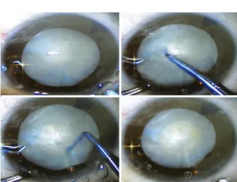
URGICALPROCEDURESThe phacoemulsification was performed by 25 different experien-ced cataract surgeons. The first step consisted of making a 2.75-mm clear corneal incision, filling the anterior chamber with Phacodyne™ (Kemin, USA), washing after 30 seconds with balanced salt solution, and filling the anterior chamber with viscoelastic. The anterior con-tinuous circular capsulorhexis (CCC) (Figure 1) was performed using an Utrata forceps. This was followed by hydrodissection and hydrode-lineation using approximately 0.1 mL BSS. Phacoemulsification was performed using the Infinity Vision System™ (Alcon, USA). Following this, the lens cortex was aspirated and a foldable intraocular lens was inserted through the 2.75-mm corneal incision (Figure 2). At the end of the surgical procedure, the viscoelastic substance was aspirated and no sutures were placed at the corneal incision. Moxifloxacyn and dexamethasone association - Vigadexa™ (Alcon, USA) eye drops were used during the immediate post-operative period.
Figure 1. Dark green color of the dye.
1 7 5 Arq Bras Oftalmol. 2014;77(3):173-7
F
OLLOW-
UPAll eyes received 0.5% moxifloxacin eye drops 4 times daily for 7 days and 0.1% dexamethasone as part of a regressive regimen. No hy-potensive eye drops were prescribed. The eyes underwent an ophthal-mologic examination at days 1, 7, and 30 by expert ophthalmologists.
Q
UESTIONNAIRESSurgeons’ evaluation of the use of PhacodyneTM (Kemin, USA) for the CCC procedure was reported using a questionnaire.
S
TATISTICALMETHODSAll data were presented as the mean and standard deviation for quantitative variables, and absolute (n) and relative (%) values for qua-litative variables. The prevalence of intense staining of the anterior lens capsule was defined. The confidence interval was set at 95%.
RESULTS
I
NTRAOPERATIVEFINDINGSThe lutein-based dye produced a green solution (Figure 1) that stained the anterior capsule and facilitated the CCC procedure (Fi-gure 2). The phacoemulsification followed by IOL implantation was performed as traditionally reported by cataract surgeons. No sutures were necessary.
P
HYSICALEVALUATIONANDOCULAREXAMINATIONSATBASELINEAND FOLLOW-
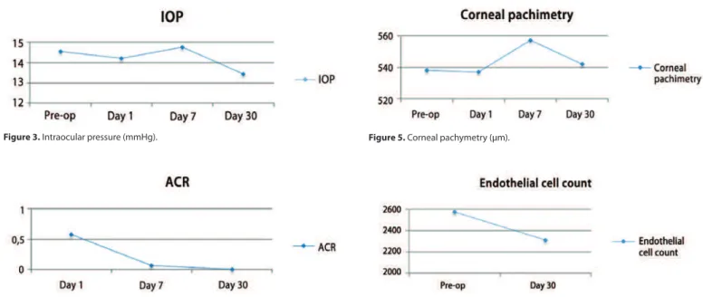
UPNuclear cataract classification (according to LOCS III) was 3.24 ± 1.12 and changed from 2 to 6 in both groups. Preoperative BCVA (logMAR) was 0.89 ± 0.59 and improved to 0.23 ± 0.22 at day 30 after the surgery. The intraocular pressure (IOP) remained stable and similar to the pre-operative measurements during the post-operative visits (p=0.004) (Figure 3). A mild inflammatory reaction (grade 0.5+ accor-ding to SUN Working Group)(16) at the anterior chamber was observed within the first 7 days (Figure 4).
The pre-operative corneal pachymetry average was 538.8 ± 40.0 and that on post-operative follow-up day 30 was 542.5 ± 39.8 (Figure 5). The preoperative endothelial cell count average was 2573.5 ± 235.9 and that on postoperative follow-up day 30 was 2311.36 ± 490.7 (Figure 6).
There were no significant differences between the results of pre- and post-operative fundus biomicroscopy.
Q
UESTIONNAIRESThe questionnaires showed that surgeons considered Phacodyne™ to be efficient for staining the anterior capsule and that the staining facilitated the CCC procedure in all eyes (Table 1).
DISCUSSION
A previous study showed that a solution containing 1% soluble lutein-zeaxanthin and 0.04% Brilliant Blue G efficiently stained the anterior capsule and facilitated the CCC in cadaveric eyes(11). Additio-nally, clinical, histological, and electroretinographic evaluations per-formed after the intravitreal injection of the dye in rabbit eyes showed no signs of toxicity,(14) indicating the enhanced safety in case of accidental leak of the dye into the vitreous. Based on these reported we decided to evaluate the application of s the new dye for cataract surgery in humans.
Based on the nuclear cataract classification, LOCS III, the eyes enrolled in this study showed varying grades of cataract from 2 to 6, demonstrating that advanced cases of hypermature cataract were also present(6). The surgeons classified the visualization of the anterior capsule with the dye as “good” and considered it as a useful tool for cataract surgery (Table 1). The green colored dye (Figure 1) stained the anterior capsule of the lens, which facilitated the CCC procedure in all cases, including the hypermature cataracts.
The surgeons observed that in 4 eyes, the cornea, the incision, or iris were stained by the dye. Residual dye was found only in 2 eyes at the end of the surgical procedure (Table 1). The dye was not seen in any eye on the first postoperative day. A mild anterior chamber reaction of grade 0.5+ (according to SUN Working Group)(16) was observed during the first week after surgery (Figure 4). These results were similar to those of earlier clinical studies, where trypan blue was used during phacoemulsification to aid the CCC procedure or when a dye was not used.(3,4,17-19) In agreement with other reports, the IOP remained stable and similar to the pre-operative measurements during the post-operative period (Figure 3)(20).
The pre-operative BCVA (logMAR) was 0.89 ± 0.59 and improved to 0.23 ± 0.22 at day 30 after surgery, clearly showing that there was an improvement in BCVA from 20/160 to approximately 20/32 in
Figure 3. Intraocular pressure (mmHg).
Figure 4. Post-operative anterior chamber reaction (+).
Figure 5. Corneal pachymetry (µm).
Table 1. Questionnaire returned by surgeons showing the efectiveness of the dye
Surgeons questionnaire response % p
How often do the surgeon use dyes in cataract surgery? Never 00 000%
Rarely 08 032%
Frequently 13 052%
Always 04 016%
Which dye does the surgeon use? Trypan blue 25 100%
Indocyanine green 00 000%
Others 00 000%
How was the visualization of the anterior capsule with Phacodyne? Good 25 100% p<0.001
Bad 00 000%
How was the degree of staining of the anterior lens capsule by the dye? Nothing 00 000% p<0.001
Adequate 20 080%
Intense 05 020%
Did it color any other ocular tissues? No 21 084% p<0.001
Yes 04 016%
Conjunctiva 00 000%
Iris 01 004%
Posterior capsule 00 000%
Incision 02 008%
Cornea 01 004%
What is the surgeon’s opinion about the usefulness of the dye? Good 25 100%
Bad 00 000%
Is the color of the dye suitable for dying the anterior lens capsule? Yes 25 100%
No 00 000%
Were there any signs of the dye at the end of the surgery? No 23 092% p<0.001
Yes 02 008%
Incision 02 008%
majority of the eyes. No signs of toxicity were observed during the 30-day follow-up period(21).
Corneal pachymetric changes were observed soon after the sur-gical procedure and peaked at one week during the follow-up. The values returned to the baseline levels on the 30th postoperative day. The pre-operative corneal pachymetry average was 538.8 ± 40.0 and the post-operative value was 542.5 ± 39.8 (Figure 5), demonstrating that anatomical changes of the cornea were temporary and reversi-ble within the first 30 days after the surgery(21,22).
The baseline endothelial cell count average was 2573.5 ± 235.9 and that post-operatively was 2311.36 ± 490.7 (Figure 6), showing an average decrease of 10.18% in the first month after the surgery. Similar endothelial cell loss during phacoemulsification procedures has been reported by others(21,22).
Pre-and post-operative fundus biomicroscopy evaluations showed no significant differences. Pre-and post-operative macular thickness measurements are needed to assess the effect on the retina.
To our knowledge, this is the first study that used a lutein-based dye (PhacodyneTM) for visualizing anterior capsulorhexis during ca-taract surgery by phacoemulsification. However, our study had limi-tations. This include, the small sample size, the absence of a control group, the large number of surgeons (that may affect the outcomes, although it is suitable for analyzing the efficacy), and the limited follow up period of 30 days. The goal of this study using lutein and zea xanthin combined with trypan blue was to evaluate the safety and efficacy of the dye for CCC. Further studies are warranted to eva-luate the effectiveness of the new dye and its advantages over those that are currently being used. We hypothesize that use of lutein and
zeaxanthin in combination with trypan blue may have two favorable effects: 1) The lower concentration of trypan blue (that can also be tested in more reduced concentrations in future studies(23)), and 2) the antioxidant effect of lutein and zeaxanthin molecules that may quench the singlet oxygen generated by the exposure of trypan blue to light. Additionally, since lutein is a natural product, the manufactu-ring costs could be reduced. The use of lutein and zeaxanthin without trypan blue will be investigated in future studies.
CONCLUSION
The newly formulated lutein-based dye facilitated the CCC during phacoemulsification procedures and showed a good safety profile.
REFERENCES
1. Gimbel HV, Neuhann T. Development, advantages and methods of continuous cir-cular capsulorhexis technique. J Cataract Refract Surg. 1990;16(1):31-7. Comment in: J CataractRefract Surg. 2001;27(9):1346-7.
2. Assia EI, Apple DJ, Barden A, Tsai JC, Castaneda VE, Hoggart JS. An experimental study comparing various anterior capsulectomy techniques. Arch Ophthalmol. 1991; 109(5):642-7. Comment in: Arch Ophthalmol. 1992;110(2):170.
3. Marback EF, Freitas LL, Fernandes FP, Branco BC, Belfort Junior R. Anterior capsule staining using 0.025% trypan blue in cataracts without red reflex. Arq Bras Oftalmol. 2001;64(4):333-5.
1 7 7 Arq Bras Oftalmol. 2014;77(3):173-7
5. Brown SM, Graham WA, McCartney DL. Trypan blue in pediatric cataract surgery. J Cataract Refract Surg. 2004;30(10):2033. Comment in: J Cataract Refract Surg. 2003; 29(9):1733-7.
6. Hoffer KJ, McFarland JE. Intracameral subcapsular fluorescein staining for improved visualization during capsulorrhexis in mature cataracts. J Cataract Refract Surg. 1993; 19(4):566.
7. Horiguchi M, Miyake K, Ohta I, Ito Y. Staining of the lens capsule for circular continuous capsulorrhexis in eyes with white cataracts. Arch Ophthalmology. 1998;116(4):535-7. 8. Horiguchi M, Nagata S, Yamamoto N, Kojima Y, Shimada Y. Kinetics of indocyanine
green dye after intraocular surgeries using indocyanine green staining. Arch Ophthal-mol. 2003;121(3):327-31.
9. Chang YS, Tseng SY, Tseng SH, Chen YT, Hsiao JH. Comparison of dyes for cataract surgery. Part 1: cytotoxicity to corneal endothelial cells in a rabbit model. J Cataract Refract Surg. 2005;31(4):792-8.
10. Chang YS, Tseng SY, Tseng SH. Comparison of dyes for cataract surgery. Part 2: efficacy of capsule staining in a rabbit model. J Cataract Refract Surg. 2005;31(4):799-804. 11. Sousa-Martins D, Maia M, Moraes M, Lima-Filho AA, Rodrigues EB, Chen J, et al. Use
of lutein and zeaxanthin alone or combined with brilliant blue to identify intraocular structures intraoperatively. Retina. 2012;32(7):1328-36.
12. Sato Y, Kobayashi M, Itagaki S, Hirano T, Noda T, Mizuno S, et al. Pharmacokinetic pro-perties of lutein emulsion after oral administration to rats and effect of food intake on plasma concentration of lutein. Biopharm Drug Dispos. 2011;32(3):151-8. 13. Ma L, Lin XM. Effect of lutein and zeaxanthin on aspects of eye health. J Sci Food Agric.
2010;90(1):2-12.
14. Furlani BA, Barroso L, Sousa-Martins D, Maia M, Moraes-Filho MN, Badaro E, et al. Lutein and zeaxanthin toxicity with and without brilliant blue in rabbits. J Ocul Phar macol Ther. 201;30(7):559-66.
15. Chylack LT Jr, Wolfe JK, Singer DM, Leske MC, Bullimore MA, Bailey IL, et al. The Lens Opacities Classification System III. The Longitudinal Study of Cataract Study Group. Arch Ophthalmol. 1993;111(6):831-6.
16. Jabs DA, Nussenblatt RB, Rosenbaum JT; Standardization of Uveitis Nomenclature (SUN) Working Group. Standardization of uveitis nomenclature for reporting clinical data. Results of the First International Workshop. Am J Ophthalmol. 2005;140(3): 509-16. Review.
17. Stock G, Ahlers C, Dunavoelgyi R, Kahraman G, Schauersberger J, Schmidt-Erfurth U, et al. Evaluation of anterior-segment inflammation and retinal thickness change following cataract surgery. Acta Ophthalmol. 2011;89(4):369-75.
18. Chung CF, Liang CC, Lai JS, Lo ES, Lam DS. Safety of trypan blue 1% and indocyanine green 0.5% in assisting visualization of anterior capsule during phacoemulsification in mature cataract. J Cataract Refract Surg. 2005;31(5):938-42.
19. Jacob S, Agarwal A, Agarwal A, Agarwal S, Chowdharly S, Chowdhary R, et al. Trypan blue as an adjunct for safe phacoemulsification in eyes with white cataract. J Cataract Refract Surg. 2002;28(10):1819-25.
20. Shrivastava A, Singh K. The effect of cataract extraction on intraocular pressure. Curr Opin Ophthalmol. 2010;21(2):118-22. Review.
21. Hengerer FH, Dick HB, Buchwald S, Hütz WW, Conrad-Hengerer I. Evaluation of corneal endothelial cell loss and corneal thickness after cataract removal with light-ad justable intraocular lens implantation: 12-month follow-up. J Cataract Refract Surg. 2011;37(12):2095-100.
22. Rosado-Adames N, Afshari NA. The changing fate of the corneal endothelium in cataract surgery. Curr Opin Ophthalmol. 2012;23(1):3-6.
23. Yetik H, Devranoglu K, Ozkan S. Determining the lowest trypan blue concentration that satisfactorily stains the anterior capsule. J Cataract Refract Surg. 2002;28(6): 988-91.