Vol.53, n. 2: pp. 473-480, March-April 2010
ISSN 1516-8913 Printed in Brazil BRAZILIAN ARCHIVES OF BIOLOGY AND TECHNOLOGY
A N I N T E R N A T I O N A L J O U R N A L
Biodegradation of Erythrosin B Dye by Paramorphic
Neurospora crassa 74A
Gisele Jane de Jesus
*Carlos Renato Corso, Adriana de Campos and Sandra Mara Martins
Franchetti
Faculdade de Ciências Biológicas e Ambientais; Universidade Federal da Grande Dourados; Rua Mozart Calheiros, 1400; 79811-010; Dourados - MS - Brasil
ABSTRACT
The present work used paramorphic forms of Neurospora crassa 74A to remove erythrosine. The fungus culture was grown in medium containing the dye, as only carbon source for 2 and 90 h of interaction. A washing process using distilled water isolated the cellular mass mycelia was dried for 12 h at 105ºC and transformed in fine powder and analyzed in FTIR. The supernatant was analyzed through spectrophotometer UV-Vis and FTIR. Significant
differences in the spectrum of UV-VIS and FTIR were observed between the control and the supernatant and
between wall control and the walls colored by red, in FTIR for 2 and 90 h. Some significant bands were modified, suggesting the possibility of enzymatic biodegradation in proportion to the time of contact between the dye and fungal biomass.
Key words: Biodegradation, FTIR, Neurospora crassa, Paramorphic, Erythrosin
*Author for correspondence:[email protected]
INTRODUCTION
Some proposals for the effluent and water treatment exist in literature, amongst these, Performance of the constructed wetland system for the treatment of water from the Corumbataí River (Borges et al., 2008). The hypothesis that effluents treated through activated sludge process cause changes in nutrient biogeochemistry of receiving water bodies was investigated in Vieira creek, southern Brazil (Santos et al. 2008). Toxic compounds release represents serious environmental problems, due to their chemical composition, which usually involves aromatic rings, azoic bounds, amines and sulphonics groups. Textile effluents containing dyes show a slow biodegradation process (Jesus, 2002). The
world’s ever increasing population and progressive adoption of an industrial – based lifestyle has inevitably led to an increased anthropogenic impact on the biosphere. In textile production, opportunities exist for the release into the ecosystem of operation. These pollutants are produced in an effort to improve the human standard of living and fashion but ironically, their unplanned intrusion into the environment can reverse the same standard of living by impacting negatively on the environment (Lefebvre et al., 2005).
Adsorption and degradation of three synthetic dyes with representative chromophores (azo, anthraquinone and indigo) were investigated on living mycelium of white rot fungus, Trametes
adsorption affinity of the dead and living fungal mycelia to the three dyes were measured and estimated by using the Langmuir model. Fungal mycelium was saturated by the dyes in one hour and its adsorption capacity was regenerated at different rates depending on dye structure and enzymes (Wang and Yu, 1998). The biodegradation and the discoloration of azoic dyes were investigated by Chu (2001).
A study evaluated Leprous florida to biodegrade red 40, a dye used in foods. Mycelia grown in nutrient-rich and poor liquid mediums were used in different concentrations of dye and with various pH. Spectrophotometric readings indicated that the dye had biodegraded in concentrations of 1.10-5 and 2.10-5 mol.dm-3, with the best results obtained at a pH of 4.5. It was also found that the mycelia grown in both rich and nutrient-poor media presented red 40 biodegrading activity (Espíndola et al., 2007).
Lefebvre et al. (2005) studied the bioremediation of Reactive Yellow KD-3G, Red reactive 24, Blue Cationic X-GRL as indicator in aqueous solution, achieved up to 90% of discoloration at 20-30 minutes of photolytic treatment. The biochemical oxygen demand (DOB) and total organic carbon (TOC) decreased. The photo-treatment association and biodegradation showed to be efficient in the color removal.
Biodegradation of triphenylmethane dyes by bacteria, actinomycetes, yeast and fungi have been discussed in detail by Azmi et al., (1998). The disadvantages of physical and chemical treatment process of dye wastewater have also been discussed. This group of dyes is toxic depending on the concentration used.
Phlebia tremellosa was used in the discoloration
of 8-synthetic-dye (200 mg/L), achieving reduction higher than 96% within 14 days under stationary incubation conditions. Results indicated that black reazol was degraded by the fungus; however, the complete mineralization did not occur in the supernatant. The lacase activity also occurred in the supernatant, when the fungus was cultivated in the presence of an artificial textile effluent (Kirby et al., 2000).
Fungal cell wall, mainly composed of chitin, an
N-acetyglucosamine polymer, is known to participate in heavy metal detoxification the studies revealed that the conformation of chitin did not alter after
Biochemical and biophysical studies with
Neurospora crassa suggested that cellular defense
against copper toxicity involved the binding of the metal ion to wall associated polyphenols. The growth of N. crassa in the presence of toxic amounts of copper (0.63 nM) resulted in the formation of blue colored mycelia and cell walls wherein most of the copper was wall-associated. Thin layer chromatography and FTIR spectra of polyphenols isolated from blue cell confirmed the presence of nitrophenols. Inhibited nitrate utilization and enhanced tyrosinase activity, occurring under conditions of copper toxicity, seem to contribute to the formation of (poly) nitro – phenols on cell surface through simple physico-chemical mechanisms catalyzed by copper (Suresh and Subramanyam, 1998).
A cellulargrowth of Azospirillum brasiliense, was observed, in the presence of high amounts of heavy metals. The structural analyses of the cells wall were obtained, together with results provided by the atomic absorbance chromatography (Kamnev et al., 1997). A new strain, exhibiting an intriguing pink-colored cell phenotype, was obtained after an encoding α-glucosidase gene from an archaebacteria Thermococcus
hydrothermalis was cloned by functional
complementation. Modifications in the major components of cell wall did not jeopardize the cell viability. Such rapid optical spectroscopic method can be used to screen a wide range of yeast mutants (Galichet et al., 2001).
In this work paramorphogenic form of N. crassa
was used for the removal and biodegradation of the Erythrosin B dye in aqueous solution, to verify the end of the biosorption and the beginning of the biodegradation, keeping the best conditions of the fungi culture.
MATERIAL AND METHODS
The paramorphogenic form of N. crassa was observed by following the method described by (Marcanti-Contato et al., (1997) and Tatum et al., (1949). After 24 hours, the pellets were ready to be submitted to biodegradation tests.
The 90 h was chosen as his maximum time the biomass was in state of decomposition.
The UV-Vis (Ultraviolet – Visible) and IV of the samples of the original dye and from the supernatant (post-treated) were analyzed, and the IV not colored the cell wall and the samples of the cellular walls with the observed dyes (biomass-dye and supernatant dye after drying with 2 and 90 h contact).
For the FTIR (Fourier transform infrared spectroscopy) analyses KBr capsule were
prepared, that is the powder of the triturated cell wall was mixed (1mg) with fresh KBr (149mg). This mixture was compacted during 5 minutes, at 30 Kn pressure. The same treatment was preformed for the dye of the supernatant. The lecture range of IV was 4000 – 400 cm-1 and it was performed in IR FTIR, Shimadzu 8300, with a resolution of 4 cm-1. For the preparation of the supernatant capsule, 1.5 mL of the dye solution were collected and mixed in 3 mg of KBr, after that they were put in the stove at 105oC for 12 h.
I
I I
I
O O
COONa NaO
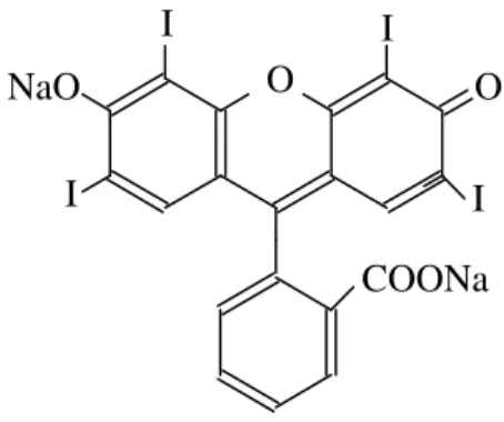
Figure 1 - The Structure of the Dye Erythrosin B.
RESULTS
The results this figures show in the process of biodegradation of the dye Erythrosin B, alterations in the molecular structure of this dye, as well as, new products generated for the biodegradation.
DISCUSSION
A molecule is not a rigid association of atoms. It can compare a molecule to a system of balls with variable masses, corresponding to the molecule atoms, and springs of different lengths, corresponding to the chemical links of the molecule. There are two fundamental modes of molecule vibration: stretching, in which the distance between the atoms increase or decrease, but the atoms are at same linking axe, deformation, in which the position of the atom changes relating to the original linking axe (Dyer, 1969).
Form the FTIR spectra carried out with
Neurospora crassa 74A, the control showed bands
at 578, 666, 1036, 1376, 1425, 1654, 2924 and 3418 cm-1 (Fig. 6), which was a characteristic of the fungus, also determined by Bhanoori and Venkateswerlu, (2000) and Jesus, et al.,( 2002). The analysis of biomass lecture in FTIR cultivated in diversified medium showed band at 2343 cm-1, witch was characteristic of the presence of CO2
was also confirmed by Orsini et. al. (2000) and Jesus (2002).
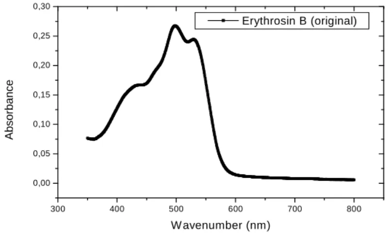
In the analyses of the Erythrosin B dye in UV-Vis spectrophotometer, in the original solution, the group kinolic showed peaks at 245 and 435 nm (Fig. 2 and 3). After 90 h the interaction the results showed that practically the bands were not more visible indicating a possible active site of an enzimatic modification (Fig. 4 and 5).
180 200 220 240 260 280 300 320 340 360 0,0
0,2 0,4 0,6 0,8 1,0 1,2 1,4 1,6 1,8
Erythrosin B (original)
A
bs
or
ba
nc
e
W avenumber (nm)
Figure 2 - Absorption spectra UV-Vis of the dye Erythrosin B original, at the range 180-350nm.
300 400 500 600 700 800
0,00 0,05 0,10 0,15 0,20 0,25 0,30
Erythrosin B (original)
A
b
s
o
rb
a
n
c
e
W avenumber (nm)
Figure 3 - Absorption spectra UV-Vis of the dye Erythrosin B original, at the range 350–800nm.
180 200 220 240 260 280 300 320 340 360
0,1 0,2 0,3 0,4 0,5 0,6 0,7 0,8 0,9
Erythrosin B (Biotreaty)
A
b
s
o
rb
a
n
c
e
300 400 500 600 700 800 0,02
0,04 0,06 0,08 0,10
0,12 Erythrosin B (Biotreaty)
A
b
s
o
rb
a
n
c
e
Wavenumber (nm)
Figure 5 - Absorption spectra UV-Vis of the dye Erythrosin B biotreaty (after 90 hours of interaction), at the range 300-800nm.
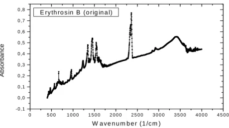
FTIR spectra of the natural cell wall of the fungus compared with the one that was in contact with the dye, indicated that the Erythrosin B caused a increase in the signals, mainly between the regions
400 and 900 cm-1 and 1100 and 1750 cm1 (Fig. 6, 8 and 9), and an increase at 2750 and 3.450 cm1 after 2 and 90-h interaction, was also informed by Galichet et al., (2001).
0 5 0 0 1 0 0 0 1 5 0 0 2 0 0 0 2 5 0 0 3 0 0 0 3 5 0 0 4 0 0 0 4 5 0 0 0 ,0 0
0 ,0 5 0 ,1 0 0 ,1 5 0 ,2 0 0 ,2 5
0 ,3 0 N e u ro s p o ra c ra s s a 7 4 A
A
b
s
o
rb
a
n
c
e
W a v e n u m b e r (1 /c m )
0 5 0 0 1 0 0 0 1 5 0 0 2 0 0 0 2 5 0 0 3 0 0 0 3 5 0 0 4 0 0 0 4 5 0 0 -0 ,1
0 ,0 0 ,1 0 ,2 0 ,3 0 ,4 0 ,5 0 ,6 0 ,7 0 ,8
E ry th ro s in B (o rig in a l)
A
b
s
o
rb
a
n
c
e
W a ve n u m b e r (1 /c m )
Figure 7 - Erythrosin B (1mg dry weight – after the mixture dye/KBr and subject to FTIR.
0 5 0 0 1 0 0 0 1 5 0 0 2 0 0 0 2 5 0 0 3 0 0 0 3 5 0 0 4 0 0 0 4 5 0 0 0 ,0 0
0 ,0 5 0 ,1 0 0 ,1 5 0 ,2 0 0 ,2 5 0 ,3 0 0 ,3 5
N . c ra s s a a n d E ry th ro s in B
(2 h o u rs )
A
b
s
o
rb
a
n
c
e
W a v e n u m b e r (1 /c m )
Figure 8 - FTIR cell walls spectra of N. crassa (1mg dry weight) and Erythrosin B mycelia after 2 hours of dye treatment were ground with KBr.
0 500 1000 1500 2000 2500 3000 3500 4000 4500 0,30
0,32 0,34 0,36 0,38 0,40 0,42 0,44 0,46 0,48 0,50
N. crassa and Erythrosin B (90 hours)
A
b
s
o
rb
a
n
c
e
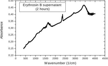
In the FTIR spectra, Erythrosin B showed that aromatics nucleus mono and di halogenics strongly absorve in this region from 1350 to 1120 cm –1 probably these sites are occupied by biodegradation of the iodum (Fig. 7), after 2 and
90 hours the bands decrease significantly in this region (Fig. 10 and 11) In the kinolic nucleous 1690-1635 cm –1, this fact also occurred, this modification confirms an adaptation period dye/cell wall (Kamnev et al., 1997).
0 500 1000 1500 2000 2500 3000 3500 4000 4500 0,10
0,15 0,20 0,25 0,30 0,35 0,40 0,45 0,50
Erythrosin B supernatant (2 hours)
A
b
s
o
rb
a
n
c
e
W avenumber (1/cm)
Figure 10 - FTIR spectra of Erythrosin B supernatant (1mg dry weight) after 2 hours of dye treatment were ground with KBr.
0 500 1000 1500 2000 2500 3000 3500 4000 4500
-0,05 0,00 0,05 0,10 0,15 0,20 0,25 0,30 0,35 0,40
Erythrosin B supernatant (90 hours)
A
b
s
o
rb
a
n
c
e
W avenum ber (1/cm )
Figure 11 - FTIR spectra of Erythrosin B supernatant (1mg dry weight) after 90 hours of dye treatment were ground with KBr.
CONCLUSIONS
Significant differences in the spectrum of UV-VIS were observed between the control and the supernatant. The FTIR spectra revealed modifications both in the cellular wall of N. crassa
and Erythrosin B, supernatant fraction. Significant differences in the FTIR spectra were observed
possibility of biodegradation, and this was in proportion to the contact time dye with the fungus biomass.
ACKNOWLEDGMENT
Capes, CNPq, Fapesp and Fundunesp.
RESUMO
O presente trabalho utilizou formas paramorfogênicas de Neurospora crassa 74A linhagens, na remoção do corante “Erythrosin B”. O fungo, induzido química e fisicamente em forma de “pellets”, foi usado no estudo da biodegradação deste corante. A cultura fúngica foi crescida em meio contendo o corante, como única fonte de carbono por 2 e 90h de interação. As paredes celulares foram isoladas por um processo de lavagem em água destilada e o micélio fresco foi secado por 12 h a 105ºC, e transformado num pó fino, e analisado em FTIR. O sobrenadante foi analisado através de espectrofotômetro UV-VIS e FTIR. Diferenças significativas no espectro UV-VIS e no FTIR foram observadas entre o controle e o sobrenadante e entre o controle e as paredes coloridas de vermelho e em FTIR no tempo de 2 e 90 h. Algumas bandas foram modificadas sugerindo a possibilidade de uma biodegradação enzimática em função do tempo de contato entre o corante e a biomassa fúngica.
REFERENCES
Azmi, W., Sani, R. J., Banerjee, U. C. (1998), Biodegradation of triphenylmryhane dyes. Enzyme
and Microbiol Technology, 22, 185-191.
Bhanoori, M. e Venkateswerlu, G. (2000), In vivo chitin-cadmium complexation in cell wall of
Neurospora crassa. Biochimica et Biophysica Acta,
1519, 21-28.
Borges, A.K.P.; Tauk-Tornisielo, S.M.; Domingos, R.N.; Angelis, D.F. de. (2008), Performance of the Constructed Wetland System for the Treatment of Water from the Corumbataí River. Brazilian Archives
biology and Technology, 51 (6), 1279-1286,
Chu, W. Dye Removal from Textile Dye (2001), Wastewater using recycled Alum Sludge. Water
Dayer, J.R. (1969), Aplicações da espectroscopia de absorção aos compostos orgânicos, São Paulo: Ed. Edgard Blücher LTDA, 155p.
Espíndola, L. H. S., Espíndola, F. S., Freitas, G. R., Brandeburgo, M. A. M. (2007), Biodegradation of Red 40 by the Mushroom Pleorotus sp florida. Biosci. J. 23 (3), 90-93.
Lefebvre, D. D., Chenaux, P., Edwards, M. (2005), Dye Degradation by fungi: An Exercise in Apllied Science for Biology Students. Bioscene, 31 (3), 13-16. Galechet, A., Sockalingum, G. D., Belarbi, A., Manfait,
M. (2001), FTIR spectroscopic analysis of
Saccharomyces cerevisiae cell wall: study of an
anomalous strain exihibiting a pink-colored cell phenotype. FEMS Microbiology Letters, 197, 179-186.
Jesus, G. J., Franchetti, S. M. M., Corso, C. R. (2002), Estudo da biodegradação de compostos azóicos de efluentes da indústria têxtil. Arquivos Instituto
Biológico, São Paulo, 69, 159-162,.
Kammev, A., A., Ristié, M., Antonyuk, L. P., Chernychev, A V., Ignatov, V. V. (1997), Fourier transform infrared spectroscop study of intact cells on the nitrogen-fixing bacterium Azozpirillum
brasiliense. Journal of Molecular Structure, 408-409,
201-205.
Kirby, N.; Marchant, R.; Mcmullan, G. (2000), “Decolourisation of synthetic textile dyes by Phlebia
tremellosa”Fems Microbiology Letters, 188, 93-96.
Marcanti-Contato, I., Corso, C. R., Oliveira, J.E. (1997), Induction of physical paramorphogenesis in
Aspergillus sp. Revista de Microbiologia , 28, 65-67.
Orsini, F.; Ami, D.; Villa, A. M.; Sala, G.; Bellotti, M.G.; Doglia, S. M. (2000), FT-IR microspectroscopy for microbiological studies.
Journal of Microbiological Methods, 42, 17-27.
Santos, I.R.; Costa, R.C.; Freitas, U.; Fillmann, G. (2008), Influence of effluents from a wastewater treatment plant on nutrient distribution in a coastal creek from southern Brazilian Archives Biology and
Technology,51 (1), 153-162.
Suresh, K. e Subramanyam, C. P. (1998), Polyphenols are involved in copper binding to cell walls of
Neurospora crassa. Journal of Inorganic
Biochemistry,69, 209-215.
Tatum, E. I.; Barratt, R. W.; Cutter, V .M.-Jr. (1949), “Chemical induction of colonial paramorphs in
Neurospora crassa and Syncephalastrum”. Science.
109, 509-511.
Wang, Y., YU, J. (1998), Adsorption and degradation of synthetic dyes on the mycelium of Trametes
versicolor. Water Science Technology