Pregnancy and lower limb varicose veins: prevalence and risk
factors
Gestação e varizes de membros inferiores: prevalência e fatores de risco
Newton de Barros Junior1, Maria Del Carmen Janeiro Perez2, Jorge Eduardo de Amorim2, Fausto Miranda Junior3
Abstract
Background: During and after pregnancy, lower limb varicose disease presents speciic features that have inluenced the conduction of studies designed to provide a better understanding of the condition. Such features include the appearance of lower limb varicose veins, their early development and intensity, and their rapid regression after delivery.
Objective: To assess the prevalence of lower limb varicose disease during pregnancy and to identify the main associated risk factors. Prevalence of varicose disease during pregnancy is high, afecting almost 70% of pregnant women considering all types of varicose disease. his high prevalence is mainly caused by the increase in the estrogen and progesterone levels during pregnancy.
Material and method: We analyzed 352 pregnant women during prenatal follow-up. he subjects were randomly selected during a 14-month period. Varicose disease was clinically identiied and classiied according to Widmer’s criteria: trunk varicose veins, reticular varicose veins, and telangiectasias; being reclassiied according to the criteria of the CEAP clinical classiication. he results of prevalence and risk factors were statistically analyzed using univariate and multivariate analyses.
Results: Considering all types of varicose veins, prevalence of varicose disease was 72.7% (256 pregnant women). Only 27.3% (96) of pregnant women did not have varicose disease (C0), and this group was considered the control group. After multivariate analysis, the main risk factors were: family history and pregnant women’s age.
Conclusion: he high prevalence of varicose disease and the associated risk factors suggest the need of providing the health professionals involved in women’s health care, especially during the fertile period, with information on this disease.
Keywords: Pregnancy, varicose veins, epidemiology, veins.
Resumo
Contexto: Durante e após a gestação, as varizes dos membros inferiores têm aspectos peculiares, tais como o seu aparecimento, a precocidade de seu desenvolvimento, a intensidade e, no puerpério, a rapidez com que regridem. Esses aspectos têm inluenciado os estudos para a compreensão dessa patologia.
Objetivo: Veriicar a prevalência das varizes dos membros inferiores em gestantes e os fatores de risco mais relevantes envolvidos. A prevalência na gestação é alta, atingindo cerca de 70%, quando se consideram todos os tipos de varizes. Essa alta prevalência decorre principalmente do aumento nas taxas dos estrógenos e progestágenos que ocorre durante a gravidez.
Material e método: Foram avaliadas 352 gestantes no período pré-natal, durante 14 meses, escolhidas ao acaso. A doença varicosa foi diagnosticada clinicamente e classiicada segundo os critérios de Widmer em varizes tronculares, reticulares e telangiectasias e reclassiicadas pela classiicação CEAP, segundo o critério clínico. Os resultados de prevalência e fatores de risco foram submetidos às análises univariada e multivariada.
Resultados: A prevalência da doença varicosa, quando considerados todos os tipos de varizes, foi de 72,7% (256 gestantes). As 96 gestantes (27,3%) que não apresentaram doença varicosa foram consideradas, para análise estatística, como controle. Os fatores de risco de signiicância, após análise multivariada, foram: antecedente familiar positivo e idade.
Conclusão: A prevalência da doença varicosa durante a gestação e os fatores de risco envolvidos indicam a necessidade de divulgação dessa patologia entre os proissionais envolvidos na prevenção e manutenção da saúde da mulher, especialmente aquelas em período fértil.
Palavras-chave: Varizes, gestação, epidemiologia, veias varicosas.
Study approved by the Research Ethics Committee of the Universidade Federal de São Paulo (UNIFESP), resolution n. 196, Sept 10 1996, on researches involving human beings, DOU 1996 Out 16, nº 201, section 1:21082-21085.
1 Doutor. Professor adjunto e Chefe, Disciplina de Cirurgia Vascular, Departamento de Cirurgia, Escola Paulista de Medicina (EPM), Universidade Federal de São Paulo (UNIFESP), São Paulo, SP. 2 Doutores. Professores adjuntos, Disciplina de Cirurgia Vascular, Departamento de Cirurgia, EPM, UNIFESP, São Paulo, SP.
3 Professor titular, Disciplina de Cirurgia Vascular, Departamento de Cirurgia, EPM, UNIFESP, São Paulo, SP
Introduction
Lower limbs varicose disease in pregnant women for decades have been drawing researchers’ attention. he appearance of varicose veins during pregnancy and its precocity, the intensity of its development, the uncommon symptoms and mainly the rapidity of regression ater puer-perium are peculiar aspects to lower limbs varicose disea-se during pregnancy which inluence the development of studies about the subject. he reversibility of this disease is the most typical phenomenon; they may decrease or vanish ater delivery. Around half of the world population carries lower limbs varicose disease, afecting 50-55% of women and 40-50% of men if minor forms of varicose disease (re-ticular varicose veins and telangiectasias) are considered. Considering larger and more visible varicose veins, the
di-sease afects less than 1/4 of the population, assailing 2-25% of women and 10-15% of men.1
Researchers have been observing the correlation be-tween pregnancy and varicose disease for a long time. he appearance of venous dilatations in lower limbs or in bre-asts of women in reproductive age is considered a sign of pregnancy, and some women attribute the appearance of varicose veins to pregnancy and its worsening to successive pregnancies.2
According to the literature (Table 1), the prevalence of varicose veins during pregnancy varies widely, due the use of diverse concepts, classiications and even the type of epidemiological analysis performed, in addition to regional and racial diferences. Many studies on this subject present only an estimative of the prevalence of varicose disease du-ring pregnancy (Table 1). his estimative varies from 20 to 50% of pregnant women and, when all the types of varicose veins are included, e.g. telangiectasias, the number may re-ach 70%.
We have found no epidemiological studies on varicose disease during pregnancy in Brazil, and many authors sim-ply repeat prevalence data presented in previous publica-tions when addressing the subject.
Material and method
Aiming at assessing the prevalence of varicose dise-ase during pregnancy we have conducted this study with
Authors Year Prevalence (%) Type of varicose disease
Bassi3 1967 10* trunk varicose veins
Boivin & Hutinel4 1987 30-40* trunk varicose veins
Griton et al.5 1987 63 all types
Valdevenito et al.6 1989 14.4 trunk varicose veins
Barile et al.7 1990 50-60* all types
Dindelli et al.8 1990 57.9 all types
Sciannameo et al.9 1993 50-60* all types
Table 1 - Prevalence of varicose disease during pregnancy, according to the authors
*data presented as estimative by the authors.
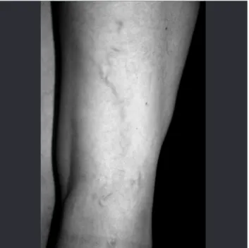
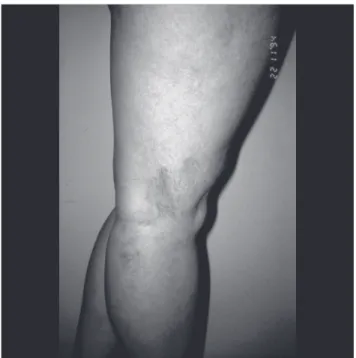
352 pregnant women, randomly selected, at the Pre-Natal Assistance Program of the Escola Paulista de Medicina, UNIFESP, in 1994. For evaluation the CEAP criterion was used,10 which classiies varicose disease in: a) varicose veins
– dilated subcutaneous vein with 3 mm or more of diame-ter, in supine position, possibly involving saphenous veins, its tributaries, or non saphenous supericial veins (Figure 1); b) reticular veins – dilated subdermic vein, 1-3 mm of diameter and tortuous (Figure 2); c) telangiectasias – con-luent intradermic venulae with less than 1 mm of diameter (Figure 3). Patients without visible or palpable signs of va-ricose disease were considered as CEAP C0, which formed the control group for statistical analysis.
Results
A high prevalence of varicose disease was observed in this sample in comparing data for presence and absence of varicose disease (Table 2).
In studying the 256 (72.7%) pregnant women carrying varicose disease, we observed that 72 (20.5%) presented varicose veins (CEAP C2) and 184 (52.2%) presented re-ticular veins and/or telangiectasias (CEAP C1). Ninety-six women from the sample (27.3%) did not present any type or varicosities (CEAP C0) (Table 3).
he prevalence of vulvar varicose veins in 14 pregnant women was also observed (4%) (Figure 4). We have no-ted that in all the cases there was an association with lo-wer limbs varicose disease, and in 12 patients (85.7%) this association was with severer forms (varicose veins) and in two (14.3%) the association was with reticular veins and telangiectasias.
Multivariate analysis of risk factors revealed that age, number of pregnancies and positive family history for vari-cose disease were associated with the presence of the disea-se. In multivariate analysis only two risk factors, age over 22 years and positive family history, were signiicant for vari-cose veins disease (CEAP C2) with odds ratio above 1. Ater this analysis, the number of pregnancies was no longer con-sidered a signiicant risk factor, although it characterized an association trend (Table 4).
Discussion
Varicose veins are classiied in two groups, according to their etiology: primary (essential) and secondary (post-thrombotic, due to congenital or acquired arteriovenous is-tulae). Etiopathogeny of primary varicose veins is still con-troversial, multiple and present unknown etiopathogenic
Varicose disease Number of patients %
CEAP C1 e C2 256 72.7
CEAP C0 96 27.3
Total 352 100.0
Table 2 – Prevalence of varicose disease in the 352 patients studied
Figure 3 - Telangiectasias (CEAP C1)
CEAP Classiication of varicose veins N %
C2 72 20.5
C1 184 52.2
C0 96 27.3
Total 352 100.0
Table 3 - Prevalence of varicose disease according to types of varicose veins
factors. In general population, the following etiopathoge-nic factors are highlighted: family predisposition, sex, age, number of pregnancies, endocrine alterations, obesity, pregnancy, habits and profession, congenital valve altera-tions and others. Nevertheless, several theories try to ex-plain the appearance or worsening of varicose disease du-ring pregnancy. hose theories are exposed below.
a) Mechanical theory – the oldest and the most widely known. Lower limbs varicose disease would be caused by mechanic compression exerted by the pregnant ute-rus on pelvic and iliac veins. Nowadays this mechanical concept was abandoned, because clinical evidence has shown that venous dilatations begin their development in the irst weeks of pregnancy, when the increase in uterine volume is still insigniicant. In case of fetal de-ath, venous dilatation rapidly and linearly recede, even before fetal expulsion; in twin pregnancies, the deve-lopment of varicose veins is big, but not as expected; venous dilatations are not limited to tributary veins of the inferior vena cava, observable in the arm, abdo-minal lank and breasts; uterine tumors of a similar or even higher volume than that of the pregnant uterus do not provoke the formation of varicose veins, neither an increase in pre-existing varicose veins’ intensity. here is evidence, however, of mechanic compression of the uterus on the iliac veins and inferior vena cava, especially in the last trimester of pregnancy.11,12 hese
compressions may possibly explain the etiopathogeny of vulvar varicose veins that frequently emerge in this period of pregnancy, as we could observe in this se-ries. hrough a duplex scanning, phlebography and even computed tomography, it was demonstrated that the speed of blood low in femoral veins progressively decreases, proportionally to the increase of the uterine volume, until diminishing in 50% in the third trimes-ter. In some cases, the uterus completely occludes the inferior vena cava with the patient in dorsal or right lateral decubitus.13
b) Hormonal theory – currently, the most widely
accept-ed.14,15 The most important piece of evidence
support-ing the hormonal theory in varicose veins development
was obtained in 1943 by McLennam.16 This author has
compared the measures of antecubital and femoral ve-nous pressure in pregnant women in dorsal decubitus and observed a progressive increase in femoral pres-sures, while antecubital venous pressures remained un-changed even in the initial stages of pregnancy, when the uterine volume was small and could lead to com-pression of the inferior vena cava or even of the iliac veins (Figure 4). These alterations in venous pressure would be caused by hormonal increase, both estrogenic and progestogenic. Indeed in the secretory phase of the menstrual cycle, progesterone rises from 30 mg/24h to 75 mg/24 h in the 20th week of pregnancy and peaks 250 mg/24h in the end of pregnancy, representing an 8 times increment. Estrogens also suffer a great increase, rising from 0.02 µg/24 in the proliferative phase of the menstrual cycle to 5.0 µg/24 h at the end of preg-nancy, representing an increase of up to 250 times.16
Progesterone increase results in hypotonia of smooth
muscle ibers and myocells (joint muscle framework of
the venous wall), reducing excitability, electric activity and increasing venous distensibility, which reaches up to 150%, returning to normal values in 8 to 12 weeks after delivery.17 On its turn, estrogenic secretion causes
an increase in arterial low in uterus and pelvis, and
this increment in the venous return low toward hypo
-gastric venous system would cause a functional obsta-cle in external iliac veins, transmitted to lower limbs veins. The classical theory of Piulachs et al.18 claimed
that the increase in progesterone and hypophysary hor-mones would result in a massive opening of arteriove-nous anastomoses, causing vearteriove-nous hypertension in the lower limbs. Some facts support this theory, because there is an atypical distribution of varicose veins in the lower limbs, ‘hyperoxygenation ‘ of venous blood and rapid contrastation of the venous network during arte-riographies.19,20 On the other hand, this theory would
not explain the appearance of varicose veins in only one side, a fact routinely observed in clinical practice. More recently, Boivin et al.21 have shown, through
du-plex scanning, the diameter’s increase in competent
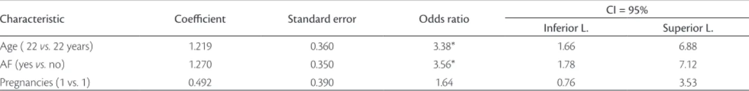
Characteristic Coeicient Standard error Odds ratio CI = 95%
Inferior L. Superior L.
Age ( 22 vs. 22 years) 1.219 0.360 3.38* 1.66 6.88
AF (yes vs. no) 1.270 0.350 3.56* 1.78 7.12
Pregnancies (1 vs. 1) 0.492 0.390 1.64 0.76 3.53
Table 4 - Logistic regression multivariate analysis for risk factors of lower limbs trunk varicose veins in the 352 patients
and incompetent veins, comparing the values in the irst
and third semesters of pregnancy and the decrease of these values after birth. When the diameters observed
in the irst trimester and in puerperium were compared,
no statistic difference was found, which shows that the veins had returned to initial values.
c) Increase in pelvic circulation – in pregnancy, there is an increase in uterine blood debit (500 ml/min of total blood low22), resulting in an addition to pelvic venous
pressure and venous engorgement of iliac veins and re-duction in draining capacity of lower extremities’ col-lecting veins.
d) Hereditary predisposition – for most authors it is a necessary and indispensable condition for varicose disease development,8,12,14,23 an issue disputed by some
authors. Ludwig,24 for instance, did not ind positive
fa-mily history in 56% of varicose patients, and Bertone et al.25 found only 35% positivity in 700 cases investigated.
Nevertheless, in a careful study, Cornu-henard et al.26
evaluated 134 patients – 67 with varicose disease and 67 normal – and their parents, concluding that family factor is of great importance in the genesis of varicose disease.
e) Increase in volemia – blood volume during pregnancy is increased in more than 30%; this occurs mainly due to plasma activity.27
f) Mesodermic deiciency – Arruda14 considers that
con-genital mesodermic deiciency is an important factor in etiopathogeny of essential varicose disease. he expres-sion of this deiciency would be the association, in the same patient, of lower limbs varicose veins with ingui-nal or muscle hernias, cutaneous stretch marks and lat feet, relatively common.
g) Structural alterations of the venous wall – there is a reduction of smooth muscle ibers of the venous wall and qualitative and quantitative alterations of the joint tissue in the wall of the varicose vein. Fibers are deformed and immersed in joint tissue, with collagen, reduced and disorderly disposed, with an excess of ‘proteoglycans.’ In addition, there is an increase in all the activities of lysosomal enzymes (hyaluronidases, glucosaminidases, and phosphatases).9 According to
Silveira,28 in our milieu varicose saphenous vein
pre-sents signiicant structural modiications in its wall, occurring, in addition to a greater intimal thickening, deep modiications in the structure of the tunica me-dia, with interposition of elastic ibers to smooth mus-cle clusters, consequently altering the resistance of the damaged venous wall.
h) Venous valve anatomic alterations – through agenesis or hypoplasia of the iliac-femoral valve, which supports the hydrostatic pressure of a blood column from the heart to the inguinal region. In 8% of the persons, this valve is not present bilaterally, and in 30% it is present only in one of the sides.29 Barile et al.7 referred that this
valve is inefective in 64% of varicose disease carriers. Agenesis or incompetence of these valves would occa-sion an increase in hydrostatic pressure of the saphe-nous-femoral ostium and consequent relux in the su-pericial system.
Risk factors
he most important risk factors for the development of varicose disease during pregnancy are:
1) Age – most authors agree that age is one of the main risk factors1,9,11,23,26,29. Widmer,30 in his Basle Study III,
observed that age is the most important risk factor, with a 6-10 times higher prevalence in 70-year-old persons than in 30-year-old persons. Mafei23 also observed an
increase in prevalence of varicose disease and chronic venous insuiciency with age, reaching 78.2% of exa-mined women older than 70 years. During pregnancy, there is a predominance of trunk varicose veins in age groups between 21 and 40 years old.31 In our study we
have observed that 65% of the 352 patients were betwe-en 20 and 29 years old, predominantly 20-24 (41.2%). When we performed the multivariate analysis, odds ra-tio was 3.38 times higher in the occurrence of trunk va-ricose veins in 23-year-old patients than in those who were 22 or younger (Table 4).
2) Number of pregnancies – another important risk fac-tor in the development of varicose disease in women is pregnancy.3,15,20,23,30,32-34 Basellini et al.35 have observed a
higher prevalence of varicose disease in patients who had undergone more than one pregnancy in compa-rison to nulliparae, in a 1:5 proportion, but have not observed a higher incidence with the increase in the number of pregnancies. Boivin & Hutinel4 have
refer-red that the prevalence of varicose veins in men and women could be classiied in two diferent orders: be-tween men and nulliparae a proportion of 1:1.2 was found, whereas between men and multiparae, it is 1:4.6. Dindelli et al.36, in a series of 611 women, have
analysis and prevalence of pregnant women with trunk varicose veins was signiicantly higher in secundiges-tae. In performing a multivariate analysis, with age cor-rection, this factor became non-signiicant, revealing that the age factor was more important than the num-ber of pregnancies in the prevalence of trunk varicose veins (Table 4). hese corrections and adjustments had already been highlighted by some authors that did not obtain signiicance ater age adjustments.32,33 he mean
number of pregnancies in this casuistic (2.4 per pa-tient) was lower than in other series, and this may have inluenced the results. Mafei,23 in his study, indicates
that there was a positive correlation between the pre-valence of varicose veins and number of pregnancies, even with age adjustment. Of 68 women with varicose disease, 66 (9.9%) were nulligestae, 44 (6.6%) were pri-migestae, 76 (11.4%) were secundigestae, and the other 482 (72.2%) had three or more pregnancies.
3) Family history – the importance of heredity in varico-se divarico-seavarico-se prevalence still prevarico-sents some controversial opinions, because some factors may inluence heredity analysis. Varicose disease is very frequent in the popu-lation, causing a high family positivity. Moreover, it is easier for persons carrying varicosities to remember relatives who have the disease than for those who do not carry it.1 While some authors have airmed they
had not observed any inluence of heredity in varico-se divarico-seavarico-se prevalence,11,36,37 others have found a higher
prevalence of varicose disease in persons with positive family history.5,10,24,38 Dindelli et al.36 have found a
re-lative risk 6.2 times higher of venous disease in preg-nant women with positive family history than in those who did not have a family history of the disease. In this study, only irst-degree relatives were considered, and the relation between venous disease and family history remained consistent, even ater age adjustment. In a ca-reful study evaluating men and women between 30 and 40 years old and their parents through physical exam, Cornu-henard et al.26 have reported that the risk of
developing varicose veins was 90% when both parents presented the disease and 25% for males and 62% for females when only one of the parents was afected. In patients whose parents did not present varicose veins, the risk of developing this disease reached 20%. In our study, in comparing pregnant women carrying varicose disease with positive family history with those who had no family history, we observed a signiicantly higher prevalence of pregnant women with varicose disease and family history, with an odds ratio 2.48 higher than
those with the disease but no family history. In multi-variate analysis for trunk varicose veins, we observed that the family history was the most important factor; pregnant women with family history of the disease have 3.56 times more chances of acquiring it than those who do not have it.
Conclusion
he high prevalence of varicose disease during preg-nancy, etiopathogeny and risk factors involved (mainly age and family history) in the development of this disease indi-cate the necessity of using efective prophylactic measures that should be indicated since the beginning of pregnancy and since the irst pregnancy, thus promoting the mainte-nance of the pregnant woman’s health and, consequently, of the newborn.
Acknowledgements
We would like to thank Dr. Neil Ferreira Novo and Yara Juliano for the analysis of statistic data.
References
1. Callam MJ. Epidemiology of varicose veins. Br J Surg. 1994;81:167-73.
2. Bowes K, Riterband SH, Andrews JE. Demonstration by infra-red photography of the supericial veins in the pregnant and nonpreg-nant woman. J Obstet Gynaecol Br Emp. 1948;55:285-92.
3. Bassi G. Les varices des membres inférieurs. 12ª ed. Paris: Doins; 1967.
4. Boivin P, Hutinel B. [Varices and pregnancy]. J Mal Vasc. 1987;12:218-21.
5. Griton P, Escalier-Imbert M, Cuit A. [Varicose disease. Epidemiologic study apropos of 1600 cases]. Phlebologie. 1987;40:923-9.
6. Valdevenito R, Silva C, Yañez N, et al. Estudio epidemiologico de pacientes operados de varices en el Hospital Sanatorio de Valparaiso. Bol Hosp San Juan de Dios. 1989;36:3-11.
7. Barile C, Merlo M, Buzzacchino A, Pegoraro M. [Physiopathology of varices during pregnancy]. Minerva Ginecol. 1990;42:117-21.
8. Dindelli M, Basellini A, Rabaiotti E, et al. [Epidemiological analysis of the incidence of varicose pathology in pregnancy]. Ann Ostet Ginecol Med Perinat. 1990;111:257-64.
9. Sciannameo F, Ronca P, Alberti D, Madami C. [Varicose veins in pregnancy: physiopathology and therapeutic approach]. Minerva Ginecol. 1993;45:539-43.
10. Eklöf B, Rutherford RB, Bergan JJ, et al. Revision of the CEAP classi-ication for chronic venous disorders: consensus statement. J Vasc Surg. 2004;40:1248-52.
12. Samuel E. he inferior vena cavogram in pregnancy. Radiological aspects. Proc R Soc Med. 1964;57:702-4.
13. Sumner DS. Venous dynamics: varicosities. Clin Obstet Gynecol. 1981;24:743-60.
14. Arruda S. Aspectos etiopatogênicos das varizes na gravidez. Rev Bras Cardiovasc. 1966;2:125-34.
15. Basellini A, Agus GB, Antonucci E, Papacharalambus D. [Varices in pregnancy (an up-date)]. Ann Ostet Ginecol Med Perinat. 1985;106:337-41.
16. McLennan CE. Antecubital and femoral venous pressure in normal and toxemic pregnancy. Am J Obstet Gynecol. 1943;45:568-91.
17. Rezende J, Linhares E. Endocrinologia do ciclo gravídico. In: Obstetrícia. 3ª ed. Rio de Janeiro: Guanabara Koogan; 1974. p. 116-29.
18. McCausland AM, Holmes F, Trotter Junior AD. Venous distensi-bility during the menstrual cycle. Am J Obstet Gynecol. 1963;86: 640-5.
19. Piulachs P, Vidal-Barraquer F, Biel JM. [Pathogenesis of varicose veins in pregnancy.]. Lyon Chir. 1952;47:236-78.
20. Charles-Edouard Otrante D, Zacca Pena E, Ariosa Coloma MC, Robaina Jorge F. [Relative risk in varicose veins and pregnancy]. Angiologia. 1980;32:66-9.
21. Grismondi GL. [Treatment of phlebopathies caused by stasis in pregnancy]. Minerva Ginecol. 1981;33:221-3.
22. Boivin P, Cornu-henard A, Charpak Y. Pregnancy-induced chan-ges in lower extremity supericial veins: an ultrasound scan study. J Vasc Surg. 2000;32:570-4
23. Nahoum JC, Barcellos JM. Placenta. Cordão umbilical. Sistema am-niótico. In: Rezende J. Obstetrícia. 3ª ed. Rio de Janeiro: Guanabara-Koogan; 1974. p. 44.
24. Mafei FHA, Magaldi C, Pinho SZ, et al. Varicose veins and chronic venous insuiciency in Brazil: prevalence among 1755 inhabitants of a country town. Int J Epidemiol. 1986;15:210-7.
25. Ludwig H. [Pregnancy varicoses]. Zentralbl Phlebol. 1964;126:22-8.
26. Bertone C. [he puerperal varicose syndrome]. Minerva Med. 1961;52:655-9.
27. Cornu-henard A, Boivin P, Baud JM, De Vicenzi I, Carpentier PH. Importance of the familial factor in varicose disease. Clinical stu-dy of 134 families. J Dermatol Surg Oncol. 1994;20:318-26
28. Rezende J, Coslovsky S. Repercussões na gravidez sobre o or-ganismo. In: Rezende J, editor. Obstetrícia. 3ª ed. Rio de Janeiro: Guanabara Koogan; 1974. p. 130-58.
29. Silveira PRM. Estudo estrutural da veia safena magna normal e varicosa. Rev Angiol Cir Vasc. 1993;2:116-33.
30. Beaglehole R. he epidemiology of venous disease. Phlebology. 1995;(Suppl 1):25-8.
31. Widmer LK. Peripheral venous disorders; prevalence and socio-medical importance; observations in 4529 apparently healthy persons; Basle Study III. Bern: Hans Huber Publishers; 1978.
32. Guerrini S, Marietta G, Ferreri G. [Varices and pregnancy. Statistical evaluation]. Minerva Ginecol. 1987;39:503-10.
33. Lake M, Pratt GH, Wright IS. Arteriosclerosis and varicose veins: occupational activities and others factors. J Am Med Assoc. 1942;119:696-701
34. Mullane DJ. Varicose veins in pregnancy. Am J Obstet Gynecol. 1952;63:620-8.
35. Donato VM, Nejamkim J. [Varices and pregnancy; their treat-ment]. Prensa Med Argent. 1956;43:551-7.
36. Dindelli M, Parazzini F, Basellini A, Rabaiotti E, Corsi G, Ferrari A. Risk Factors for varicose disease before and during pregnancy. Angiology 1993;44:361-7.
37. Drury M. Varicose veins in pregnancy. Br Med J. 1965;2:304.
38 Guberan E, Widmer LK, Glaus L, et al. Causative factors of va-ricose veins: myths and facts. An epidemiological study of 610 women. Vasa. 1973;2:115-20.
39. Burkitt DP. Varicose veins, deep vein thrombosis and he-morrhoids: epidemiology and suggested aetiology. Br Med J. 1972;2:556-61.
Correspondence:
Newton de Barros Júnior R. Coronel Lisboa, 690 CEP 04020-041 - São Paulo, SP, Brazil Tel.: (11) 5904.4429 Fax: (11) 5579.9814 E-mail: nbj032.dcir@epm.br
Author contributions