Thermally activated exchange narrowing of the Gd
3+ESR fine structure in a single crystal
of Ce1-
xGd
xFe4P12
(
x
≈
0
.
001) skutterudite
F. A. Garcia,1P. A. Venegas,2P. G. Pagliuso,1C. Rettori,1,3Z. Fisk,4P. Schlottmann,5and S. B. Oseroff6
1Instituto de F´ısica “Gleb Wataghin,” C.P. 6165, UNICAMP, Campinas-SP 13083-970, Brazil
2UNESP-Universidade Estadual Paulista, Departamento de F´ısica, Faculdade de Ciˆencias, C.P. 473, Bauru-SP 17033-360, Brazil
3Centro de Ciˆencias Naturais e Humanas, Universidade Federal do ABC, Santo Andre-SP 09210-170, Brazil
4University of California, Irvine, California 92697, USA
5Department of Physics, Florida State University, Tallahassee, Florida 32306, USA
6San Diego State University, San Diego, California 92182, USA
(Received 24 March 2011; revised manuscript received 12 July 2011; published 9 September 2011) We report electron spin resonance (ESR) measurements in the Gd3+doped semiconducting filled skutterudite compound Ce1-xGdxFe4P12 (x≈0.001). As the temperature T varies from T ≃ 150 K toT ≃ 165 K, the
Gd3+ ESR fine and hyperfine structures coalesce into a broad inhomogeneous single resonance. At T ≃ 200 K the line narrows and as T increases further, the resonance becomes homogeneous with a thermal broadening of 1.1(2) Oe/K. These results suggest that the origin of these features may be associated with a subtle interdependence of thermally activated mechanisms that combine: (i) an increase withT of the density of activated conduction carriers across theT-dependent semiconducting pseudogap; (ii) the Gd3+ Korringa relaxation process due to an exchange interactionJf dS.sbetween the Gd3+localized magnetic moments and the thermally activated conduction carriers; and (iii) a relatively weak confining potential of the rare earth ions inside the oversized (Fe2P3)4cage, which allows the rare earths to becomerattlerEinstein oscillators aboveT ≈
148 K. We argue that therattlingof the Gd3+ions, via a motional narrowing mechanism, also contributes to the coalescence of the ESR fine and hyperfine structure.
DOI:10.1103/PhysRevB.84.125116 PACS number(s): 71.27.+a, 75.20.Hr, 76.30.−v
I. INTRODUCTION
The filled skutterudite RT4X12 compounds, where R is a rare earth or actinide, T is a transition metal (Fe, Ru, Os), and X is a pnictogen (P, As, Sb) have attracted great attention due to their broad range of physical properties. In particular, they are of interest to those investigating basic mechanisms of strongly correlated electronic systems1–3 and also to those
seeking for more efficient thermoelectric materials.4,5
These compounds crystallize in the LaFe4P12structure with space group I m3 and local point symmetry Th for the R
ions. The R ions are guests in the oversized rigid (T2X3)4 cages.6The dynamics of the guest R ions is believed to be of great importance in the damping of the thermal conductivity observed in the filled skutterudite compounds.7,8 Moreover, they may also play an important role in the appearance of heavy fermion behavior and superconductivity.1,9
Electron spin resonance (ESR) is a sensitive and powerful microscopic tool that provides information about crystal field (CF) effects, site symmetries, valencies of paramagnetic ions, g values, and fine and hyperfine parameters.10 In a recent work our group11found ESR to be a sensitive and useful tool to study the dynamics of the R ions in this family of filled skutterudites. The weak confining potential on the R ions at the center of the oversized cage allows them to easily get off-center and experience a slightly different local strength and symmetry of the CF which may lead to (i) a distribution of the ESR parameters and (ii) arattlingof the R ions that, due to motional narrowing effects,12 may cause remarkable
changes in the observed ESR spectra. In our previous ESR experiments on Yb3+in Ce
1-xYbxFe4P12,11these two features were observed and the coexistence of two distinct Yb3+sites was confirmed.
Ogita et al.,13 performing Raman scattering experiments
on several metallic skutterudite compounds of the RT4X12 (T=Fe,Ru,Os; X=P,Sb) series, found resonant2nd order phonon modes associated with the vibrations that change the bond length of the R-X stretching mode. However, in semiconducting CeFe4P12 the 2nd order phonon modes were found to benonresonant. Based on their results Ogitaet al.13
concluded that there should be a strong coupling between the R-X stretching modes and the conduction electrons (ce). Most reports on the T dependence of the dc resistivity in CeFe4P12 present a semiconductorlike behavior.14 However, the resistivity is strongly sample dependent, and only in some cases it show metallic behavior belowT ≈ 200 K.15
Nevertheless, for most of the reported samples the conductivity due to thermally activated carriers predominates aboveT ≈ 200 K. Thus, for the semiconductor CeFe4P12 with a gap of≃0.15 eV and an estimated Debye temperature ofD ≃
500 K,16at least a weak coupling of the R-X stretching mode and the ce should be expected. This compound experiences a huge increase in the density of thermally activated conduction carriers atT ≈150 K.14 Also, evidences forrattlingof the Yb3+and Ce4+ions were found in ESR11and extended x-ray
absorption fine structure (EXAFS) experiments, respectively. The aim of this work is to learn if the presence of thermally activated conduction carriers andrattlingof the R ions can be observed by the ESR technique. For that reason we measured the evolution of the Gd3+ ESR spectra in Ce1-xGdxFe4P12 withT. To compare our data with a nonrattling compound, we have also studied the evolution of the Gd3+ESR spectra in
in Sn flux as described in Ref. 14. The cubic structure (I m3) and phase purity were checked by x-ray powder diffraction. Crystals of ∼2×2×2 mm3 of naturally grown crystallographic faces were used in the ESR experiments. Single crystals of Ca1-xGdxB6 (x0.001) were grown as described in Ref.18. The cubic structure (space group 221, Pm3m, CsCl type, and local point symmetry Td for the
R ions) and phase purity were checked by x-ray powder diffraction and the crystals orientation was determined by Laue x-ray diffraction. Most of the ESR experiments were done in∼2×1×0.5 mm3 single crystals. The ESR spectra were taken in a Bruker X-band (9.48 GHz) spectrometer using appropriated resonators coupled to a T controller of a helium gas flux system for 4.2T 300 K. The Gd concentrations were determined from theHandTdependence of the magnetizationM(H,T), measured in a Quantum Design superconducting quantum interference device (SQUID) dc magnetometer. In both systems the magnetic susceptibility follows a Curie-Weiss behavior. Also, in both compounds the T dependence of the Gd3+ ESR intensity presents a
Curie-Weiss-like behavior within the accuracy of the experiments.
III. RESULTS AND DISCUSSION
In both compounds at lowT the Gd3+ESR spectra show the full resolved fine structure corresponding to the spin Hamiltonian for the Zeeman and cubic CF interactions,H= gβH S+b4O4+b6O6.10 The angular andT dependence of the spectra were taken mostly with the applied magnetic fieldH in the (1,−1,0) plane. The fitting of the data to the spin Hamiltonian shows that the parameters are, within the experimental accuracy,T independent for the entire studied T range. The measured parameters were g =1.986(3) and b4 = 7(1) Oe for Ce1-xGdxFe4P12 and g = 1.992(3) and b4=13.8(5) Oe for Ca1-xGdxB6, in agreement with previous low-T reports.19,20 The accuracy of the data was not enough to estimate the value of b6. Theg shift measured for Gd3+ in Ce1-xGdxFe4P12 is negative: g=1.986(3)−1.993≈ −0.007. An additional term to the spin Hamiltonian,Jf dS.s,
due to a covalent exchange hybridization between the Gd3+
4f electrons and ce withd character would be responsible for this negative g shift.21 For the Ce1-xGdxFe4P12 crystal, careful measurements of the spectra were taken fromT ≃150 toT ≃200 K for various directions ofH. In thisT interval, the fine structure coalesces into a single broad line and its line shape changes from Lorentzian (insulator) to Dysonian (metallic).22Notice that these features are independent of the field orientation and none of them is observed in Ca1-xGdxB6.
(b)
FIG. 1.T dependence of theX-band ESR spectra forH[001]: (a) Ce1-xGdxFe4P12and (b) Ca1-xGdxB6.
Figures1–3display the evolution withT(4.2T 300 K) of the normalized ESR spectra of Gd3+in the Ce
1-xGdxFe4P12 and Ca1-xGdxB6 (x 0.001) crystals forH in the (1,−1,0) plane along [001], 30◦ from [001], and [110], respectively.
These data show that forT 150 K theT dependence of the Gd3+fine structure is quite different in both compounds.
For Ce1-xGdxFe4P12 the central transition (12 ↔ −12) at 4.2 K andH along [001] is narrow enough to observe the hyperfine satellites lines of the isotopes 155,157Gd3+ (I =
3/2)(see Fig. 4). The measured hyperfine parameter is A = 5.5(2) Oe.23This hyperfine structure is also observed, although
not so clearly, for the other transitions in the spectrum. For the angle where the fine structure collapses (29.6◦ from [001])
and the various transitions overlap, a small misorientation of H by 2◦ away from this direction affects the overall line
shape, and the hyperfine structure is then strongly blurred (see Fig.2). For Ca1-xGdxB6Fig.4shows that, due to its higherg value and broader linewidth, the (12 ↔ −12) transition is shifted to lowerH and the hyperfine structure is not well resolved. However, the hyperfine parameter can still be estimated to be A≃7(1) Oe.
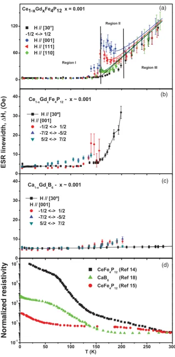
Figure5presents for both compounds the evolution withT (4.2T 300 K) of the linewidth (H) for the various Gd3+
ESR transitions at severalHorientations. For Ce1-xGdxFe4P12 Fig.5(a)shows theT dependence ofH for the (12 ↔ −12) transition andHalong [001], [111], and [110] directions and at∼30◦ from [001] in the (1,−1,0) plane for the collapsed
(a)
(b)
FIG. 2.T dependence of theX-band ESR spectra forH along
θ≈30◦ from [001] in the (1,−1,0) plane: (a) Ce
1-xGdxFe4P12and
(b) Ca1-xGdxB6.
H is nearly T independent and very narrow at ≃5(1) Oe; Region II, for 165 T 200 K, where the full fine structure dramatically coalesces into a broad inhomogeneous
(a)
(b)
FIG. 3.T dependence of theX-band ESR spectra forH [110]: (a) Ce1-xGdxFe4P12and (b) Ca1-xGdxB6.
FIG. 4. (Color online) X-band ESR (12 ↔ −12) transition for
H [001]. The arrows show the satellite hyperfine structure for both samples ofx=0.001.
single resonance with anisotropicH; and Region III, for T 200 K, whereH is again isotropic and homogeneous. It corresponds to a single coalesced resonance and has a linear thermal broadening of ≃1.1(2) Oe/K, reminiscent of a Korringa-like relaxation process via the ce.24
Figures5(b)and5(c)show theT dependence ofH for the various transitions in Ce1-xGdxFe4P12 and Ca1-xGdxB6 forH along [001] and 30◦ from [001], respectively. A timid
broadening starts to be observed onHfor the fine structure components atT ≃60 K for Ce1-xGdxFe4P12and atT ≃120 K for Ca1-xGdxB6. Presumably this broadening is caused by a phonon spin-lattice relaxation process.25 The fact that such a
phonon contribution starts at lowerT in Ce1-xGdxFe4P12than in Ca1-xGdxB6is consistent with the lower Debye temperature for the former compound. Alternatively, the Gd3+ions produce
bound states in the gap, which in the case of CaB6 are donor states. Carriers bounded at low T in these states can be promoted into the conduction band asT increases and produce a faster relaxation. However, asT increases in Region I a small local distribution of the CF cannot be excluded as the reason for the small broadening of the fine structure lines. The large voided space and concomitant increase of the carrier density as T increases may thermally activate slow motions of the Gd3+
ions inside the oversized (Fe2P3)4 cage which could slightly alter, in an inhomogeneous way, the local CF at the Gd3+site.
As already mentioned, for Ce1-xGdxFe4P12 above T ≃ 160 K a dramatic broadening mechanism drives the whole Gd3+ resolved ESR fine structure in Region I to coalesce
into the broad inhomogeneous and unresolved anisotropic spectrum of Region II [see Fig.5(a)]. This striking result occurs at about the sameT where (i) the density of thermally activated mobile carriers increases by several orders of magnitude [see Fig. 5(d)], (ii) the rattling of the filler R atom is confirmed by EXAFS experiments,16 (iii) the existence of a
T dependent semiconducting pseudogap is observed forT
300 K in ultraviolet and x-ray photoemission spectroscopies (UPS, XPS),26 and (iv) where the change from Lorentzian
(c)
(d)
Normaliz
e
d resistivity
FIG. 5. (Color online)T evolution (4.2T 300 K) of the Gd3+ESR linewidthHfor both compounds and various transitions at several H orientations. Notice that in Fig.5(a)the behavior of
H(T) clearly characterizes three different regions (I–III). The solid lines in Regions II and III correspond to the calculatedH(T) for the coalesced ESR spectra using the Plefka-Barnes27exchange narrowing
mechanism (see text). Figure5(d)presents the generalT dependence reported for the resistivity in these compounds. The resistivity of our crystals is similar to that of Ref.14.
ESR spectra using the Plefka-Barnes27 exchange (Jf dS.s)
narrowing theory of the fine structure. In the calculation we used a Korringa relaxation of 1.1(2) Oe K that is“switched-on” at 157(2) K, a fourth-order CF parameterb4=7(1) Oe, and a residual linewidthH(T =0)=5(1) Oe.
Figure 6 presents the T dependence of the hyperfine structure for the (12 ↔ −12) transition. The data show that the
FIG. 6. (Color online)T dependence of the hyperfine structure for the (1
2 ↔ − 1
2) transition. Notice that the apparent difference in
the field for resonance between the low- and high-Tspectra is mainly due to the change from Lorentzian to Dysonian, and also to a small change in the frequency of the microwave cavity due to the temperature (0.05%).
coalescence of the hyperfine structure is already observed at T ≈150 K, i.e., at≈15 K below the coalescence of the fine structure atT ≈165 K. This is expected since the exchange interaction would act first on the hyperfine structure due to its much smaller spectral splitting.
Figure7shows the angular dependence ofHat different T corresponding to Regions II and III. Following the analysis of Urbanet al.28
for the exchange narrowing of the Gd3+ESR fine structure, the anisotropy ofHin Region II can be fitted to the general expression for the intermediate coupling regime:
H =A(T)+B(T)p2(θ), (1)
where
p2(θ)=1−5[sin2(θ)+(3/4) sin4(θ)]. (2)
Figure 5(a) shows that there is narrowing ofH for T approaching 200 K and that the anisotropy decreases, i.e.,B → 0 asT →200 K. ForT 200 K,Hbecomes isotropic (see Fig.7) and increases linearly at a rate of 1.1(2) Oe/K [see Fig.5(a)]. This linear increase is an evidence for the presence of a Korringa relaxation process, i.e., the Gd3+ions relax to
the lattice via an exchange interaction Jf dS.s between the
FIG. 7. (Color online) Angular dependence of theH for the Gd3+ ESR for temperatures in the Regions II and III of Fig.5(a). The solid line corresponds to the fitting of the data to Eq. (1) for
FIG. 8. (Color online) Dependence of the Gd3+ESR intensity at lowT on the microwave power for both materials.
Gd3+localized magnetic moment and the thermally activated conduction carriers.24
Agshift ofg= −0.007 has been measured for the entire range of T studied for Ce1-xGdxFe4P12. This is surprising because in Region I there are no conduction carriers that could be polarized. However, the host is a Kondo insulator with a finite Van Vleck susceptibility due to the crystalline field splitting of the Ce ions. This Van Vleck susceptibility is larger than the susceptibility of the thermally excited electrons in Regions II and III and provides the polarization to produce the gshift. This effect is of course not present in the Ca1-xGdxB6 sample, since CaB6has no significant susceptibility. There is a second unusual issue with thegshift. In a simple metallic host Gd3+ ions are expected to have a ferromagnetic Heisenberg
exchange. However, the g shift is negative, indicative of a hybridization mechanism. The overlap of the Gd 4f electrons with the hybridized Ce 4f band forming the valence and conduction bands of the Kondo insulator could give rise to an antiferromagnetic exchange.
It is interesting to note that in Gd3+ doped simple metals
the Korringa relaxationd(H)/dT would be related to theg shiftgby29
d(H)/dT =(π k/gμB)(g)2. (3)
Using our experimental value of 1.1(2) Oe/K ford(H)/dT and 2.34×10⁴Oe/K forπ k/gμ
B, we estimate a
correspond-ing g of ≈|0.007|. These results and Eq. (3) suggest that (i) in Region I, where there is no Korringa relaxation, the exchange coupling due to covalent hybridization gives rise to just polarization effects,Jf d(q=0);30 (ii) the trigger of the
Korringa mechanism in Regions II and III is due to the presence of mobile activated conduction carriers at the Fermi level, which are responsible for the momentum transfer between the conduction carriers and the localized magnetic moment via the exchange couplingJf d(q =0);29,30and (iii) in the metallic
Regions II and III, there is noqdependence of the exchange interaction, i.e.,Jf d(q=0)≡ Jf d2 (q)
1/2
EF.
29,30
ForT 10 K Fig.8shows that, due to the long spin-lattice relaxation time T1 in these materials, the Gd3+ (12 ↔ −12) transition saturates as a function of the microwave power.31
From H = (γ T2)−1 the spin-spin relaxation time can be estimated to be T2 ≃0.1μs in both compounds. From the
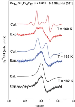
FIG. 9. (Color online) Experimental and calculated27Gd3+ESR spectra in Ce1-xGdxFe4P12forx=0.001 at the transition from Region
I to Region II. The parameters used to calculate the theoretical spectra were the same as those used for the calculation ofH(T) in Fig.5(a).
saturation factor,S=[1+(1/4)H12γ 2T
1T2]−1 and microwave powerP =(1/4)H12, we estimate the spin-lattice relaxation time to beT1≃10 ms andT1 ≃4 ms for Ce1-xGdxFe4P12and Ca1-xGdxB6, respectively. Notice that a Korringa relaxation is absent in Region I where the compound behaves as an insulator. Moreover, fromH atT ≃300 K in Fig.5(a)we estimateT1≃0.002μs which is much shorter than the lowT value ofT2. Therefore, at highT , T2≃T1and, as far as ESR is concerned, this is another evidence that Ce1-xGdxFe4P12 behaves as a regular metal in Regions II and III.
Finally, according to the Raman results and conclusions about the rattling modes in metallic skutterudites,13 it is plausible that the huge increase in the metallic character of CeFe4P12 activates the R-X stretching mode and triggers the rattling of the R ions inside the oversized (Fe2P3)4 cage. Hence, via a motional narrowing mechanism12 the rattling
Region II and then, in Region III, to a homogeneous linewidth with a broadening which is linear in T, resembling the Korringa-like relaxation process in a metallic host.24Point (b)
indicates that, at our microwave frequency and betweenT ≃ 150 andT ≃165 K, there is also a clear and strong change in the ac conductivity of the material. We associate this change to asmooth crossoverfrom insulator to metal which was only possible to be detected due to the high sensitivity that the ESR line shape has in a metallic media.
Our ESR observations in Ce1-xGdxFe4P12, along with those of Raman, EXAFS, UPS, and XPS for CeFe4P12, suggest
filled skutterudite compounds. In particular, our work supports the idea that some metallic character is always needed to set up the necessary conditions for therattlingof the R ions in these materials.
ACKNOWLEDGMENTS
This work was supported in part by FAPESP, CNPq, CAPES, and NCC from Brazil. P.S. is supported by the US Department of Energy through Grant No. DE-FG02-98ER45707.
1T. Goto, Y. Nemoto, K. Sakai, T. Yamaguchi, M. Akatsu,
T. Yanagisawa, H. Hazama, K. Onuki, H. Sugawara, and H. Sato,
Phys. Rev. B69, 180511(R) (2004).
2E. D. Bauer, A. Slebarski, E. J. Freeman, C. Sirvent, and M. B.
Maple,J. Phys. Condens. Matter13, 4495 (2001).
3N. R. Dilley, E. J. Freeman, E. D. Bauer, and M. B. Maple,Phys.
Rev. B58, 6287 (1998).
4G. J. Snyder and E. S. Toberer,Nat. Mater.7, 105 (2008). 5B. C. Sales, D. Mandrus, and R. K. Williams,Science272, 1325
(1996).
6W. Jeitschko and D. Braun,Acta Crystallogr. Sect. A33, 3401
(1977).
7C. H. Lee, I. Hase, H. Sugawara, H. Yoshizawa, and H. Sato,
J. Phys. Soc. Jpn.75, 123602 (2006).
8R. P. Hermann, R. Jin, W. Schweika, F. Grandjean, D. Mandrus,
B. C. Sales, and G. J. Long, Phys. Rev. Lett. 90, 135505 (2003).
9T. Yanagisawa, P-C. Ho, W. M. Yuhasz, M. B. Maple, Y. Yasumoto,
H. Watanabe, Y. Nemoto, and T. Goto,J. Phys. Soc. Jpn.77, 074607 (2008).
10A. Abragam and B. Bleaney,EPR of Transition Ions(Clarendon,
Oxford, 1970).
11F. A. Garcia, D. J. Garcia, M. A. Avila, J. M. Vargas, P. G. Pagliuso,
C. Rettori, M. C. G. Passeggi Jr., S. B. Oseroff, P. Schlottmann, B. Alascio, and Z. Fisk,Phys. Rev. B80, 052401 (2009).
12P. W. Anderson,J. Phys. Soc. Jpn.9, 816 (1954).
13N. Ogita, R. Kojima, T. Haegawa, Y. Takasu, M. Udagawa,
T. Kondo, N. Narazu, T. Takabatake, N. Takeda, Y. Ishikawa, H. Sugawara, T. Ikeno, D. Kikuchi, H. Sato, C. Sekine, and I. Shirotani, J. Phys. Soc. Jpn. Suppl. A77, 251 (2008).
14G. P. Meisner, M. S. Torikachvili, K. N. Yang, M. B. Maple, and
R. P. Guertin,J. Appl. Phys.57, 3073 (1985).
15H. Sato, Y. Abe, H. Okada, T. D. Matsuda, K. Abe, H. Sugawara,
and Y. Aoki,Phys. Rev. B62, 15125 (2000).
16D. Cao, F. Bridges, P. Chesler, S. Bushart, E. D. Bauer, and M. B.
Maple,Phys. Rev. B70, 094109 (2004).
17P. Vonlanthen, E. Felder, L. Degiorgi, H. R. Ott, D. P. Young, A. D.
Bianchi, and Z. Fisk,Phys. Rev. B62, 10076 (2000).
18D. P. Young, D. Hall, M. E. Torelli, J. L. Sarrao, Z. Fisk, J. D.
Thompson, H. R. Ott, S. B. Oseroff, R. G. Goodrich, and R. Zysler,
Nature (London)397, 412 (1999).
19R. N. de Mesquita, G. E. Barberis, C. Rettori, M. S.
Torikachvili, and M. B. Maple, Solid State Commun. 74, 1047 (1990).
20R. R. Urbano, C. Rettori, G. E. Barberis, M. Torelli, A. Bianchi,
Z. Fisk, P. G. Pagliuso, A. Malinowski, M. F. Hundley, J. L. Sarrao, and S. B. Oseroff, Phys. Rev. B 65, 180407(R) (2002).
21G. E. Barberis, D. Davidov, J. P. Donoso, C. Rettori, J. F. Suassuna,
and H. D. Dokter,Phys. Rev. B19, 5495 (1979).
22G. Feher and A. F. Kip, Phys. Rev. 98, 337 (1955); F. J.
Dyson, ibid. 98, 349 (1955); G. E. Pake and E. M. Purcell, ibid. 74, 1184 (1948);N. Bloembergen,J. Appl. Phys. 23, 1383 (1952).
23F. A. Garcia, J. G. S. Duque, P. G. Pagliuso, C. Rettori, Z. Fisk, and
S. B. Oseroff,Phys. Status Solidi B247, 647 (2010).
24J. Korringa,Physica16, 601 (1950); H. Hasegawa, Prog. Theor.
Phys. (Kyoto)21, 1093 (1959).
25R. Orbach,Proc. R. Soc. London A264, 458 (1961).
26P. A. Rayjada, A. Chainani, M. Matsunami, M. Taguchi, S. Tsuda,
T. Yokoya, S. Shin, H. Sugawara, and H. Sato,J. Phys. Condens.
Matter22, 095502 (2010).
27T. Plefka,Phys. Status Solidi B51, K113 (1972); 55, 129 (1973);
28P. Urban, D. Davidov, B. Elschner, T. Plefka, and G. Sperlich,Phys.
Rev. B12, 72 (1975).
29C. Rettori, S. B. Oseroff, D. Rao, P. G. Pagliuso, G. E. Barberis,
J. L. Sarrao, Z. Fisk, and M. F. Hundley,Phys. Rev. B55, 1016 (1997).
30D. Davidov, K. Maki, R. Orbach, C. Rettori, and E. P. Chock,Solid
State Commun.12, 621 (1973).
31Charles P. Poole Jr.,Electron Spin Resonance: A Comprehensive

![FIG. 2. T dependence of the X-band ESR spectra for H along θ ≈ 30 ◦ from [001] in the (1,−1,0) plane: (a) Ce 1-x Gd x Fe 4 P 12 and (b) Ca 1-x Gd x B 6 .](https://thumb-eu.123doks.com/thumbv2/123dok_br/16345641.189201/3.911.117.399.100.521/fig-dependence-band-esr-spectra-plane-ce-gd.webp)