during infection. Importantly,ZNF2overexpression abolishes fungal virulence in murine models of cryptococcosis. Thus, Znf2 bridges the sex-independent morphotype transition and fungal pathogenicity. The impacts of Znf2 on morphological switch and pathogenicity are at least partly mediated through its effects on cell adhesion property. Cfl1, a Znf2 downstream factor, regulates morphogenesis, cell adhesion, biofilm formation, and virulence. Cfl1 is the first adhesin discovered in the phylum Basidiomycota of the Kingdom Fungi. Together with previous findings in other eukaryotic pathogens, our findings support a convergent evolution of plasticity in morphology and its impact on cell adhesion as a critical adaptive trait for pathogenesis.
Citation:Wang L, Zhai B, Lin X (2012) The Link between Morphotype Transition and Virulence inCryptococcus neoformans. PLoS Pathog 8(6): e1002765. doi:10.1371/journal.ppat.1002765
Editor:Robin Charles May, University of Birmingham, United Kingdom
ReceivedJanuary 26, 2012;AcceptedMay 7, 2012;PublishedJune 21, 2012
Copyright:ß2012 Wang et al. This is an open-access article distributed under the terms of the Creative Commons Attribution License, which permits unrestricted use, distribution, and reproduction in any medium, provided the original author and source are credited.
Funding:We gratefully acknowledge financial support from the American Heart Association (grant 0BGIA3740040 to XL), Norman Hackerman Advanced Research Program (grant 01957 to XL), the National Institute of Allergy and Infectious Diseases (grant R01AI097599 to XL), and the Department of Biology of Texas A&M University (startup fund to XL). The funders had no role in study design, data collection and analysis, decision to publish, or preparation of the manuscript.
Competing Interests:The authors have declared that no competing interests exist.
* E-mail: [email protected]
Introduction
Adaptation to the host environment by many eukaryotic pathogens is often companied by transition in cellular morphology [1,2,3,4,5,6,7,8,9]. The ubiquitous fungal pathogen Cryptococcus neoformanscauses more than half a million deaths each year [10]. It can grow in the yeast form as well as the filamentous form. Earlier pre-genetic studies indicate an inverse relationship between filamentation and virulence [11,12,13,14,15,16,17]. These studies also point to the potential of filament-specific antigens as vaccines againstCryptococcusinfections [18,19,20].
BecauseCryptococcus typically grows in the yeast form and the morphological transition from the yeast form to the filamentous form appears to be coupled with mating, signaling pathways that lead to bisexual mating (a-amating) and unisexual mating (mostly a-amating) have been intensively investigated [21,22,23,24]. The roles of these signaling components in fungal pathogenicity are also scrutinized in animal models. However, accumulating evidence indicates that key signaling components that specifically lead to mating, such as those in the pheromone sensing pathway, have no or minimal direct effect on virulence [25,26,27,28]. Furthermore, conditions relevant to host physiology (e.g. aqueous environment, high temperatures, and high levels of CO2) are mating-suppressive, suggesting sex-independent mechanisms in orchestrating morphotype and virulence in Cryptococcus [29].
Therefore, the existence and the nature of the link between morphological transition and virulence in Cryptococcus remain enigmatic.
Results
Activation of Pheromone Signaling Is Insufficient to Drive Filamentation under Mating-Suppressing Conditions
Although Cryptococcus morphological transition from the yeast form to the filamentous form is historically associated with mating, the observations that filamentation can be achieved in strains in the absence of key pheromone signaling components or meiotic genes [30,31,32,33], lead us to hypothesize that pheromone signaling pathways are not essential or sufficient for filamentation per se, but they are critical in stimulating filamentation in response to mating cues. To test this hypothesis, we decided to examine the effect of constitutive activation of the pheromone signaling circuit on morphogenesis under mating-inducing and mating-suppressing conditions.
It is known that the expression of genes in the pheromone signaling pathway, such as those encoding the pheromone Mf1a,
the pheromone receptor Ste3a, the pheromone transporter Ste6,
shown) [30,32]. We found that the expression level of these pheromone signaling genes in wildtypeastrain H99 alone was low
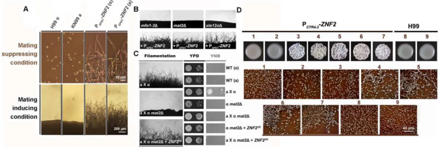
when cells were cultured on either mating-inducing condition (V8 agar) or mating-suppressing conditions (YPD agar and serum) (Figure 1B, C, D, E and F). This is consistent with the well-noted poor ability of the H99 strain to undergo unisexual mating. In fact, filamentation has never been observed when H99 was cultured alone under mating-inducing conditions (Figure 2A) [34]. We chose this strain to study the link between morphogenesis and virulence because H99 is one of the most virulent clinical strains tested in various animal models and it is also widely used as a reference strain inCryptococcusresearch.
When we placed the MAT2 gene under the control of the constitutively active promoter of GPD1 (glycerol-3-phosphate dehydrogenase 1) and introduced this construct to H99, the transcript level ofMAT2was dramatically increased under mating-inducing as well as mating-suppressing conditions (Figure 1C). As expected for a key regulator of the pheromone signaling, overexpression ofMAT2led to high expression ofMF1a,STE3a, and STE6 under both mating-inducing and mating-suppressing conditions (Figure 1D, E and F). This result indicates that constitutively overexpression of MAT2 is sufficient to induce pheromone signaling circuit independent of mating cues.
We next tested the effect of activation of pheromone signaling on filamentation under different conditions. The PGPD1-MAT2 conferred filamentation to H99 during unisexual mating (a cells
alone) and it significantly enhanced filamentation during bisexual mating (a-a coculture) under the mating-inducing condition
(Figure 2A). However, under mating-suppressing conditions, overexpression of MAT2failed to stimulate filamentation in the
aalone culture or in thea-acoculture (Figure 2A), and this was not due to insufficient activation of pheromone signaling. Effective activation of pheromone signaling in the PGPD1-MAT2 strain is supported by both the high expression levels of genes involved in pheromone signaling (Figure 1D, 1E, and 1F) and the formation of shmoo-like cells under both mating-inducing and mating-sup-pressing conditions (Figure 2B). Shmoo-like cells are typically observed when cells respond to mating signals prior to cell fusion. These observations indicate that activation of pheromone signal-ing alone is not sufficient to initiate filamentation under matsignal-ing- mating-suppressive conditions, including conditions relevant to host physiology. Thus mating signaling is unable to coordinate the yeast-filament morphological transition and virulence during infections.
Filamentation Can Be Independent of Sex and Is Controlled by the Transcription Factor Znf2
We previously showed that the deletion ofZNF2, which encodes a zinc-finger transcription factor, locked cells in the yeast form during mating without impairing pheromone signaling [28]. This suggests that Znf2 is not essential for mating signal relay; rather, it is crucial for filamentation. Although Znf2 functions downstream of Mat2 during mating [28] and its gene expression was significantly induced byMAT2overexpression under the mating-inducing condition (Figure 2C and 2D), activation of the pheromone signaling pathway was unable to induce ZNF2 expression in the absence of mating stimuli. This was evidenced by the low expression level of ZNF2in the PGPD1-MAT2 strain under mating-suppressing conditions (Figure 2C and 2D). The ability ofCryptococcusto undergo filamentation correlates with the expression level of ZNF2, but not that of MAT2 (Figure 1C, Figure 2A, 2C and 2D). Thus, Znf2 could be a master regulator that dictatesCryptococcusmorphotype irrespective of environmental stimuli or mating type.
To test this hypothesis, we constructed the PGPD1-ZNF2strains. Indeed, the PGPD1-ZNF2 triggered filamentation in Cryptococcus strains of either mating types a or a in both serotype A and serotype D backgrounds under all tested conditions, including those that are inducing or suppressive to mating (Figure 3A and Figure S1). In contrast to the PGPD1-MAT2 strain, filaments produced by the PGPD1-ZNF2 strain under mating-inducing condition maintained their filamentous morphology after being transferred to mating-suppressive conditions (Figure S2). However, it is notable that the PGPD1-ZNF2 strain produces more robust hyphae under mating-inducing condition, suggesting that other factors induced under mating-inducing condition could further activate Znf2.
The PGPD1-ZNF2also conferred filamentation to mutants that harbor deletions in the key mating components under various conditions tested (Mfa1-3, Mat2, or Ste12 functioning in a
branching pathway in pheromone signaling) (Figure 3B). To confirm that filamentation conferred by Znf2 activation is not due to some cryptic restoration of mating ability, we measured the efficiency of cell fusion of the wildtype, themat2Dmutant, and the
mat2D+PGPD1-ZNF2 strain during bisexual a-a mating. Indeed,
overexpression ofZNF2did not rescue the cell fusion defects of the mat2Dmutant (Figure 3C). Consistently, gene ontology analyses of our previous transcription data indicate that Znf2, unlike Mat2, does not regulate genes involved in the cell fusion event critical for mating (Figure S3) [28]. Taken together, the results indicate that filamentation can be independent of mating and Znf2 is one key determinant of this sex-independent morphogenesis.
Author Summary
Expression Level of Znf2 Mediates Bi-directional Morphological Transition
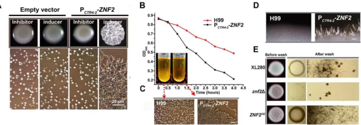
To verify the correlation ofZNF2 expression andCryptococcus morphology, we constructed the ZNF2 gene driven by two inducible promoters: the galactose-inducible GAL10 promoter (data not shown) [35] or the copper transporterCTR4promoter (Figure 3D) (copper deprivation–on; copper repletion–off) [36]. Transformation of the PGAL10-ZNF2 or the PCTR4-2-ZNF2 construct into wildtype either the serotype D reference strain JEC21 or the serotype A reference strain H99 conferred filamentous growth under promoter-inducing conditions. These strains grew as yeasts under promoter-repressive conditions (Figure 3D and data not shown). Increasing the concentration of the copper chelator BCS (inducer) increased the frequency of filamentation in the PCTR4-2-ZNF2 strain (Figure 3D), indicating that the expression level of ZNF2 dictates Cryptococcus cellular morphology.
To examine the effect ofZNF2 on the dynamic morphological transition, we incubated the PCTR4-2 -ZNF2 strain in H99 background in liquid YPD medium containing 200mM BCS (inducer) and examined cell morphology over time. Morphological transition from the yeast form to the filamentous form completed by 60 hours (Figure 4). At this time, the hyphae were transferred to YPD medium containing copper sulfate (inhibitor).Cryptococcuscells then switched from the filamentous form to the yeast form over time (Figure 4). The control of bi-directional morphological transition by Znf2 is also observed when cells were cultured in serum (data not shown), indicating that this control is independent of environmental cues. These results demonstrate that (i) the expression level ofZNF2 determinesCryptococcus cell morphology: high expression level of ZNF2drives the cells to the filamentous form and low expression level ofZNF2renders cells unicellular yeast; (ii) Znf2 is necessary and sufficient to initiate morphological transition; (iii) High Znf2 activity is required to maintain cells in the filamentous morphotype.
Figure 1. Overexpression ofMAT2causes constitutive activation of pheromone signaling.(A) TheC. neoformanspheromone signaling pathway. Pheromone signaling is triggered by environmental cues (mating cues) and it turns on the master regulator Mat2, which in turn activates pheromone signaling, thereby constituting a self-reinforcing system. Activated pheromone signaling determines the output mating-relevant behaviors (e.g. formation of shmoo cells and mating projections, and initiation and cell contact and cell fusion). (B)MF1a(pheromone) and other
mating signal genes (not shown here) were highly induced inaxacocultures under the inducing condition (V8), but not under mating-suppressing conditions (YPD and Serum). Because unisexual mating is not observed in the wildtype strain, the induction of pheromone was evaluated during bisexual mating with the coculture of H99aand its congenic partner KN99aincubated on different media for 72 hr. The expression level ofMF1aduring bisexual mating on V8 medium was arbitrarily set as 1 for comparison. (C, D, E and F) In order to analyze the effect of the
deletion or the overexpression of MAT2on the key elements of the pheromone pathway and to avoid potential complication due to higher expression of these elements in the presence of a compatible mating partner under mating-inducing conditions, onlyastrains alone in the H99
background were used in the these assays. Overexpression ofMAT2constitutively activated pheromone signaling in single strain cultures under all the conditions tested. The expression patterns ofMAT2(C),MF1a(D),STE3(pheromone receptor gene) (E), andSTE6(pheromone transporter gene) (F) in wildtype (H99), and its derivedmat2Dmutant and the PGPD1-MAT2strain were shown. Gene expression levels in the wildtype H99 grown on V8 medium were arbitrarily set as 1 for comparison. Cells were cultured on different media for 72 hr.
Znf2 Controls Fungal Ability to Cause Disease
The relationship between morphotype and pathogenicity is typically defined through studying morphological mutants that are otherwise isogenic to the wildtype strains and are able to maintain given morphotype under host relevant conditions, even though mutants with such extreme phenotypes are unlikely to be encountered clinically due to natural selections in the host [1,2,3,4]. For Cryptococcus, host physiological environment (e.g. high body temperature, aqueous environment, and high levels of CO2) is extremely inhibitory to mating. Consistently, constitutively activated mating signaling induced filamentation under mating-inducing conditions, such filaments could not be maintained when transferred toin vitroconditions that mimicked host physiological environment (Figure S2). In contrast, the PGPD1-ZNF2 strain can readily initiate and maintain filamentous growth under such host-relevant conditions (Figure S2). ThusZNF2overexpression strains could serve as a model to investigate the relationship between morphotype and pathogenicity.
We tested the virulence of the wildtype H99 and the PGPD1-ZNF2 strain in the murine inhalation model of cryptococcosis. The PGPD1 -ZNF2 strain exhibited heterogeneity in cell morphology and a mixture of cell types is always present in this strain. To obtain accurate inoculation and to avoid potential problems caused by differences in cell types at initial infection, only cells in the yeast form were used for animal inoculation. Remarkably, the PGPD1 -ZNF2strain was completely avirulent (Figure 5A). By day 60 post infection (DPI 60) when the study was terminated, the PGPD1-ZNF2 cells were either completely cleared from animal lungs or existed in very low numbers (1000 fold lower than the original inocula). We further examined the fungal burden in the lungs and the brain of animals infected with H99, theznf2Dmutant, and the PGPD1-ZNF2
strain at DPI 10 before any animal succumbed to cryptococcosis. Consistent with the animal survival rates, the lung fungal burden in animals infected with theznf2Dmutant and the PGPD1-ZNF2strain
was 236% and 0.6% respectively compared to those infected with the wildtype (Figure 5B). The brain fungal burden showed a similar
Figure 2. Activated pheromone signaling triggers formation of shmoo cells or mating projections, but not filaments under mating-suppressing conditions.(A) Overexpression ofMAT2drove filamentation during unisexual mating (acell culture alone) or bilateral mating (a-a
cocultures) only under mating-inducing condition. (B) The pheromone overexpression strain PGPD1-MAT2 was able to form shmoo-like cells
irrespective of culture conditions. White arrows indicate shmoo-like cells and the red arrow indicates a hypha cell. (C and D) Activated pheromone signaling only induced the transcription ofZNF2under mating-inducing condition either during unisexual mating (acell culture alone) (C) or during
bisexual mating (a-acocultures) (D). Transcript levels ofZNF2in the wildtype H99 grown on YPD agar were arbitrarily set as 1 for comparison. The cells were cultured on different media for 72 hr.
trend with larger variations due to individual differences in the timing of dissemination in this inhalation model (Figure S4), and no fungal cells were recovered from the brains of animals infected by the PGPD1-ZNF2strain. To examine the effects of Znf2 on fungal morphologyin vivo, we infected animals intranasally with H99, the znf2Dmutant, and the PGPD1-ZNF2strain and performed histolog-ical examination of lung tissues at DPI 1, 7, and 12. Remarkably, even though only yeast cells from the PGPD1-ZNF2strain were used in the original inoculation into animals, lungs infected by the PGPD1 -ZNF2strain containedCryptococcuscells of mixed morphology: yeast, pseudohyphae, and hyphae in all the time points examined
(Figure 5C and Figure S5). This is consistent with the morphological heterogeneity of the PGPD1-ZNF2 strain in vitro (Figure S2). In comparison, only yeast cells were observed in the wildtype H99 or theznf2Dmutant infected animals (Figure 5C and Figure S5). This histological examination indicates that activation of Znf2 can drive filamentationin vivo.
Znf2 Controls Cell Adhesion through Its Regulation of Adhesion Proteins
Tolerance of host temperatures is a pre-requisite of fungal virulence. In some fungal pathogens, morphological changes are
Figure 3. Znf2 is a master regulator of filamentation.(A) Wildtype congenic pair H99aand KN99a, and their derived PGPD1-ZNF2strains were grown at 22uC on V8 juice agar medium (mating-inducing condition, scale bar: 200mm) for 5 days or in YPD liquid medium (mating-suppressing condition, scale bar: 20mm) (see Figure S1 for images of strains in serotype D backgrounds). (B) Themfa1-3D,mat2D, andste12aDmutants in the JEC21 background and their transformants with the PGPD1-ZNF2construct were incubated on V8 agar medium for 2 days. Wildtype JEC21 can
self-filament sporadically and poorly only after prolonged incubation (.1 week) under this condition. (C) For cell fusion assays, auxotrophic parental cells of either a oramating type were cultured alone or together on V8 agar medium for 24 hrs. Cells were then collected, washed, and spotted onto rich
YPD medium for growth control or onto minimal YNB medium to select prototrophic cell-fusion products. The images of cocultures on V8 agar medium are shown to the left. See Figure S2 and 3 for the effect of Znf2 and Mat2 on hyphal maintenance under host conditions, as well as expression of genes related to cell fusion and mating projection formation during mating. (D) The PCTR4-2-ZNF2strain and the wildtype H99 were
incubated on YPD media that contain either BCS (inducer) or CuSO4(inhibitor) of varied concentrations. Cells scraped from the colony above were
examined microscopically (shown below). 1: 25mM CuSO4, 2: 0 CuSO4, 3: 5mM BCS, 4: 25mM BCS, 5: 50mM BCS, 6: 100mM BCS, 7: 200mM BCS, 4, 8: 25mM CuSO4, and 9: 200mM BCS.
doi:10.1371/journal.ppat.1002765.g003
Figure 4. Znf2 governs bi-directional morphological transition and morphotype maintenance.Yeast cells of the PCTR4-2-ZNF2strain were
incubated in YPD liquid medium supplemented with 200mM BCS at 30uC. At 60 hrs, the filaments were collected, washed, and transferred to fresh YPD liquid medium containing 25mM CuSO4. The wildtype strain remained in the yeast form under such conditions. Images were taken at indicated
time points.
often a response to temperature and some morphological defective mutants lose the ability to cause diseases in mammalian hosts due to growth inhibition by high temperaturesin vivo. To determine if alteration of virulence potential in theznf2mutants are simply due to altered sensitivity to high temperature, we compared the growth of the wildtype H99, the znf2D mutant, and the PCTR4-2-ZNF2 strain at 30uC and 37uC on a variety of mediaviathe spot assay. No apparent growth defects were observed in theznf2Dmutant or the ZNF2overexpression strain when compared to the wildtype under the conditions tested (Figure S6). Furthermore, the observation that the ZNF2overexpression strain was capable of amplification during early stages of infection based on the fungal burden time course experiment (Figure S7) also suggests that factors other than growth inhibition by high temperature are mainly responsible for the effects of Znf2 on virulence.
As morphological changes reflect changes in cell surface properties, we predict that Znf2 controls cell surface constitutes. One property likely regulated by Znf2 is cell adhesion, as supported by the following observations. First, increasing the ZNF2expression led to increasingly wrinkled colony morphology and flocculation (Figure 3D, and Figure 6A, B and C). Both phenotypes are likely caused by increased expression of flocculins (adhesins or adhesion proteins), as previously shown in bacteria and in yeasts [37,38]. Second, aerial hyphae of the ZNF2 overexpression strains formed on solid media also tended to attach to each other, forming bundles (Figure 6D), as observed in
flocculated strains of the filamentous fungusAshbya gossypii [39]. Third, deletion of ZNF2impairs agar invasion whereas overex-pression of ZNF2 remarkably promoted invasive growth (Figure 6E), and invasive growth reflects cell-substrate adhesion. The results suggest that Znf2 plays a pivotal role in morphogen-esis-associated cell flocculation inCryptococcus.
Given that Cryptococcus strains with increased flocculation are reduced in virulence [40,41], this transcription factor likely impacts pathogenicity at least partly through its effects on cell adhesion. Ontology analysis of our previous transcriptional profiling data [42] revealed that of those genes that are differentially expressed in the znf2D mutants, 23% encode secretory proteins based on WoLF PSORT prediction (http:// wolfpsort.org/) (Figure 7A). We selected 9 such genes and examined their transcript level in a ZNF2overexpression strain incubated in serum at 37uC in 5% CO2by quantitative realtime PCR. All genes tested were also differentially expressed in the ZNF2overexpression strain (Figure 7B).
We overexpressed these 9 genes using the constitutively active GPD1 promoter and examined if their overexpression could recapitulate some of the phenotypes caused by the ZNF2 overexpression (Figure 7C). Interestingly, strains with overexpres-sion of CNAG_00795 (designated as CFL1: Cell FLocculin 1) formed extremely wrinkled colonies, like ZNF2 overexpression strains (Figure 6A). Interestingly, the expression ofCFL1was also most dramatically induced by the ZNF2 overexpression
Figure 5. Znf2 links morphogenesis and pathogenicity.(A) Mice were infected intranasally with either the wildtype H99 or the PGPD1-ZNF2
strain. Survival rate was plotted against days after inoculation. (B) Mice were infected with the wildtype H99, theznf2Dmutant, and the PGPD1-ZNF2
strain. Fungal burden in the lungs and the brains (Figure S4) was determined at DPI 10. Differences among the groups are statistically significant. (C) Lung tissues were stained with the Grocott Methenamine Silver stain. Fungal cells appear black or dark brown. Only tissues at DPI 12 are shown here. See Figure S4 for the correlation between brain fungal burden and Znf2 activity; see Figure S5 for the fungal cell morphology during the course of infection.
Figure 6. Znf2 regulates cell adhesion inCryptococcus.(A) The PCTR4-2-ZNF2strain and a strain transformed with the empty vector (control)
were incubated on YPD agar medium that contains either BCS (inducer) or CuSO4 (inhibitor). Cells scraped from the colony were examined
microscopically (images below). (B) Wildtype H99 and the PCTR4-2-ZNF2strain were pre-grown in YPD medium containing 25mM CuSO4(inhibitor) for
12 hrs (no cell aggregation). The yeast cells were washed twice, inoculated into fresh YPD medium containing 200mM BCS (inducer) and grown for additional 4 hrs with shaking before they were allowed to settle. Cell concentration of the upper stagnate culture (OD600) was measured every
30 min. (C) Cells from the bottom of the cultures were examined microscopically. (D) Overexpression ofZNF2leads to the formation of hyphal bundles. Wildtype H99 and its derived PCTR4-2-ZNF2strain were grown on YPD BCS agar plate at 22uC for 5 days. Multiple hyphae were attached
together forming bundles in the PCTR4-2-ZNF2strain (scale bar: 100mm). (E) Znf2 controls invasive growth. Wildtype XL280 and its derivedznf2D mutant and the PGPD1-ZNF2strain were grown on YPD agar medium at 22uC for 5 days. The left column shows the original colonies; the middle
column shows invasive cells after surface cells were washed off; and the right column shows enlarged images of the remaining invasive cells. doi:10.1371/journal.ppat.1002765.g006
Figure 7. Znf2 regulates the expression of many extracellular Proteins.(A) Classification of genes differentially expressed inznf2Dmutants
compared with wildtype. (B) Selected genes predicted to encode extracellular proteins were also differentially expressed in theZNF2overexpression strains by qPCR. (C) Gene overexpression strains in the H99 background were grown on YPD medium at 22uC for 3 days. (D) H99, the PCTR4-2-ZNF2
strain, and the PCTR4-2-CFL1strain were pre-grown for 12 hrs in YPD liquid medium containing CuSO4(inhibitor). The yeast cells were washed twice
and then incubated on YPD agar medium containing BCS (inducer) for 3 days. Cells scraped from the colony above were examined microscopically (shown below).
(Figure 7B). Because acapsular Cryptococcus mutants also form wrinkled colony, we examined capsule production in the CFL1 overexpression strain and cfl1D mutants. No apparent defect in
capsule production was detected based on microscopic examina-tion (data not shown).
To confirm that cell adhesion is indeed caused by increased CFL1expression, we then constructed PCTR4-2-CFL1strains. These strains grew as yeast cells in liquid cultures. A sharp increase in cell aggregation was observed when PCTR4-2-CFL1cells were cultured under promoter-inducing conditions, a reminiscence of some of the phenotypes of the PCTR4-2-ZNF2strains (Figure 7D).
To further confirm that CFL1 is regulated by Znf2, we engineered a reporter strain where ZNF2expression is inducible by galactose and the fluorescent Cfl1 is driven by its native promoter. We grew the reporter strain under mating-suppressing conditions to avoid complication due to potential activation of mating signaling. Under such conditions, the colony formed by the reporter strain became fluorescent and wrinkled when theZNF2 expression was induced in the presence of galactose (Figure 8A and B), while the colony was non-fluorescent and smooth when the ZNF2 expression was inhibited in the presence of glucose (Figure 8A and B). Thus the expression of the fluorescent Cfl1 is driven by Znf2. Taken together, Znf2 triggers morphological
switch as well as flocculation (cell adhesion), and its downstream factor Cfl1 regulates cell adhesion.
Cfl1 Is Morphotype-specific and Its Secretion Is Required for Cell Adhesion
We examined the sub-localization of Cfl1 using a strain harboring the mCherry fused Cfl1 protein driven by its native promoter. Because Cfl1 is induced during mating and controlled by key components of mating signaling (Figure S8A and B), we examined microscopically the expression ofCFL1-m-cherry during mating. Cfl1 was rarely detected in yeast cells (Figure 8C), but it was highly expressed in hyphae during both unisexual mating and bisexual mating (Figure 8D). The fluorescent Cfl1 delineated the periphery of hyphal cells, consistent with the function of adhesins on the cell surface and the prediction that Cfl1 is a secretory protein based on the presence of an N-terminal signal peptide for secretion.
Secretion is required for Cfl1’s function as an adhesin. This is supported by the observation that overexpression of the fluores-cent Cfl1 that lacks the N-terminal signal peptide [Cfl1(sigPD
)-mCherry] failed to confer wrinkled colony morphology or cell aggregation toCryptococcus(Figure S9 and Figure 8E). This is not due to a failure of producing the mutant allele protein, as
Figure 8. Cfl1 is a hypha-specific adhesin regulated by Znf2.(A) Diagram of the reporter system. Theznf2Dmutant carries both the PGAL10 -ZNF2 construct (ZNF2 driven by the galactose-inducible promoter) and the PCFL1-CFL1-m-Cherryconstruct (fluorescent Cfl1 driven by its native
promoter). The output behaviors (fluorescence, wrinkled colony morphology, and filamentation) are determined by the input signals (inducer/ galactose or inhibitor/glucose). (B) The reporter strain showed wrinkled colony morphology and filamentation under the inducing condition (galactose). Under the suppressing condition (glucose), only smooth yeast colony without fluorescent was observed. (C and D) Cfl1-mCherry under the control ofCFL1native promoter was detected in hyphae but not in yeasts. It was detected during both unisexual mating (characteristic unfused clamp cells, pointed by the black arrow) and bisexual mating (characteristic fused clamp cells, pointed by red arrow). Scale bars: 10mm. (E) The coding region of the predicted N-terminal signal peptide ofCFL1was deleted in frame in the construct of PCTR4-2-CFL1(sigPD)-mCherry. Both the P CTR4-2-CFL1-mCherrystrain and the PCTR4-2-CFL1(sigPD)-mCherrystrain showed cherry fluorescence under the inducing condition in the presence of BCS.
Wrinkled colony morphology was observed in the PCTR4-2-CFL1-mCherrystrain but not in the PCTR4-2-CFL1(sigPD)-mCherrystrain. (F) The PCTR4-2
-CFL1-mCherrystrain and the PCTR4-2-CFL1(sigPD)-mCherrystrain were grown in YPD liquid medium at 22uC for 7 days. The cultures with the same cell
density were centrifuged and the supernatants were collected and filtered to remove the residual cells. Two microliters of the corresponding culture supernatants were spotted onto a glass slide and observed microscopically.
observed effects of CFL1 deletion or overexpression on hyphal development indicate the importance of this adhesin in hyphal morphogenesis. Like the ZNF2 overexpression strain, hyphae formed by the PGPD1-CFL1 strain on YPD medium (mating-suppressive) tended to attach to each other, forming bundles (Figure 6C and Figure 9A).
Cfl1 Affects Fungal Virulence
Previous studies implicate an inverse association between flocculation and virulence in Cryptococcus [40,41]. Consistently, we found that overexpression ofCFL1resulted in attenuation in virulence, indicating that Cfl1-mediated cell adhesion negatively modulates virulence (Figure 9C). Consistently, organ fungal burdens were maintained at low level in the PGPD1-CFL1 and PGPD1-ZNF2infected animals at DPI 7, whereas the wildtype H99 strain proliferated significantly (Figure 9D). Unlike the PGPD1 -ZNF2strain, the PGPD1-CFL1strain was not completely avirulent and the PGPD1-CFL1strain proliferated significantly when
exam-early mating events such as cell recognition, mating projection formation, and initiation of cell contact and cell fusion [47,48,49]. However, increasing evidence implies that filamentation in Cryptococcus is a plastic process that is not limited to mating or the production of recombinant progeny: Filamentation is occa-sionally observed under mating-suppressing conditions, even in some attenuated strains isolated from infected host tissues [50,51,52,53]; Filamentation can occur in the absence of some key components of pheromone signaling or meiosis machinery [30,32,33,54]. Thus, filamentation could be used in behaviors unrelated with mating, such as foraging nutrients or defending predation. Such sex-independent cellular differentiation likely involves signaling pathways in response to cues other than the mating signal.
Here we show that sex-independent morphogenesis is linked with virulence in this fungus. We further demonstrate that the transcription factor Znf2 plays a pivotal role in cryptococcal morphological transition, and it is necessary and sufficient to drive
Figure 9. Cfl1 plays important roles in hyphal morphogenesis and pathogenicity inCryptococcus.(A) Cfl1 affects hyphal formation in the hyper-filamentous wildtype strain XL280 under both mating-inducing and mating-suppressing conditions. (B) Deletion ofCFL1reduced filamentation during bisexual mating. (C and D) Mice were infected intranasally with either wildtype H99 or the PGPD1-CFL1strain. Survival rate was plotted against
filamentation irrespective of environmental cues, mating types, or pheromone signaling. Znf2 not only controls morphogenesis in vivo, but also the ability of this fungus to cause diseases. Thus Znf2 provides the key link between morphogenesis and virulence in Cryptococcus.
The exact mechanism by which Znf2 controls morphogenesis and links Cryptococcus pathogenicity is of great interest. Previous and this currentin vitrostudies indicate that Znf2 does not affect typical Cryptococcus virulence traits (e.g. melanization, capsule production, growth at high temperatures, growth in minimal media, and resistance to salt or H2O2 [42,55]). Although the PGPD1-ZNF2 strain is avirulent, this strain was capable of propagation during the first two weeks of infection (Figure S7). This is in contrast with other avirulent strains such as cna1 or capsule mutants, which are less fit under various stress conditions and are rapidly cleared by the host [56,57]. These lines of evidence point to new traits regulated by Znf2 that influence pathogenicity. Our observation that genes encoding secretory proteins are enriched within the regulon of Znf2 emphasizes the importance of changes in cell surface during morphogenesis. Given that Cryptococcus strains with increased flocculation have been noted to be reduced in virulence [40,41], Znf2 likely impacts pathoge-nicity at least partly through its effects on cell adhesion (flocculation). Cell adhesion mediated by microbial pathogens usually involves a repertoire of extracellular adhesion proteins. One of Znf2’s downstream factors, Cfl1, is a prominent adhesion protein which orchestrates filamentation, cell adhesion, and virulence. To our knowledge, Cfl1 is the firstCryptococcusadhesin discovered. Interestingly, Cfl1 does not resemble any known adhesins characterized in ascomycetous fungi in terms of primary sequences and functional domains based on Pfam prediction (http://pfam.sanger.ac.uk/). There are four other homologues of CFL1in the genome ofCryptococcusand in some other species in the phylum of Basidiomycota (Figure S13), in which no adhesin has been identified so far. This suggests that Cfl1 and its homologues represent a novel adhesion family specific to Basidiomycota.
Unlike Znf2, overexpression ofCFL1 attenuates but does not abolish Cryptococcus virulence in the murine model of cryptococ-cosis. This is not unexpected as studies show that microbes are typically endowed with multiple adhesins. The master regulator Znf2 likely controls additional adhesins and other morphogenesis factors, and it is the orchestrated effects of its downstream targets that give rise to its overall impact on morphogenesis and virulence. Further characterization of Cfl1, other adhesins, and morphogens downstream of Znf2 can help parse out the effects of cell morphotype and other cell properties (e.g. changes in cell surface proteins like adhesins) onCryptococcusvirulence. Such investigation may lay a foundation for future endeavors to develop vaccines or alternative therapies against cryptococcosis.
Materials and Methods
Ethics Statement
This study was performed according to the guidelines of NIH and Texas A&M University Institutional Animal Care and Use Committee (IACUC). The animal models and procedures used have been approved by the Institutional Animal Care and Use Committee (IACUC) at Texas A&M University (protocol number: 2011-22).
Strains, Mating, andin vitroPhenotypic Assays
Strains used in this study are listed in Table S1. For mating assays, parental strains (aanda) with equal number of cells were cocultured together on V8 medium in the dark at 22uC, and
mating was examined microscopically for formation of mating hyphae and spores [58]. For cell fusion assays, the coculture of marked parental strains were removed after 48 hours of incuba-tion on V8 medium, washed, and plated on selective media to select fusion products at 37uC as described previously [28,33,59]. For self-filamentation assays, cells were patched on V8 medium alone and hypha formation was examined microscopically. Phenotypical assaysin vitrowere performed as previously described [59]. The serotype A strain H99 is highly virulent and has been widely used in pathogenesis studies. Thus strains generated in this genetic background were used in the animal experiments and many of the in vitro characterization experiments. However, because wildtype H99 has not been observed to undergo unisexual mating and its bisexual mating is rather weak compared to the well-characterized but less virulent serotype D strains such as JEC21 and XL280, strains generated in these genetic backgrounds were used in some of the morphogenesis and mating assays.
Construction of Gene Deletion and Gene Overexpression Strains
Plasmids and primers used in this study are listed in Table S2 and S3. For gene deletion, overlap PCR products with an appropriate selection marker connected with the 59 and 39 flanking regions of gene of interests were introduced into Cryptococcus strains by biolistic transformation and transformants with homologous replacement were selected as described previ-ously [60]. For overexpression, genes were amplified by PCR and the amplified fragments were digested and inserted into pXL1 after theGPD1promoter [61]. The PGPD1of the resulting plasmids was replaced with either the PCTR4-2or the PGAL10to generate the copper or the galactose inducible system. The PCTR4-2 and the PGAL10were amplified from the plasmid pNAT/CTR4-2and H99 genomic DNA respectively [35,62].
Construction of Fluorescent Proteins and Microscopic Examination
Because Cfl1 contains a predicted secretory signal peptide at its N-terminus, the mCherry [63] was fused to the C-terminus. The fragment including CFL1 coding region and 1 kb upstream sequences (NCfl1) was pieced together with the mCherry by an overlap PCR. The resulting products were introduced into plasmid pXL1 to generate pXL1-NCfl1-mCherryA (for the serotype A H99 allele) and pXL1-NCfl1-mCherryD (for the serotype D JEC21 allele). The CFL1-mCherry without the CFL1 promoter was amplified and introduced into pXL1 to produce the plasmid pXL1-Cfl1-mCherry. The PGPD1in pXL1-Cfl1-mCherry was replaced with the PCTR4-2 to generate plasmid pXC-Cfl1-mCherry. To construct overexpression of the fluorescent Cfl1 that lacks the N-terminal signal peptide [Cfl1(sigPD)-mCherry],
prim-ers primprim-ers Linlab948 and Linlab864 were used to generate CFL1(sigPD)-mCherryallele and pXC-Cfl1-mCherry was used as
the template. The resulting PCR product was introduced into pXC to produce pXC- Cfl1(sigPD)-mCherry. Plasmids were linearized before introduced into relevantCryptococcus strains. To examine the sub-cellular localization of Cfl1::mCherry, strains were grown on V8 agar medium at 22uC for 72 hrs before examined with a BX50 (Olympus) microscope.
RNA Purification and qPCR Analyses
instruction.
Measurement of Cryptococcal Biofilms
The cells were cultured in 96-well microtiter plates under a variety of growth conditions. The air-liquid interface biofilm was only observed inCFL1 overexpression strains. The strains were grown in YPD liquid medium for 8 days. Crystal violet method was used for the quantitative assessment of the ability of Cryptococcusstrains to form biofilm as previously described [65].
Murine Models of Cryptococcosis
Animals were infected essentially as previously described [59,66]. Groups of 6- to 8-week-old female A/J mice (Jackson Labs) were infected intranasally with 16105 Cryptococcus cells in 50ml PBS. For the PGPD1-ZNF2 strain, the culture of cells with mixed morphotype was centrifuged briefly at a low speed to allow the enrichment of yeast cells on the top. The top culture was then centrifuged again and only yeast cells were collected for infection. Ten mice per group were used for survival studies, and four or five were used for organ fungal burden studies and histological examinations. For organ fungal burden studies, fungal CFUs from lungs, kidneys, spleen, and the brains of sacrificed mice at each time point were measured as described previously [59,67]. Dunnett’s two-tailed t test was used to test statistical differences (P#0.05). For histological examinations, organs from the sacrificed animals were fixed in 10% formalin, embedded in paraffin, sectioned at 5mm in thickness, and stained with hematoxylin and eosin (H&E) and Gomori methenamine silver (GMS) as previously described [56,68]. For mortality studies, the infected animals were monitored until all mice were sacrificed due to sickness or up to DPI 60 when the experiment was terminated. If the experiment was terminated, surviving animals were examined for the presence ofCryptococcuscells. Statistical significance (P#0.05) of the survival data between different groups was assessed by the Mantel-Cox log-rank test [69].
Accession Numbers for Genes and Proteins Mentioned in this Study
C. neoformans var. grubii (H99): ZNF2 (CNAG_03366); MAT2 (CNAG_06203); STE3a(CNAG_06808); STE6 (CNAG_03600); MF1a(CNAG_07407),CFL1(CNAG_00795) and other secretory protein encoding genes controlled by Znf2 (CNAG_00596, CNAG_00925, CNAG_01211, CNAG_05778, CNAG_07422, CNAG_06239, CNAG_06411, CNAG_05729), KEL1 (CNAG_ 01149), CDC10 (CNAG_01373), CDC12 (CNAG_01740), cnCDC11 (CNAG_02196), cnMUC1 (CNAG_03234), cnCDC24 (CNAG_04243),cnCDC3(CNAG_05925).
Figure S2 Constitutively activated pheromone signaling is insufficient to maintain hyphal growth under a host-relevant condition.Wildtype H99 and its derived PGPD1-MAT2 and PGPD1-ZNF2strains were grown on V8 agar medium at 22uC (mating-inducing condition). At the 5th day, cells were collected, washed, and transferred to serum at 37uC with 5% CO2 (host-relevant condition) and incubated for additional 5 days (scale bar: 40mm). Only cells of the PGPD1-ZNF2 strain remained in the hyphal form under such conditions.
(TIF)
Figure S3 Znf2 does not control the expression of genes involved in the early events of mating. Comparative profiling of gene expression in the wildtype, themat2D mutant,
the ste7D mutant, and the znf2D mutant [28] revealed that S. cerevisiae homologues known to be involved in early events of mating (e.g. mating projection formation and cell fusion) were regulated by Mat2 and Ste7, but not by Znf2.CDC3andCDC12 were also experimentally shown inCryptococcusto be required for full mating efficiency [63]. The transcript level change (fold) is represented by a color code. The homologues of C. neoformans genes inS. cerevisiaewere identified based on HUWU-BLASTUH program (http://amigo.geneontology.org). ‘‘Sce gene’’ shows the corresponding S. cerevisiae gene name of the C. neoformans homologue.
(TIF)
Figure S4 Znf2 controls the level of fungal burden in the brain of infected mice.Mice were infected intranasally with wildtype H99, theznf2Dmutant, and a PGPD1-ZNF2strain. Fungal
burden in the brains at DPI 10 was determined. Differences among the groups are statistically significant (p,0.05). n. d.: Not detectable. CFU: colony forming unit.
(TIF)
Figure S5 The PGPD1-ZNF2strain produces cells of the
filamentous form during infection.Lung tissues from mice infected withCryptococcusstrains (H99, theznf2Dmutant and the
PGPD1-ZNF2 strain) were fixed, sectioned, and stained with Grocot–Gomori methenamine silver to visualize fungal cells. Scale bar: 10mm.
(TIF)
Figure S6 The znf2 mutations do not cause any apparent growth defects at high temperature. (A) Diagram of the PCTR4-2-ZNF2 inducible system. (B) Cells of C. neoformans strains (H99, znf2D mutant and PCTR4-2-ZNF2) were
Three-microliters of the cell suspensions with 106serial dilutions were spotted onto media. Growth of cells on YPD, DME, and RPMI media containing either BCS or CuSO4 at 30uC in the ambient air for 3 days were compared to those at 37uC under 5% CO2. Cells grown on DME medium or RPMI medium at 37uC for 3 days appeared more mucoid due to enhanced capsule production. Capsule production was confirmed with India ink staining (data not shown).
(TIF)
Figure S7 TheZNF2overexpression strain proliferated in vivo.Mice were infected with the PGPD1-ZNF2cells in the yeast form intranasally. Fungal burden in the lungs was determined at DPI 1, 7, 12, and 16. The graph shows the changes in fungal burden over time. The average CFU at DPI 1 was 0.666105. (TIF)
Figure S8 The CFL1 expression is dependent on Znf2 during bisexual mating.(A)CFL1was highly expressed inaxa
cocultures under the mating-inducing condition (V8) but not under mating-suppressing conditions (YPD and Serum). H99a and its congenic partner KN99awere cocultured on different media for 72 hr. The expression level ofCFL1during bisexual mating on V8 medium was arbitrarily set as 1 for comparison. (B) The expression pattern ofCFL1during bilateral matings of the cocultures (aXa,a
znf2DX aznf2D,amat2DXa mat2Dand aste7DX aste7Din
JEC21 background) on V8 medium (pH = 7.0) for 24 hr. (TIF)
Figure S9 Diagram of the m-Cherry labeled wildtype CFL1 allele and the mutant CFL1 allele that lacks the secretion signal. BothCFL1alleles are constructed under the control of PCTR4-2so that the transcriptional levels ofCFL1-mCherry hybrid alleles can be readily manipulated by external addition of inducer (BCS) or inhibitor (CuSO4). The arrow points to the 54-bp DNA region predicted to code the secretory signal peptide. (TIF)
Figure S10 Overexpression ofCFL1results in complex colony morphology and formation of different biofilms.
(A) TheCFL1overexpression strain and wildtype H99 were grown on YPD agar medium for 4 days. TheCFL1overexpression strain showed an elaborate pattern of complex multicellular growth. This complex colony morphology resembles the mat biofilm formation reported inSaccharomyces cerevisiae[45]. (B and C) The overexpres-sion ofCFL1 greatly enhances the ability of Cryptococcus to form different biofilms.CFL1overexpression induced by inducer (BCS) triggers the formation of air-liquid interface biofilm (B) and it increases the formation of plastic surface-anchored biofilm (C). (TIF)
Figure S11 Overexpression of CFL1 results in reduced lung fungal burden. Mice were infected intranasally with
16105 cells of either wildtype H99 or the PGPD1-CFL1 strain. Fungal burden in the lungs at DPI 10 was measured. Differences among the groups are statistically significant (p,0.05).
(TIF)
Figure S12 The cfl1 mutations do not cause any apparent growth defects at high temperature. (A) Cells ofC. neoformansstrains (H99, thecfl1Dmutant, and the PCTR4-2
-CFL1strain) were cultured on YPD medium containing CuSO4 overnight and all strains were in the yeast form under such condition. The cells then were quantified by determining the optical density at 600 nm. Three-microliters of the cell suspensions with 106serial dilutions were spotted onto media. Growth of cells on YPD, DME, and RPMI media containing either BCS or CuSO4at 30uC for 3 days in the ambient air were compared to those at 37uC under 5% CO2. Notably, cells grown on DME or RPMI medium at 37uC under 5% CO2appeared more mucoid due to enhanced capsule production. Capsule production was confirmed with India ink staining (data not shown). (B) CFL1 overexpression leads to cell aggregation on RPMI agar.
(TIF)
Figure S13 Phylogenetic tree of Cfl1 homologs. Protein sequences were aligned using the neighbor-joining method with MEGA v5.04 program (http://www.megasoftware.net/mega4/ mega.html). Cfl1 and its paralogues (Cfl2, Cfl3, Cfl4 and Cfl5) fromCryptococcus neoformansare indicated by red dots. Organisms whose genomes contain CFL1 homologues all belong to the phylum Basidiomycota.
(TIF)
Table S1 Strains used in this study.
(DOC)
Table S2 Plasmids used in this study.
(DOC)
Table S3 Primers used in this study.
(DOC)
Acknowledgments
We thank Ms. Srijana Upadhyay for her assistance with microscopic studies and Drs. John Edward and Shuping Zhang for their assistance with histological examinations. We thank Drs. Dennis Thiele, Chen Ding, Ricky Festa, Suneng Fu, Rene Garcia, and Richard Bennett for critical reading.
Author Contributions
Conceived and designed the experiments: LW BZ XL. Performed the experiments: LW BZ XL. Analyzed the data: LW BZ XL. Contributed reagents/materials/analysis tools: XL. Wrote the paper: LW XL.
References
1. Lo HJ, Kohler JR, DiDomenico B, Loebenberg D, Cacciapuoti A, et al. (1997) NonfilamentousC. albicansmutants are avirulent. Cell 90: 939–949. 2. Nemecek JC, Wuthrich M, Klein BS (2006) Global control of dimorphism and
virulence in fungi. Science 312: 583–588.
3. Nguyen VQ, Sil A (2008) Temperature-induced switch to the pathogenic yeast form of Histoplasma capsulatum requires Ryp1, a conserved transcriptional regulator. Proc Natl Acad Sci U S A 105: 4880–4885.
4. Webster RH, Sil A (2008) Conserved factors Ryp2 and Ryp3 control cell morphology and infectious spore formation in the fungal pathogenHistoplasma capsulatum. Proc Natl Acad Sci U S A 105: 14573–14578.
5. Bibel DJ, Crumrine DA, Yee K, King RD (1977) Development of arthrospores ofTrichophyton mentagrophytes. Infect Immun 15: 958–971.
6. Lillis JV, Dawson ES, Chang R, White CR, Jr. (2010) Disseminated dermal
Trichophyton rubrum infection - an expression of dermatophyte dimorphism? J Cutan Pathol 37: 1168–1169.
7. Marconi VC, Kradin R, Marty FM, Hospenthal DR, Kotton CN (2010) Disseminated dermatophytosis in a patient with hereditary hemochromatosis and hepatic cirrhosis: case report and review of the literature. Med Mycol 48: 518–527.
8. Dubey JP (2009) History of the discovery of the life cycle ofToxoplasma gondii. Int J Parasitol 39: 877–882.
9. Matthews KR (2005) The developmental cell biology ofTrypanosoma brucei. J Cell Sci 118: 283–290.
10. Park BJ, Wannemuehler KA, Marston BJ, Govender N, Pappas PG, et al. (2009) Estimation of the current global burden of cryptococcal meningitis among persons living with HIV/AIDS. AIDS 23: 525–530.
11. Lin X (2009)Cryptococcus neoformans: morphogenesis, infection, and evolution. Infect Genet Evol 9: 401–416.
12. Zimmer BL, Hempel HO, Goodman NL (1983) Pathogenicity of the hyphae of
Amsterdam. Princeton; New York: Excerpta Medica. distributors for the USA: Elsevier North-Holland. pp. 122–124.
21. Kwon-Chung KJ (1976) Morphogenesis ofFilobasidiella neoformans, the sexual state ofCryptococcus neoformans. Mycologia 68: 821–833.
22. Alspaugh JA, Davidson RC, Heitman J (2000) Morphogenesis ofCryptococcus neoformans. Contrib Microbiol 5: 217–238.
23. Heitman J (2010) Evolution of eukaryotic microbial pathogens via covert sexual reproduction. Cell Host Microbe 8: 86–99.
24. McClelland CM, Chang YC, Varma A, Kwon-Chung KJ (2004) Uniqueness of the mating system inCryptococcus neoformans. Trends Microbiol 12: 208–212. 25. Jung KW, Kim SY, Okagaki LH, Nielsen K, Bahn YS (2011) Ste50 adaptor
protein governs sexual differentiation of Cryptococcus neoformans via the pheromone-response MAPK signaling pathway. Fungal Genet Biol 48: 154–165. 26. Davidson RC, Nichols CB, Cox GM, Perfect JR, Heitman J (2003) A MAP kinase cascade composed of cell type specific and non-specific elements controls mating and differentiation of the fungal pathogenCryptococcus neoformans. Mol Microbiol 49: 469–485.
27. Hull CM, Cox GM, Heitman J (2004) The alpha-specific cell identity factor Sxi1alpha is not required for virulence ofCryptococcus neoformans. Infect Immun 72: 3643–3645.
28. Lin X, Jackson JC, Feretzaki M, Xue C, Heitman J (2010) Transcription factors Mat2 and Znf2 operate cellular circuits orchestrating opposite- and same-sex mating inCryptococcus neoformans. PLoS Genet 6: e1000953.
29. Lin X (2009)Cryptococcus neoformans: morphogenesis, infection, and evolution. Infect Genet Evol 9: 401–416.
30. Hsueh YP, Shen WC (2005) A homolog of Ste6, the a-factor transporter in
Saccharomyces cerevisiae, is required for mating but not for monokaryotic fruiting in
Cryptococcus neoformans. Eukaryot Cell 4: 147–155.
31. Chung S, Karos M, Chang YC, Lukszo J, Wickes BL, et al. (2002) Molecular analysis of CPRalpha, a MATalpha-specific pheromone receptor gene of
Cryptococcus neoformans. Eukaryot Cell 1: 432–439.
32. Shen WC, Davidson RC, Cox GM, Heitman J (2002) Pheromones stimulate mating and differentiation via paracrine and autocrine signaling inCryptococcus neoformans. Eukaryot Cell 1: 366–377.
33. Lin X, Hull CM, Heitman J (2005) Sexual reproduction between partners of the same mating type in Cryptococcus neoformans. Nature 434: 1017–1021. 34. Wang P, Perfect JR, Heitman J (2000) The G-protein beta subunitGPB1is
required for mating and haploid fruiting inCryptococcus neoformans. Mol Cell Biol 20: 352–362.
35. Ruff JA, Lodge JK, Baker LG (2009) Three galactose inducible promoters for use inC. neoformansvar.grubii. Fungal Genet Biol 46: 9–16.
36. Ory JJ, Griffith CL, Doering TL (2004) An efficiently regulated promoter system forCryptococcus neoformansutilizing theCTR4promoter. Yeast 21: 919–926. 37. Bumgarner SL, Dowell RD, Grisafi P, Gifford DK, Fink GR (2009) Toggle
involving cis-interfering noncoding RNAs controls variegated gene expression in yeast. Proc Natl Acad Sci U S A 106: 18321–18326.
38. Schembri MA, Dalsgaard D, Klemm P (2004) Capsule shields the function of short bacterial adhesins. J Bacteriol 186: 1249–1257.
39. Grunler A, Walther A, Lammel J, Wendland J (2010) Analysis of flocculins in
Ashbya gossypiirevealsFIG2regulation byTEC1. Fungal Genet Biol 47: 619–628. 40. Li L, Zaragoza O, Casadevall A, Fries BC (2006) Characterization of a flocculation-like phenotype in Cryptococcus neoformans and its effects on pathogenesis. Cell Microbiol 8: 1730–1739.
41. Wormley FL, Jr., Heinrich G, Miller JL, Perfect JR, Cox GM (2005) Identification and characterization of an SKN7 homologue in Cryptococcus neoformans. Infect Immun 73: 5022–5030.
unusual morphology in a patient with AIDS. Mycoses 34: 377–379. 51. Bemis DA, Krahwinkel DJ, Bowman LA, Mondon P, Kwon-Chung KJ (2000)
Temperature-sensitive strain ofCryptococcus neoformansproducing hyphal elements in a feline nasal granuloma. J Clin Microbiol 38: 926–928.
52. Freed ER, Duma RJ, Shadomy HJ, Utz JP (1971) Meningoencephalitis due to hyphae-formingCryptococcus neoformans. Am J Clin Pathol 55: 30–33. 53. Williamson JD, Silverman JF, Mallak CT, Christie JD (1996) Atypical
cytomorphologic appearance ofCryptococcus neoformans: a report of five cases. Acta Cytol 40: 363–370.
54. Hsueh YP, Xue C, Heitman J (2007) G protein signaling governing cell fate decisions involves opposing Galpha subunits inCryptococcus neoformans. Mol Biol Cell 18: 3237–3249.
55. Liu OW, Chun CD, Chow ED, Chen C, Madhani HD, et al. (2008) Systematic genetic analysis of virulence in the human fungal pathogenCryptococcus neoformans. Cell 135: 174–188.
56. Wormley FL, Jr., Cox GM, Perfect JR (2005) Evaluation of host immune responses to pulmonary cryptococcosis using a temperature-sensitiveC. neofor-manscalcineurin A mutant strain. Microb Pathog 38: 113–123.
57. Moyrand F, Fontaine T, Janbon G (2007) Systematic capsule gene disruption reveals the central role of galactose metabolism on Cryptococcus neoformans
virulence. Mol Microbiol 64: 771–781.
58. Kwon-Chung KJ, Bennett JE, Rhodes JC (1982) Taxonomic studies on
Filobasidiellaspecies and their anamorphs. Antonie Van Leeuwenhoek 48: 25–38. 59. Lin X, Nielsen K, Patel S, Heitman J (2008) Impact of mating type, serotype, and ploidy on the virulence ofCryptococcus neoformans. Infect Immun 76: 2923– 2938.
60. Toffaletti DL, Rude TH, Johnston SA, Durack DT, Perfect JR (1993) Gene transfer inCryptococcus neoformansby use of biolistic delivery of DNA. J Bacteriol 175: 1405–1411.
61. Xue C, Bahn YS, Cox GM, Heitman J (2006) G protein-coupled receptor Gpr4 senses amino acids and activates the cAMP-PKA pathway in Cryptococcus neoformans. Mol Biol Cell 17: 667–679.
62. Chayakulkeeree M, Rude TH, Toffaletti DL, Perfect JR (2007) Fatty acid synthesis is essential for survival of Cryptococcus neoformans and a potential fungicidal target. Antimicrob Agents Chemother 51: 3537–3545.
63. Kozubowski L, Heitman J (2010) Septins enforce morphogenetic events during sexual reproduction and contribute to virulence ofCryptococcus neoformans. Mol Microbiol 75: 658–675.
64. Liu TB, Wang Y, Stukes S, Chen Q, Casadevall A, et al. (2011) The F-box protein Fbp1 regulates sexual reproduction and virulence in Cryptococcus neoformans. Eukaryot Cell 10: 791–802.
65. Ravi S, Pierce C, Witt C, Wormley FL, Jr. (2009) Biofilm formation by
Cryptococcus neoformansunder distinct environmental conditions. Mycopathologia 167: 307–314.
66. Cox GM, Mukherjee J, Cole GT, Casadevall A, Perfect JR (2000) Urease as a virulence factor in experimental cryptococcosis. Infect Immun 68: 443–448. 67. Lin X, Litvintseva A, Nielsen K, Patel S, Kapadia Z, et al. (2007) aADa hybrids
ofCryptococcus neoformans: Evidence of same sex mating in nature and hybrid fitness. PLoS Genet 3: e186.
68. Carlisle PL, Banerjee M, Lazzell A, Monteagudo C, Lopez-Ribot JL, et al. (2009) Expression levels of a filament-specific transcriptional regulator are sufficient to determineCandida albicansmorphology and virulence. Proc Natl Acad Sci U S A 106: 599–604.