Learning Forum
D E S C R I P T I O N o f C A S E
T
he patient, a two-month-old Gambian infant, was one of twins born in November 2003. In the latter part of her pregnancy, the patient’s mother went to stay with her family in southern Senegal, where she delivered at uncertain gestation in a village health centre. The patient, a 1.75-kg girl, and her sibling, a 2.4-kg boy, were both born alive. Soon after delivery the twins’ mother died. The cause of her death is not known.The twins were brought from Senegal to Gambia, where the second twin died, again of an unknown cause. The patient was cared for by an aunt who, being unable to lactate, attempted to feed her with an infant formula intended for use beyond six months of age, though the limited fi nancial means of the family made this diffi cult. The patient was admitted to the Royal Victoria Teaching Hospital in Gambia at seven weeks of age with a history of fever and generalised oedema.
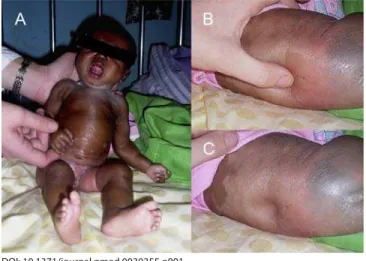
On examination the patient looked unwell, was grossly oedematous, and weighed 2.3 kg (Figure 1). She was alert and had no evidence of neurological abnormality. A low-grade pyrexia was present, but there were no other abnormal physical fi ndings.
What Are the Likely Causes of This Presentation
and Which Tests Would You Perform?
For any sick young infant in any setting, the possibility of infection must be foremost in the clinician’s mind. Neonates and young infants are particularly susceptible to infection, the more so when they are low birth weight and suffering from malnutrition, as must be suspected in a motherless child in this setting. Bacterial infections top the list, with group B streptococcus, gram-negative organisms (such as Escherichia coli), pneumococcus, Haemophilus infl uenzae type b, and Staphylococcus aureus being common pathogens. A systematic search for a focus of infection is needed because young infants localise infections poorly and present with non-specifi c symptoms and signs. While respiratory distress may indicate pneumonia, a swollen joint may indicate septic arthritis, and a bulging fontanelle, seizures, and depressed level of consciousness may indicate meningitis, these signs cannot be relied upon. A sepsis work-up in any sick young infant should include, regardless of signs, microscopy and culture of urine and cerebrospinal fl uid (CSF), blood culture, and a chest radiograph as well as swabs of suspect lesions.
In endemic countries malaria is uncommon in very young infants, in part because of the presence of maternal
antibodies and high levels of fetal haemoglobin, but nevertheless a blood fi lm is appropriate to rule out malaria.
HIV infection must be considered in this sick infant whose mother and sibling have died. Without intervention, mother-to-child transmission of HIV-1 occurs during pregnancy and delivery in 15–30 percent of infants and via breast-milk in 10– 20 percent [1]. Perinatally acquired HIV progresses rapidly in up to 25 percent of infants and usually presents with
A Gambian Infant with Fever
and an Unexpected Blood Film
Stephen Howie*, Malcolm Guy, Louise Fleming, Wendi Bailey, Harry Noyes, Joseph Axel Faye, Jacques Pepin,
Brian Greenwood, Hilton Whittle, David Molyneux, Tumani Corrah
Funding: Funding for this work was provided by the Medical Research Council Laboratories.
Competing Interests: The authors have declared that no competing interests exist.
Citation: Howie S, Guy M, Fleming L, Bailey W, Noyes H, et al. (2006) A Gambian infant with fever and an unexpected blood fi lm. PLoS Med 3(9): e355. DOI: 10.1371/ journal.pmed.0030355
DOI: 10.1371/journal.pmed.0030355
Copyright: © 2006 Howie et al. This is an open-access article distributed under the terms of the Creative Commons Attribution License, which permits unrestricted use, distribution, and reproduction in any medium, provided the original author and source are credited.
Abbreviations: CATT, card agglutination test for trypanosomiasis; CNS, central nervous system; CSF, cerebrospinal fl uid; HAT, Human African Trypanosomiasis
Stephen Howie, Malcolm Guy, Hilton Whittle, and Tumani Corrah are at the Medical Research Council Laboratories, Banjul, Gambia. Louise Fleming is at the Royal Victoria Teaching Hospital, Banjul, Gambia. Wendi Bailey and David Molyneux are at the Liverpool School of Tropical Medicine, Liverpool, United Kingdom. Harry Noyes is at the University of Liverpool, Liverpool, United Kingdom. Joseph Axel Faye is at the International Trypanotolerance Centre, Banjul, Gambia. Jacques Pepin is at Université de Sherbrooke, Quebec, Canada. Brian Greenwood is at the London School of Hygiene and Tropical Medicine, London, United Kingdom.
* To whom correspondence should be addressed. E-mail: showie@mrc.gm The Learning Forum discusses an important clinical problem of relevance
to a general medical audience.
DOI: 10.1371/journal.pmed.0030355.g001
Figure 1. Photographs of the Patient Soon after Admission, Showing Generalised Oedema
non-specifi c features. Malnutrition, diarrhoea, pneumonia, dermatitis, lymphadenopathy, hepatosplenomegaly, oral candidiasis, and parotitis are among the commoner features. The diagnosis of HIV infection in infants is complicated by the passive transfer of maternal anti-HIV antibodies. Antibody-based testing cannot reliably confi rm infection until the infant is 18 months old. However, polymerase chain reaction testing to detect the presence of HIV DNA can confi rm infection in the early stages.
The differential diagnosis of pyrexia in an infant in the tropics includes a range of other infections, including tuberculosis and salmonellosis, the likelihood of which will be infl uenced by local epidemiology and specifi c features of the presentation.
In this case, oedema was the major feature that accompanied the pyrexia and it requires explanation as it is unusual in young infants. Heart failure is a cause of generalised oedema but will be accompanied by other signs such as tachypnoea, cardiomegaly, hepatomegaly, and often a heart murmur, all of which were absent in this case. Renal causes such as nephrotic syndrome are unlikely. In this setting, an infant whose mother has died is very much at risk of malnutrition. Kwashiorkor, a less common manifestation of malnutrition in young infants than marasmus, is one possible explanation for the oedema in this case, though the severity and transience of it would be unusual. Basic anthropometry (weight, length/height, weight-for-length/height z-score) should be routine in the assessment of sick children, though anthropometry may not indicate malnutrition when the patient has oedema (as in this case where the z-score was above the median). Transient oedema can also be a feature of African trypanosomiasis.
The child was treated for malnutrition with appropriate nutritional support and broad-spectrum antibiotics. The sepsis work-up was negative, but on a routine thick blood fi lm, which was negative for malaria parasites, the microscopist was surprised to see numerous extracellular fl agellate parasites, identifi ed by a senior colleague as trypanosomes. The child was referred to the Medical Research Council Laboratories hospital in Gambia for further management. Tests for HIV-1 and HIV-2, both antibodies (Murex HIV-1.2.0, Abbott-Murex) and polymerase chain reaction, were negative.
What Are the Most Important Complications of Human
African Trypanosomiasis? How Can the Diagnosis of
Human African Trypanosomiasis Be Confi rmed?
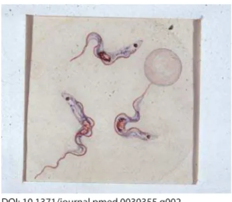
Human African Trypanosomiasis (HAT), also known as sleeping sickness, is caused by Trypanosoma brucei rhodesiense (in East and Southern Africa) or Trypanosoma brucei gambiense (in West and Central Africa). Infection with T. b. gambiense was fi rst described by Dutton in 1902 (Figure 2) in Gambia in a riverboat captain by the name of Kelly. Although it has re-emerged in several countries in sub-Saharan Africa, it has not been seen in Gambia, Senegal, or Guinea-Bissau for more than 20 years [2].Disease due to T. b. rhodesiense progresses rapidly over weeks, while disease due to T. b. gambiense tends to progress more slowly over months. In either case, infection leads to invasion of the central nervous system (CNS) and death if untreated. The two subspecies are morphologically indistinguishable. Fortunately, from the point of view of diagnosis, their geographical distribution does not overlap,
though there is now concern about geographical convergence in Uganda [3].
The diagnosis of HAT caused by T. b. gambiense presents different challenges from that caused by T. b. rhodesiense, which is usually directly detectable in the blood. A stepwise approach to the diagnosis of T. b. gambiense starts with a serological test such as the card agglutination test for trypanosomiasis (CATT), a fi eld diagnostic test of high sensitivity but lower specifi city (due to cross-reactivity with animal trypanosomes), followed by parasitological diagnosis from blood or lymph samples. In T. b. gambiense infections, detection of trypanosomes in blood is diffi cult, given the low-level and intermittent parasitaemia. In both cases, detection of parasites in blood or lymph node samples or clinical suspicion alone mandates examination of the CSF to determine the presence of CNS disease (late-stage HAT). Parasites can easily be missed on microscopy of a simple wet preparation of CSF, and sensitivity is increased by examining a doubly centrifuged specimen. The presence of CSF trypanosomes or a CSF white cell count of >5 cells per mm3 is
diagnostic of late-stage trypanosomiasis.
In this case a second blood fi lm at the Medical Research Council Laboratories hospital confi rmed the presence of numerous trypanosomes. A CSF sample was blood-stained and showed trypanosomes on a wet preparation. A CSF sample repeated fi ve days later, before any specifi c treatment was given, showed again the presence of occasional trypanosomes. This specimen was not blood-stained and showed no
pleocytosis. The fi nding of live trypanosomes in two CSF samples taken fi ve days apart suggests that the CNS had been invaded, although no neurological abnormalities were found. It is not certain whether this invasion was spontaneous or a result of seeding from the fi rst lumbar puncture.
What Treatment Is Indicated for HAT?
In the absence of CNS involvement (i.e., in early-stage HAT), pentamidine is the drug of choice for infection with T. b. gambiense while suramin is indicated for T. b. rhodesiense. Pentamidine is associated with cure rates above 90 percent in T. b. gambiense infection [4]. Hypotension is the chief immediate adverse drug reaction, occurring more frequently when administered intravenously than intramuscularly, but pentamidine also causes potentially severe adverse effects such as hypoglycaemia, hypocalcaemia, renal failure, neutropenia, and ventricular arrhythmia, all of which make it an unattractive option for treating an infant. In T. b.
DOI: 10.1371/journal.pmed.0030355.g002
rhodesiense infection, treatment failures with pentamidine are common, making suramin the drug of choice for early-stage disease.
Both pentamidine and suramin penetrate CSF poorly; therefore, given the presence of CSF trypanonosomes in this case, the alternatives for the treatment of presumptive T. b. gambiense infection were efl ornithine or melarsoprol. Efl ornithine is a trypanostatic drug associated with bone marrow suppression in up to half of recipients, though this usually resolves at the end of treatment. Melarsoprol is a trivalent arsenical drug that is highly trypanocidal, but also causes a potentially fatal encephalopathy in fi ve to ten percent of patients. Efl ornithine needs to be given six-hourly intravenously for 14 days, which is impractical in a developing-world setting, and the dose for an infant is not well established. Melarsoprol was the only practical option; therefore, despite its potential toxicity, it was given at a dose of 2.2 mg/kg daily intravenously for ten days, along with prednisolone (1 mg/kg daily) to reduce the risk of melarsoprol-induced encephalopathy [5,6].
Was This Case
T. b. gambiense
HAT?
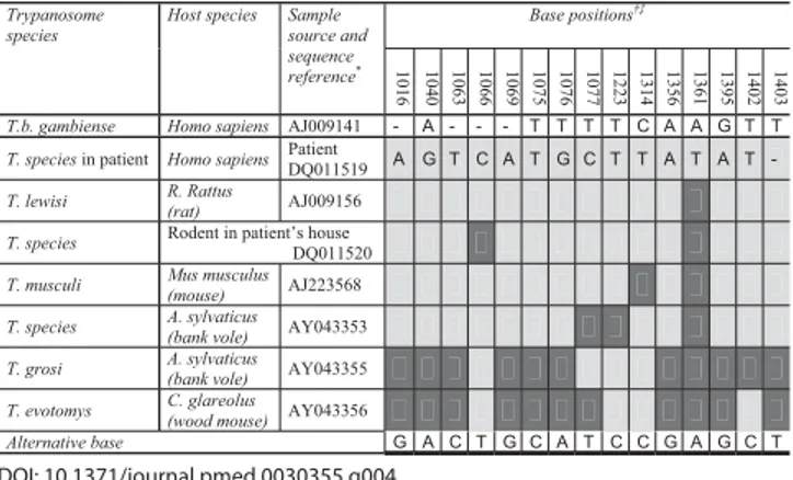
Confi rmed infection with a trypanosome should be regarded as HAT until proven otherwise. In this case, the thin blood fi lm done to examine the morphology of the trypanosome raised a doubt that was not resolved one way or the other until after the acute management of the patient. The appearance of the parasites was not typical of T. b. gambiense (Figure 2), but was typical of rodent trypanosomes of the subgenus T. (Herpetosoma) (Figure 3). Antibodies to T. b. gambiense were not detected in blood or CSF by fl uorescent antibody testing. Sequencing of part of the 18S ribosomal RNA confi rmed a genotype quite distinct from that of T. b. gambiense but very similar to that of the T. (Herpetosoma) group,
particularly T. lewisi, from which it differed by just one base (Figure 4) [7]. Molecular methods may become increasingly important in the diagnosis and management of HAT if there is, as feared, convergence of the geographical ranges of T. b. gambiense and T. b. rhodesiense [3].
Progress
The patient’s oedema resolved without specifi c treatment over the course of a week, and she remained clinically stable until therapy with melarsoprol was started. A repeat blood fi lm the day before starting treatment, 15 days after initial presentation, showed a persistent high density of trypanosomes. Three days into treatment a blood fi lm showed no evidence of trypanosomes. Treatment was well tolerated: apart from a temperature of up to 38 °C early in the course of treatment, a well-described phenomenon, the patient remained well and had no signs of encephalopathy [8]. Repeat blood fi lm and CSF samples taken at the completion of treatment and ten months later showed no evidence of parasites, and there were no white cells in the CSF. Follow-up to 13 months of age confi rmed that the child was making normal developmental progress.
Visits were made to the family homes in Gambia and Senegal. The caregiver’s house in rural Gambia was
reportedly infested with rodents, which was confi rmed by the fi nding of rodent droppings on the bed where the patient slept and a dead rodent on the porch. The mother’s home also reportedly had a rodent problem, but there was no report of this in her family’s home in Senegal.
Two live rodents of unidentifi ed species were trapped at the caregiver’s home. Examination of a blood fi lm revealed trypanosomes morphologically identical to those seen in the patient. Genotyping of a part of the 18S ribosome revealed a sequence differing by two bases from that of the patient’s parasite (Figure 4), which may well represent variation within the species rather than a different species [9]. A blood fi lm of the caregiver showed no evidence of trypanosomes.
D I S C U S S I O N
This unique case has provided the opportunity to review approaches to the unwell young infant in general and the management of suspected HAT in particular. To our knowledge, this is the fi rst documented case of an infection by trypanosomes of the subgenus T. (Herpetosoma) in humans in Africa, and the fi rst case anywhere in which this parasite has been able to infect the CNS and survive. As such, it offers the opportunity to review what is known of this parasite, and to consider the environmental, host-related, and organism-related factors that may have resulted in the child’s illness.
The subgenus T. (Herpetosoma) comprises at least 45 morphologically identical species that infect rodents throughout the world [10,11]. T. lewisi, the archetype of the subgenus, is a parasite of the genus Rattus and is transmitted via the excreta of fl eas. In West Africa there are records of T. (Herpetosoma) infection in members of the genera Cricetomys (Gambian giant rat) and Mastomys (multimammate rat) as well as in Rattus itself [12].
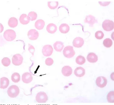
There are, to our knowledge, only three reported cases of human infection with a T. lewisi–like parasite, though in the absence of expert microscopy other cases might be DOI: 10.1371/journal.pmed.0030355.g003
Figure 3. Thin Blood Film of the Patient Showing Five Trypanosomes and Several Red Blood Cells
misdiagnosed. All reported cases occurred in Asia, and all were reliant on morphology for species identifi cation.
The fi rst patient, described in 1933, was a four-month-old Malaysian child who lived in a rat-infested dwelling [13]. The child presented with fever and lassitude associated with a heavy parasitaemia with T. lewisi. No CSF investigation was reported. The parasitaemia and accompanying symptoms resolved without specifi c treatment after fi ve days. Blood fi lms from other family members were all negative. One rat caught in the dwelling had a heavy infection with a morphologically identical parasite. This patient was followed up years later and found to have no trace of the parasite in blood or CSF [14].
The second report (of two cases) was from India in 1974 [15]. An adult couple who lived in a rat-infested village both presented with fever and one had malaise. Both were found to have heavy T. lewisi parasitaemia. Symptoms resolved without specifi c treatment after two to three days. Repeat blood fi lms taken eight weeks later were negative.
In the present case, the parasitaemia persisted unabated for more than two weeks until specifi c therapy was started, unlike the Malaysian infant reported by Johnson [13]. Neither was oedema a feature of previous cases, though it is characteristic of HAT and it is conceivable that T. lewisi infection caused the oedema in this case.
It is unclear in this case whether infection was acquired antenatally or postnatally. Congenital infections with T. b. gambiense and T. b. rhodesiense have been documented [10]. The usual duration of the incubation period of T. b. gambiense is in the order of months, whereas T. (Herpetosoma) infections develop detectable parasitaemias typically within seven to 14 days of inoculation in rodents. There is no evidence that T. (Herpetosoma) trypanosomes can be transmitted transplacentally in rodents [16]. This suggests that in our patient, transmission occurred after delivery through exposure to the excreta of infected fl eas in the environment in which the child was living.
Host factors are likely to have been important in this case: the relative immune vulnerability of early infancy together with malnutrition probably increased this child’s susceptibility to this unusual infection. Humans naturally resist infection by animal trypanosomes such as T. brucei, T. lewisi, T. congolense, and T. vivax. What drives this resistance of human serum to animal trypanosomes has been debated, but recent research suggests that for T. brucei it is an apolipoprotein, either a haptoglobin-related protein or apolipoprotein L-I [17,18]. Whether similar factors are important in controlling infections with T. lewisi is unknown.
Organism factors may also have played a part in this case. It is possible that this patient’s parasite has become better adapted to human infection than its forebears, though we cannot say this with certainty. Virulence varies between subspecies of T. brucei: T. b. rhodesiense is more virulent than T. b. gambiense, arguably killing its host too rapidly for its own interest. This is related to the serum resistance–associated SRA gene, which is now used for the molecular epidemiology of T. b. rhodesiense infections and, as its name implies, is associated with resistance to human serum in vitro and in vivo [3]. The continuing adaptation of organisms to human infection will be infl uenced by changing host factors, and in this respect there is concern that the HIV pandemic might create a new window of opportunity for organisms to make this transition [19].
Conclusion
The lessons from this learning forum are several. Firstly, infection is a major cause of mortality in young infants and must be managed diligently with a thorough search for its cause and the institution of appropriate treatment. Secondly, HAT remains a potentially fatal disease that presents challenges in the diffi culty of its diagnosis and in the toxicity of its treatments. Additionally, given the right combination of environmental, host-related, and organism-related factors, a normally harmless organism can emerge as a human pathogen. This fi rst report of invasive disease from T. lewisi shows that this parasite cannot be dismissed as a harmless cause for false positives in the work-up of suspected cases of HAT. We believe that on current evidence this infection should be treated when diagnosed. However, the data on which to make this decision are limited, so in asymptomatic confi rmed cases without CNS invasion a case can be made for careful follow-up without treatment. Nevertheless, the fi nal message must be that if there is any evidence of invasive disease, any doubt concerning the identity of the parasite, or any concern regarding the effectiveness of follow-up, standard treatment for HAT should not be delayed.
Acknowledgments
Dr. Robin Bailey and colleagues at the Hospital for Tropical Diseases, London, United Kingdom, contributed advice regarding the management and investigation of this case. Dr. Jean Jannin of the World Health Organization, Geneva, Switzerland, provided melarsoprol for treatment of the patient. Mr. Abdou Bah of the Royal Victoria Teaching Hospital fi rst detected the abnormal blood fi lm. Our sincere thanks go to the patient’s guardian for allowing her photograph to be used in this report.
Author contributions. SH supervised management of the case, undertook the fi eldwork, and wrote the paper. All authors contributed to the writing of the paper. MG did the laboratory work in Gambia and contributed to the fi eldwork. LF managed the case at
DOI: 10.1371/journal.pmed.0030355.g004
Figure 4. Genotypes of the Parasite Isolated from the Patient, the Parasite Isolated from a Rodent Captured in the Patient’s Home, T. (Herpetosoma) Species and T. b. gambiense at Positions on the 18S Ribosome that Are Polymorphic within the Subgenus T. (Herpetosoma)
the Royal Victoria Teaching Hospital. WB did the antibody testing. HN undertook genotyping of the parasites. JAF advised on the parasitology and assisted with fi eldwork. JP, BG, HW, and DM advised on the parasitology and management of the case. TC advised on management and assisted with fi eldwork.
References
1. World Health Organization (2006) Mother-to-child transmission of HIV (MTCT). Available: http:⁄⁄www.who.int/reproductive-health/stis/mtct/ index.htm. Accessed 3 August 2006.
2. Sterner G, Nasander L (1977) African trypanosomiasis: A danger for tourists visiting Gambia? Scand J Infect Dis 9: 154–156.
3. Picozzi K, Fevre E, Odiit M, Carrington M, Eisler MC, et al. (2005) Sleeping sickness in Uganda: A thin line between two fatal diseases. BMJ 331: 1238– 1241.
4. World Health Organization (1998) Control and surveillance of African trypanosomiasis. Geneva: World Health Organization. 119 p. 5. Burri C, Nkunku S, Merolle A, Smith T, Blum J, et al. (2000) Effi cacy of
new, concise schedule for melarsoprol in treatment of sleeping sickness caused by Trypanosoma brucei gambiense: A randomised trial. Lancet 355: 1419–1425.
6. Pepin J, Milord F, Guern C, Mpia B, Ethier L, et al. (1989) Trial of prednisolone for prevention of melarsoprol-induced encephalopathy in gambiense sleeping sickness. Lancet 1: 1246–1250.
7. Noyes H, Stevens J, Teixeira M, Phelan J, Holz P (1999) A nested PCR for the ssrRNA gene detects Trypanosoma binneyi in the platypus and
Trypanosoma sp. in wombats and kangaroos in Australia. Int J Parasitol 29: 331–339.
8. Whittle H, Pope H (1972) The febrile response to treatment in Gambian sleeping sickness. Ann Trop Med Parasitol 66: 7–14.
9. Noyes H, Ambrose P, Barker F, Begon M, Bennet M, et al. (2002) Host specifi city of Trypanosoma (Herpetosoma) species: Evidence that bank voles
(Clethrionomys glareolus) carry only one T. (H.) evotomys 18S rRNA genotype but wood mice (Apodemus sylvaticus) carry at least two polyphyletic parasites. Parasitology 124: 185–190.
10. Barrett M, Burchmore R, Stich A, Lazzari JO, Frasch AC, et al. (2003) The trypanosomiases. Lancet 362: 1469–1480.
11. Linardi P, Botelho J (2002) Prevalence of Trypanosoma lewisi in Rattus norvegicus from Belo Horizonte, State of Minas Gerais, Brazil. Mem Inst Oswaldo Cruz 97: 411–414.
12. Bray R (1964) A checklist of parasitic protozoa of West Africa with some notes on their classifi cation. Bull Inst Fr Afr Noire 26: 238–315.
13. Johnson P (1933) A case of infection by Trypanosoma lewisi in a child. Trans R Soc Trop Med Hyg 26: 467–468.
14. Molyneux D, Ashford R (1983) The biology of Trypanosoma and Leishmania, parasites of man and domestic animals. London: Taylor and Francis. 294 p. 15. Shrivastra K, Shrivastra G (1974) Two cases of Trypanosoma (Herpetostoma)
species infection of man in India. Trans R Soc Trop Med Hyg 68: 143–144. 16. Molyneux D (1976) Biology of trypansomes of subgenus Herpetosoma. In:
Lumsden W, Evans D, editors. Biology of the Kinetoplastida. New York, San Francisco, London: Academic Press. pp. 285–325.
17. Smith A, Esko J, Hadjuk S (1995) Killing of trypanosomes by the human haptoglobin-related protein. Science 268: 284–286.
18. Vanhamme L, Paturiaux-Hanocq F, Poelvoorde P, Nolan DP, Lins L, et al. (2003) Apoliporotein L-I is the trypanosome lytic factor of human serum. Nature 422: 83–87.
19. Weiss R (2001) Gulliver’s travels in HIVland. Nature 410: 963–967.
Learning Points
• Infection is a major cause of mortality in young infants and requires a thorough search for its cause and the institution of appropriate treatment.
• HAT remains a potentially fatal disease that can be challenging to diagnose.
• Treatments for HAT are associated with toxicities (e.g., pentamidine can cause hypotension, efl ornithine can cause bone marrow suppression, melarsoprol can cause encephalopathy).
• Given the right combination of environmental, host-related, and organism-related factors, a normally harmless organism can emerge as a human pathogen.