Published online August 29th, 2008 © http://www.ijav.org
Case Report
International Journal of Anatomical Variations (2008) 1: 19–20
Introduction
Sutural bones or Wormian bones are small irregular shaped bones found at the cranial sutures. Their size, shape and number vary from skull to skull. A sutural bone is occasionally present at the pterion or junction of the parietal, frontal, greater wing of the sphenoid, and squamous portion of the temporal bones. This bone is called “pterion ossicle” or “epipteric bone” or Flower’s bone. We found three sutural bones at the pterion.
Case Report
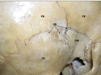
During the routine osteology demonstration class for undergraduate medical students, three Wormian bones were noted at the pterion in an Indian adult skull (Figures 1 and 2.). The abnormal bones were found only on the right pterion region. Among the three bones, the smallest one was at the meeting point of frontal, sphenoid and parietal bones; the medium sized bone was between parietal, sphenoid and temporal bones and the largest was between the temporal and parietal bones. These three bones together gave a fractured look to the pterion. There were no other abnormalities in the skull.
Discussion
Wormian bones (sutural bones) are very commonly found in the skull. According to Bergman et al., [1], nearly 40% of skulls have sutural bones in the vicinity of the lambdoid suture. The next most common is the epipteric bone (pterion ossicle) found near the former anterolateral fontanelle. There can be another Wormian bone called
preinterparietal bone or inca bone at the lambda [2, 3, 4]. The presence of sutural bones is usually associated with cranial and central nervous system anomalies [5, 6]. But there are cases where there were no anomalies associated with the sutural bones [7]. The presence of the sutural bones may be regulated by a genetic factor [8]. The incidence of epipteric bone is high in Indians. A study Satheesha NAYAK B [1]
Soumya KV [2]
Department of Anatomy, Melaka Manipal Medical College (Manipal Campus) Manipal [1], Department of Mathematics, Manipal Institute of Technology, Manipal [2], INDIA.
Satheesha Nayak B., MSc. PhD. Associate Professor of Anatomy Melaka Manipal Medical College (Manipal Campus) International Centre for Health Sciences Madhav Nagar, Manipal Udupi District
Karnataka State, 576 104 INDIA. +91 820 2922519 +91 820 2571905 [email protected]
Received July 17th, 2008; accepted August 27th, 2008
ABSTRACT
The existence of Wormian (sutural) bones in the skull is well known. We found three unusual Wormian bones at the right pterion in an adult Indian skull. The variation noted was unilateral. This type of variation has not been reported yet. © IJAV. 2008; 1: 19–20.
Key words [pterion] [skull] [wormian bone] [sutural bone] [epipteric bone] [pterion ossicle]
eISSN 1308-4038
Unusual sutural bones at pterion
20 Nayak et al.
References
[1] Bergman RA, Afifi AK, Miyauchi R. Skeletal systems: Cranium. In: Compendium of human anatomical variations. Baltimore, Urban and Schwarzenberg. 1988; 197–205.
[2] Malhotra VK, Tewari PS, Pandey SN, Tewari SP. Interparietal bone. Acta Anat. (Basel). 1978; 101: 94–96.
[3] Saxena SK, Chowdhary DS, Jain SP. Interparietal bones in Nigerian skulls. J. Anat. 1986; 144: 235–237.
[4] Pal GP. Variations of the interparietal bone in man. J. Anat. 1987; 152: 205–208.
[5] Pryles CV, Khan AJ. Wormian bones. A marker of CNS abnormality? Am. J. Dis. Child. 1979; 133: 380–382.
[6] Das S, Suri R, Kapur V. Anatomical observations on os inca and associated cranial deformities. Folia Morphol. (Warsz). 2005; 64: 118–121.
[7] Jeanty P, Silva SR, Turner C. Prenatal diagnosis of wormian bones. J. Ultrasound Med. 2000; 19: 863–869.
[8] El-Najjar M, Dawson GL. The effect of artificial cranial deformation on the incidence of Wormian bones in the lambdoidal suture. Am. J. Phys. Anthropol. 1977; 46: 155–160.
[9] Saxena SK, Jain SP, Chowdhary DS. A comparative study of pterion formation and its variations in the skulls of Nigerians and Indians. Anthropol. Anz. 1988; 46: 75–82.
[10] Ersoy M, Evliyaoglu C, Bozkurt MC, Konuskan B, Tekdemir I, Keskil IS. Epipteric bones in the pterion may be a surgical pitfall. Minim. Invasive Neurosurg. 2003; 46: 363–365.
Figure 2. Closer view of the pterion with the sutural bones. (PB: parietal bone, FB: frontal bone, TB: temporal bone, GW: greater wing of sphenoid bone, SB: sutural bones)