the Causative Gene for LGMD1F
Annalaura Torella1,2., Marina Fanin3., Margherita Mutarelli1
, Enrico Peterle3, Francesca Del Vecchio Blanco2, Rossella Rispoli1,4, Marco Savarese1,2, Arcomaria Garofalo2, Giulio Piluso2, Lucia Morandi5, Giulia Ricci6, Gabriele Siciliano6, Corrado Angelini3,7, Vincenzo Nigro1,2*
1TIGEM (Telethon Institute of Genetics and Medicine), Napoli, Italy,2Dipartimento di Biochimica Biofisica e Patologia Generale, Seconda Universita` degli Studi di Napoli, Napoli, Italy,3Dipartimento di Neuroscienze, Universita` degli Studi di Padova, Padova, Italy,4Cancer Research UK, London, United Kingdom,5Fondazione IRCCS Istituto Neurologico C. Besta, Milano, Italy,6Dipartimento di Medicina clinica e sperimentale, Universita` degli Studi di Pisa, Pisa, Italy,7IRCSS S. Camillo, Venezia, Italy
Abstract
Limb-girdle muscular dystrophies (LGMD) are genetically and clinically heterogeneous conditions. We investigated a large family with autosomal dominant transmission pattern, previously classified as LGMD1F and mapped to chromosome 7q32. Affected members are characterized by muscle weakness affecting earlier the pelvic girdle and the ileopsoas muscles. We sequenced the whole exome of four family members and identified a shared heterozygous frame-shift variant in the Transportin 3 (TNPO3) gene, encoding a member of the importin-bsuper-family. TheTNPO3gene is mapped within the LGMD1F critical interval and its 923-amino acid human gene product is also expressed in skeletal muscle. In addition, we identified an isolated case of LGMD with a new missense mutation in the same gene. We localized the mutant TNPO3 around the nucleus, but not inside. The involvement of gene related to the nuclear transport suggests a novel disease mechanism leading to muscular dystrophy.
Citation:Torella A, Fanin M, Mutarelli M, Peterle E, Del Vecchio Blanco F, et al. (2013) Next-Generation Sequencing Identifies Transportin 3 as the Causative Gene for LGMD1F. PLoS ONE 8(5): e63536. doi:10.1371/journal.pone.0063536
Editor:Paul McNeil, Medical College of Georgia, United States of America ReceivedFebruary 15, 2013;AcceptedMarch 25, 2013;PublishedMay 7, 2013
Copyright:ß2013 Torella et al. This is an open-access article distributed under the terms of the Creative Commons Attribution License, which permits unrestricted use, distribution, and reproduction in any medium, provided the original author and source are credited.
Funding:This study was mainly supported by grants from Telethon, Italy (TGM11Z06 to V.N. and GTB12001 to C.A.) and Telethon-UILDM (Unione Italiana Lotta alla Distrofia Muscolare) (GUP 10006 and GUP11006 to V.N.). This work was also supported by grants from the Association Franc¸aise contre les Myopathies (13859 to M.F. and 14999/16216 to C.A.). The funders had no role in study design, data collection and analysis, decision to publish, or preparation of the manuscript. Competing Interests:The authors have declared that no competing interests exist.
* E-mail: [email protected]
.These authors contributed equally to this work.
Introduction
Limb girdle muscular dystrophies (LGMDs) are characterized by a progressive weakness that begins from the proximal limb muscles, due to a number of independent genetic defects that are distinct from the X-linked Duchenne and Becker muscular dystrophies [1,2]. In addition to the genetic heterogeneity, the different forms are clinically heterogeneous, with the age at onset of symptoms varying from early childhood to late adulthood [3]. The milder the symptoms are, more difficult is the LGMD diagnosis. Magnetic resonance imaging is helpful to characterise the severity and pattern of muscle involvement [4,5], but recognition of LGMD type might be hard [6].
Muscle biopsy of LGMD patients generally shows a diffuse variation in fiber size, necrosis, regeneration and fibrosis, but the degree of these factors is variable and does not parallel the clinical severity. Based on the histological features alone, there is scarce, if any, possibility of diagnosing a specific LGMD form, but western blot and immunofluorescence can address to the true defect that can be demonstrated by the finding of a mutation in the corresponding gene.
The primary distinction is made between the autosomal dominant (LGMD1) and the autosomal recessive forms (LGMD2), with an alphabet letter indicating the order of gene mapping [2]. Eight LGMD1 loci have been identified so far. At present, only
sequenced the whole exome of four affected individuals and identified a number of new variations, one of which was completely new, shared by all affected subjects, and mapped to 7q32.
Methods
Ethics Statement
This study adhered to the tenets of the Declaration of Helsinki. Subjects for this study were recruited at Padua University and exome analysis was performed at the Second University of Naples and at the Telethon Institute of Genetics and Medicine. Participants were informed of the nature and risks of the study, and signed consent forms were obtained. The institutional review board of the Second University of Napoli (SUN) reviewed and approved this study (prot. AOP-SUN 862).
Patients
Nineteen patients were included in the clinical study, and they all fulfilled the diagnostic criteria for LGMD that include a characteristic pattern of muscular weakness primarily affecting pelvic girdle, assessed according to MRC Scale and a modified Gardner-Medwin & Walton scale for proximal LGMD. Age at onset was assessed as described. We collected blood from 19 patients and 8 healthy relatives. Skeletal muscle biopsy from the deltoid orvastus lateraliswas taken from 2 affected individuals.
Exome sequencing and analysis
Enrichment was performed by hybridization of shotgun fragment (average size 141 bp) libraries to Agilent SureSelect Human All Exon 50 Mb (Agilent Technologies, Santa Clara, CA, USA) in-solution capture assays. Using the SOLiD system v4 (Life Technologies), we generated an average of 4.2 Gb of mappable sequence data per sample to achieve,20x mean coverage of the targeted exome. The sequences were analyzed using an automated custom pipeline designed to perform every step of the analysis with the appropriate program or custom script. Sequencing reads were first colour-corrected using SOLiD Accuracy Enhancer Tool (SAET), then mapped to the reference genome (UCSC, hg19 build) using the software BioScope v1.3 (Life Technologies, Carlsbad, CA, USA) and duplicate reads were removed using Picard (http://picard.sourceforge.net). Single nucleotide variations (SNV) and in-del mutation calling analyses were carried out using the diBayes algorithm with medium stringency settings and the SOLiD Small Indel Fragment Tool (www3.appliedbiosystems. com), respectively.
One of the samples was sent to a commercial provider (Otogenetics Corporation, Norcross, GA, USA) who performed both whole exome enrichment with the SeqCap EZ Human Exome Library v2.0 (Roche NimbleGen, Inc, Madison, WI, USA) and sequencing with the HiSeq2000 platform (Illumina inc., San Diego, CA, USA). The sequences were analyzed using another automated pipeline designed to handle Illumina data with custom scripts and publicly available software. Paired sequencing reads were aligned to the reference genome (UCSC, hg19 build) using BWA [17] and post-alignment process and duplicate removal was performed using SAMtools [18] and Picard. Further processing
(http://evs.gs.washington.edu/EVS) and 1000 genomes large scale projects (http://www.1000genomes.org) [23], conservation and different prediction algorithms of damaging effect on protein activity [24,25,26,27] and conservation scores [28,29]. The annotated results were then imported into an in-house variation database, also used to make comparisons among samples and filter results. The alignments at candidate positions were visually inspected using the Integrative Genomics Viewer (IGV)[30]. The accession number of the dataset of this study is ERP002413 (Sequence Read Archive – EBI at www.ebi.ac.uk).
Mutation Detection
We designed both cDNA and intronic primers to amplify the cDNA and the 22 coding exons plus the 3’UTR exon of the TNPO3 gene (MIM 610032; NM_012470.3, NM_001191028.2) (Table S2). In addition, we sequenced all the other exons at the disease interval that were inadequately covered (,10x) (Table S3). We also designed additional primers to map the alternatively spliced products. We purified the amplicons and sequenced them by using the fluorescent dideoxyterminator method on an automatic sequencer (ABI 3130XL).
Immunoblotting Analysis
For TNPO3 immunoblotting, muscle samples were homoge-nized in a lyses assay buffer (Urea 8 M, SDS 4%, 125 mM Tris HCl pH 6.8). The samples were separated on sodium dodecyl sulphate –9% polyacrylamide gel electrophoresis and transferred to nitrocellulose membrane. After blocking in 10% no fat dry milk in Tween-Buffered Saline (TTBS-1X) buffer (10 mM Tris-HCl, 150 mM NaCl, 0.05% TWEEN 20) for 1h, the membranes were incubated with primary antibodies in TTBS 1X at room temperature for 2 h. The monoclonal antibody, recognizing a recombinant fragment (Human) from near the N terminus of TNPO3, was used in this experiment with a 1:100 dilution (AbcamH). We also used the rabbit monoclonal antibody Anti-TNPO3 antibody [EPR5264] (ab109386) that recognizes a synthetic peptide corresponding to residues near the C terminal of Human TNPO3. This was used for WB at 1:300 dilution.
Following primary antibody incubation and rinses, the mem-branes were incubated with the secondary antibody, goat anti-mouse immunoglobulin conjugated with horseradish (Sigma), with 1:10,000 dilution in 0.5% dry milk and TTBS 1X. After 45 minutes of antibody incubation and five washes with TTBS 1X buffer, the TNPO3 protein band was visualized with a chemiluminescence reagent (Supersignal, WestPico, Pierce) and exposed to X-ray film.
To perform this analysis, Coomassie blue staining was used for the evaluation of the myosin protein expression to understand the variations in the levels of the proteins loaded.
Transfection
Plasmid pcDNA6/A encoding N-terminal HA-tagged TNPO3
into the pCS2HA plasmid : 1) Wt TNPO3 isoform with 22 exons; 2) TNPO3 isoform with 22 exons containing del A p.X924C; 3) Wt TNPO3 isoform with 23 exons; 4) TNPO3 isoform with 23 exons containing del A p.X924C. We used 500 ng for transient trasfection of HeLa cells (26105) cells using PolyFect Transfection Reagent (Qiagen) according to manifacturer’s instruction. Cells were grown on glass coverslip put into 12 well plates. They were cultured in Dulbecco’s modified eagle’s medium (DMEM) supplemented with 10% (v/v) foetal bovine serum and penicil-lin-streptomycin (GIBCO-Invitrogen) and maintained in a 5% CO2 incubator at 37uC. 48 hours after transfection, cells were fixed with 4% paraformaldehyde in PBS for 10 min at RT, permeabilized in 0,2% Triton X-100 in PBS for 5 min at RT, and blocked for 1 h in Blocking solution (BSA 6%, Horse Serum 5% in PBS). Cells were incubated for 1 h at RT with primary antibodies, followed by 1 h incubation at RT with FITC-conjugated anti-rabbit and/or Cy3-conjugated anti-mouse antibodies.
Results
Exome analysis
The original LGMD1F family has been extended (Figure 1) to include additional family members in seven generations starting from subject II, 2. The updated pedigree includes 64 LGMD patients of both sexes and five non-penetrant carriers (93% penetrance). To perform an informative exome sequencing analysis, we selected four affected family members (VII-5, VI-53, V-28, and VI-36) with a manifest LGMD phenotype separated by the largest number of meioses. Interestingly, two family members (VI-53 and V-28) were absent from the original family used for the linkage analyses. DNA samples of three individuals (V-28, VI-53, VII-5) were fragmented, enriched using the SureSelect whole exome kit and sequenced by SOliD. DNA, muscle RNA and proteins were extracted for the studies. We found,20,000 exonic variations for each sample, 5,722 of which were common to all three (Table 1 and Table S1) of which 2,471 were non
synonymous. Considering the dominant mode of inheritance of LGMD1F, we focused on the heterozygous calls and discarded all variants present with a frequency higher than 1% in the NHLBI Exome Variant Server (http://evs.gs.washington.edu/EVS) or 1000genomes [32] large scale projects. The resulting filtered list of 273 variants was composed of 253 missense, 14 stopgain, 2 frameshift deletions, 2 nonframeshift insertions/deletions and 2 stoploss variations. Only two variants were mapped into the disease interval between D7S1822 and D7S2519 (positions: 126,287,140-129,964,025) [11]: a nonsynonymous SNV in the gene IRF5 and a frame-shift deletion that modify the termination codon in the exon 22 (stoploss) in the TNPO3 on chromosome 7q32.1 at position 128,597,310 (GRCh37/hg19). To verify whether we could have missed by NGS other shared variants, we resequenced by the dideoxy-chain termination method all the coding exons and flanking introns of the full 7q32 region with lower/absent coverage (Table S3). No other shared unknown variant was found. In addition, the DNA sample of VI-36 was sent to a commercial provider for exome sequencing using the Illumina platform HiSeq2000. Among 153 variations that were shared by all, the only one in the disease interval was that in the TNPO3 gene (Table 1). Interestingly, this was the only variation of the whole exome that resulted absent in dbSNP137. We also refined the interval: the SNP rs45445295 at the SMO gene at position 128,845,555 was present in some affected members (V-8, VI-60, V-14, VI-11, V-25, V-12), but it was absent in other affected members (VI-57, VI-27, VI-56) and in all non-affected individuals. Therefore, the linked region associated with disease locus was ,1.1 Mb smaller (126,287,140-128,845,555) than that reported by Palenzuela [11].
To confirm the complete co-segregation of the nonstopTNPO3
variant with LGMD1F, we analyzed all available family members, affected and non-affected. We sequenced by the Sanger method all the samples and, in addition, we took advantage of an AluI
restriction site that was lost upon mutation. We observed the Figure 1. LGMD1F family pedigree.Squares represent male; circles represent female; white figures symbolize normal individuals; black figures indicate individuals with clinical muscular dystrophy. The original LGMD1F family has been extended from subject II,2 and now includes 64 LGMD patients of both sexes and five non-penetrant carriers (IV-4, V-26, V-29, V-33, and VI-68). The whole-exome sequencing was performed in four patients indicated by arrows (V-28, VI-36, VI-53, VII-5).
doi:10.1371/journal.pone.0063536.g001
Table 1.Total and Shared Variants in Patients with LGMD1F.
Patient variant type V-28 VI-53 VII-5
Shared by all
SOLiD VI-36 Shared by all four
exonic/splicing 21,105 21,366 17,123 5,722 17,183 4,212
non synonymous 11,852 11,713 9,051 2,471 7,831 1,687
heterozygous 9,348 9,138 6,812 644 4,693 153
frequency in EVS and 1000genomes,1% 6,102 5,785 3,860 273 486 10
Within LGMD1F interval 13 11 5 2 1 1
complete co-segregation of the TNPO3 variant with the disease
(Figure 2a and Table S4).
We extended the analysis to additional 64 samples from LGMD1 and isolated LGMD cases, using a next generation sequencing approach. In particular, we performed a custom enrichment of exons of genes involved in muscular dystrophies, includingTNPO3.
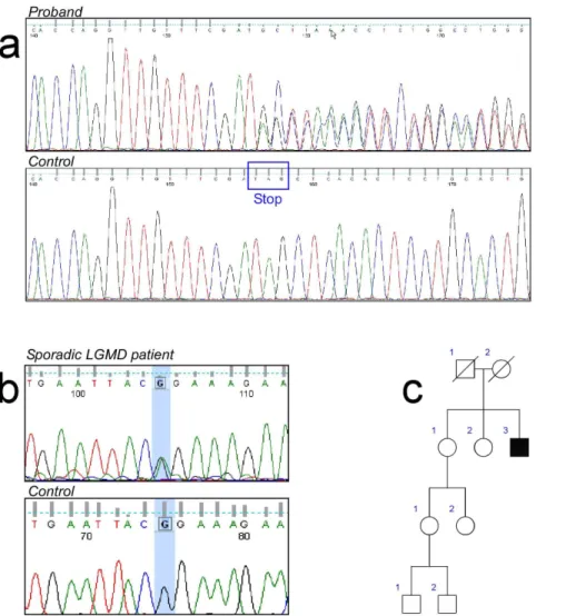
In a single individual, we found a heterozygous G.A transition (c.G2453A) in exon 21 of theTNPO3gene. This point mutation changes the Arginine in position 818 with a Proline (Figure 2b). This is an extreme conserved residue that is predicted to be damaging by all the used bioinformatic tools (SIFT, PolyPhen, Mutation Taster and LRT). Moreover, the variation is not listed in dbSNP and in the other recently developed databases collecting NGS data (Exome Variant Server and 1,000 genomes database) neither in our internal database of 150 samples whose exomes have been sequenced in our lab.
This variation has not been found in the healthy sister (Figure 2c). In addition, this patient bears no other major mutation in other 98 ‘‘muscular-disease’’ genes, but a single heterozygous ANO5 variation (Glu95Lys), without a clear significance. Young adult onset has been observed in this patient, showing a
Figure 2. Sequence analyses of the TNPO3 mutations.a) Heterozygous delA mutation in Exon 22 of theTNPO3gene in Proband VII-5. Aligned
electropherograms show mutated (top) and wild-type (bottom) sequences; b) Heterozygous. c.G2453A) in exon 21 of theTNPO3gene; c) Pedigree of
the isolated case.
doi:10.1371/journal.pone.0063536.g002
Figure 3. Western blot analysis of skeletal muscle tissue with antibodies to TNPO3. Equal amounts of muscle proteins from a LGMD1F patient and a control were run in each lane (10mg) on a 9%
characteristic LGMD phenotype. Muscular histopathological data evidenced dystrophic features and, in addition, discrete mitochon-drial alterations, with sporadic ragged-red fibers and cytochrome c
oxidase negative fibers. Mutations in the mitochondrial DNA were excluded.
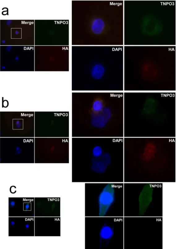
Figure 4. Indirect immunofluorescence analysis of the wt-hTNPO3 compared with delA p.X924C -hTNPO3. Following transient transfections, HeLa cells were incubated for 48 h with normal DMEM and detected by anti-HA immunofluorescence. Nuclei are stained with DAPI (blue). The endogenous protein is recognized using a rabbit monoclonal anti-TNPO3 antibody (green), while the transfected TNPO3 proteins were HA-tagged (red).a) An accumulation around the nucleus is usually observed using the mutant delA p.X924C -hTNPO3.b) The typical intranuclear staining pattern can be observed in cells transfected with wt-hTNPO3 (in red) orc)in non transfected HeLa cells.
These forms encode the same protein, when the DNA sequence is normal, because the stop codon is in exon 22. However, the LGMD1F mutation eliminates this stop and, for both forms, the muscle protein product is extended by the frame-shift. Form A should be 15 amino acids longer (CSHSCSVPVTQECLF), while form B should contain additional 95 amino acids.
We then performed immunoblotting analyses of the skeletal muscle biopsy using the anti TNPO3 antibody. While mutant form A is virtually overlapping with wild-type form A, a mutant form B can be appreciated by western blot analysis of muscle samples as a higher molecular weight band (Figure 3).
We generated a construct expressing the WT and del A p.X924C allele. HeLa cells were transfected with either the Wt or the mutant TNPO3. The transfected proteins were distinguished from the endogenous TNPO3 by adding a HA-tag. Figure 4 shows that the WT TNPO3 entered the nucleus, while the mutant was usually around the periphery of the nucleus.
Discussion
Here, we report the identification of a frame-shift variant del A p.X924C at the TNPO3/Transportin-SR2 gene on chromosome 7q32.1 at position 128,597,310 (GRCh37/hg19) in all patients with limb girdle muscular dystrophy 1F. No other variant was shared by four affected members of the family. The variant modifies the true stop codon and encode for two elongated proteins of 15 and 95 amino acids. Interestingly, one flanking SNP (rs12539741 at 128,596,805) has been identified in association with others in the region as a susceptibility locus for primary biliary cirrhosis [33]. Considering that the affected family members may share a ,2.6 Mb-region on chromosome 7q32, there is the possibility that any other rare heterozygous variant could co-segregate inciswith the true LGMD1F mutation. Thus,
we searched in a large collection of patients independentTNPO3
disease-associated variations. We found a missense Arg818Pro in an isolated LGMD case co-segregating with the disease in this family. This variation was predicted as causative by the nature of the change and the conservation.
Transportin 3 is a member of the importinbsuper-family that imports numerous proteins to the nucleus, including serine/ arginine-rich proteins (SR proteins) that control mRNA splicing [34,35]. Transportin 1 (TNPO1), also known as karyopherinb-2, mediates the nuclear import of M9-bearing proteins[36], while TNPO2 (karyopherinb-2B) participates directly in the export of a large proportion of cellular mRNAs[37]. There are two main TNPO3 proteins: variant 1 that is 923 amino acids long and variant 2 composed of 859 amino acids, while a longer variant of the 3’ terminus (hTNRSR1[35]) was probably due to a sequence artifact. The 923-amino acid protein is found in the skeletal muscle, translated from two equivalent messengers that include or not the 3’ noncoding exon.
TheTNPO3nonstop allele hindered the nuclear localization of
the protein in HeLa cells. Given the role of TNPO3 protein in the
LGMD1F. Additional functional studies in model organisms are, however, necessary to understand whether the dominant role of these mutations is due to haploinsufficiency or to a dominant-negative mechanism. This should be possible by the use of antisense morpholino oligos inDanio rerio(Zebrafish) where a single
and conserved TNPO3 ortholog is present with 792/923 (86%) amino acid identity (Table S5).
Advances in the knowledge of limb-girdle muscular dystrophies have been made in the last few years. With LGMD1F, five different autosomal dominant LGMD genes have been so far recognized. The use of NGS technologies promises a revolution in diagnostics and a more rapid characterization of patients.
Supporting Information
Table S1 Exome sequencing data. (DOC)
Table S2 Primers designed forTNPO3amplification and Sanger sequencing.
(DOC)
Table S3 Exons inadequately covered by NGS exome sequenc-ing and primers designed for Sanger sequencsequenc-ing.
(DOC)
Table S4 Co-segregation study. (DOC)
Table S5 Alignment of Human andDanio r.TNPO3 proteins. (DOC)
Table S6 Shared SNVs in the four samples sequenced by NGS. (XLSX)
Acknowledgments
We thank all the patients and their families for their contribution to this work. We acknowledge the Neuromuscular Bank of Tissues and DNA samples (NMTB) for collecting samples (C.A. and M.F.), Stefania Crispi and Luigi Leone at the IGB Facility of Next Generation Sequencing using the SOLID platform, and Anna Cuomo and Rosalba Erpice for Sanger sequencing. The authors would like to thank the NHLBI GO Exome Sequencing Project and its ongoing studies which produced and provided exome variant calls for comparison: the Lung GO Sequencing Project (HL-102923), the WHI Sequencing Project (HL-102924), the Broad GO Sequencing Project 102925), the Seattle GO Sequencing Project (HL-102926) and the Heart GO Sequencing Project (HL-103010). We also thank Gopuraja Dharmalingam and the TIGEM Bioinformatics Core for support in exome data analysis and Giuseppina Di Fruscio for the analysis of isolated cases of LGMD and Marina Mora for helpful suggestions.
Author Contributions
Conceived and designed the experiments: VN MM RR CA. Performed the experiments: AT AG MF MM EP FDVB MS GP LM VN. Analyzed the data: CA VN RR MM. Contributed reagents/materials/analysis tools: AT MF EP GR LM GS CA VN. Wrote the paper: VN.
References
1. Nigro V (2003) Molecular bases of autosomal recessive limb-girdle muscular dystrophies. Acta Myol 22: 35–42.
3. Fanin M, Nascimbeni AC, Aurino S, Tasca E, Pegoraro E, et al. (2009) Frequency of LGMD gene mutations in Italian patients with distinct clinical phenotypes. Neurology 72: 1432–1435.
4. Mercuri E, Bushby K, Ricci E, Birchall D, Pane M, et al. (2005) Muscle MRI findings in patients with limb girdle muscular dystrophy with calpain 3 deficiency (LGMD2A) and early contractures. Neuromuscul Disord 15: 164– 171.
5. Wattjes MP, Kley RA, Fischer D (2010) Neuromuscular imaging in inherited muscle diseases. Eur Radiol 20: 2447–2460.
6. ten Dam L, van der Kooi AJ, van Wattingen M, de Haan RJ, de Visser M (2012) Reliability and accuracy of skeletal muscle imaging in limb-girdle muscular dystrophies. Neurology 79: 1716–1723.
7. Sarparanta J, Jonson PH, Golzio C, Sandell S, Luque H, et al. (2012) Mutations affecting the cytoplasmic functions of the co-chaperone DNAJB6 cause limb-girdle muscular dystrophy. Nat Genet 44:450–455, S451–452
8. Harms MB, Sommerville RB, Allred P, Bell S, Ma D, et al. (2012) Exome sequencing reveals DNAJB6 mutations in dominantly-inherited myopathy. Ann Neurol 71: 407–416.
9. Gamez J, Navarro C, Andreu AL, Fernandez JM, Palenzuela L, et al. (2001) Autosomal dominant limb-girdle muscular dystrophy: a large kindred with evidence for anticipation. Neurology 56: 450–454.
10. Cenacchi G, Peterle E, Fanin M, Papa V, Salaroli R, et al. (2012) Ultrastructural changes in LGMD1F. Neuropathology.
11. Palenzuela L, Andreu AL, Gamez J, Vila MR, Kunimatsu T, et al. (2003) A novel autosomal dominant limb-girdle muscular dystrophy (LGMD 1F) maps to 7q32.1-32.2. Neurology 61: 404–406.
12. Vorgerd M, van der Ven PF, Bruchertseifer V, Lowe T, Kley RA, et al. (2005) A mutation in the dimerization domain of filamin c causes a novel type of autosomal dominant myofibrillar myopathy. Am J Hum Genet 77: 297–304. 13. Kley RA, Serdaroglu-Oflazer P, Leber Y, Odgerel Z, van der Ven PF, et al.
(2012) Pathophysiology of protein aggregation and extended phenotyping in filaminopathy. Brain 135: 2642–2660.
14. Metzker ML (2009) Sequencing technologies - the next generation. Nat Rev Genet 11: 31–46.
15. Mamanova L, Coffey AJ, Scott CE, Kozarewa I, Turner EH, et al. (2010) Target-enrichment strategies for next-generation sequencing. Nat Methods 7: 111–118.
16. Ng SB, Buckingham KJ, Lee C, Bigham AW, Tabor HK, et al. (2009) Exome sequencing identifies the cause of a mendelian disorder. Nat Genet 42: 30–35. 17. Li H, Durbin R (2009) Fast and accurate short read alignment with
Burrows-Wheeler transform. Bioinformatics 25: 1754–1760.
18. Li H, Handsaker B, Wysoker A, Fennell T, Ruan J, et al. (2009) The Sequence Alignment/Map format and SAMtools. Bioinformatics 25: 2078–2079. 19. DePristo MA, Banks E, Poplin R, Garimella KV, Maguire JR, et al. (2011) A
framework for variation discovery and genotyping using next-generation DNA sequencing data. Nat Genet 43: 491–498.
20. Wang K, Li M, Hakonarson H (2010) ANNOVAR: functional annotation of genetic variants from high-throughput sequencing data. Nucleic Acids Res 38: e164.
21. Pruitt KD, Tatusova T, Brown GR, Maglott DR (2012) NCBI Reference Sequences (RefSeq): current status, new features and genome annotation policy. Nucleic Acids Res 40: D130–135.
22. Sherry ST, Ward MH, Kholodov M, Baker J, Phan L, et al. (2001) dbSNP: the NCBI database of genetic variation. Nucleic Acids Res 29: 308–311. 23. Abecasis GR, Altshuler D, Auton A, Brooks LD, Durbin RM, et al. (2010) A
map of human genome variation from population-scale sequencing. Nature 467: 1061–1073.
24. Kumar P, Henikoff S, Ng PC (2009) Predicting the effects of coding non-synonymous variants on protein function using the SIFT algorithm. Nat Protoc 4: 1073–1081.
25. Adzhubei I, Jordan DM, Sunyaev SR (2013) Predicting Functional Effect of Human Missense Mutations Using PolyPhen-2. Curr Protoc Hum Genet Chapter 7: Unit7 20.
26. Liu X, Jian X, Boerwinkle E (2011) dbNSFP: a lightweight database of human nonsynonymous SNPs and their functional predictions. Hum Mutat 32: 894– 899.
27. Schwarz JM, Rodelsperger C, Schuelke M, Seelow D (2010) MutationTaster evaluates disease-causing potential of sequence alterations. Nat Methods 7: 575– 576.
28. Pollard KS, Hubisz MJ, Rosenbloom KR, Siepel A (2010) Detection of nonneutral substitution rates on mammalian phylogenies. Genome Res 20: 110– 121.
29. Goode DL, Cooper GM, Schmutz J, Dickson M, Gonzales E, et al. (2010) Evolutionary constraint facilitates interpretation of genetic variation in resequenced human genomes. Genome Res 20: 301–310.
30. Thorvaldsdottir H, Robinson JT, Mesirov JP (2012) Integrative Genomics Viewer (IGV): high-performance genomics data visualization and exploration. Brief Bioinform.
31. Logue EC, Taylor KT, Goff PH, Landau NR (2011) The cargo-binding domain of transportin 3 is required for lentivirus nuclear import. J Virol 85: 12950– 12961.
32. Consortium GP (2010) A map of human genome variation from population-scale sequencing. Nature 467: 1061–1073.
33. Hirschfield GM, Liu X, Han Y, Gorlov IP, Lu Y, et al. (2010) Variants at IRF5-TNPO3, 17q12-21 and MMEL1 are associated with primary biliary cirrhosis. Nat Genet 42: 655–657.
34. Lai MC, Lin RI, Huang SY, Tsai CW, Tarn WY (2000) A human importin-beta family protein, transportin-SR2, interacts with the phosphorylated RS domain of SR proteins. J Biol Chem 275: 7950–7957.
35. Kataoka N, Bachorik JL, Dreyfuss G (1999) Transportin-SR, a nuclear import receptor for SR proteins. J Cell Biol 145: 1145–1152.
36. Pollard VW, Michael WM, Nakielny S, Siomi MC, Wang F, et al. (1996) A novel receptor-mediated nuclear protein import pathway. Cell 86: 985–994. 37. Shamsher MK, Ploski J, Radu A (2002) Karyopherin beta 2B participates in