202
All the authors declare that there is no potential conflict of interest referring to this article.
1. Universidade Federal de São Carlos (Ufscar), São Carlos, SP, Brazil.
2. Escola de Medicina, Universidade Estadual Paulista Júlio de Mesquita Filho, Campus Botucatu (FMB-UNESP), Botucatu, SP, Brazil. 3. Faculdade de Medicina, Universidade de São Paulo (FMRP-USP), Ribeirão Preto, SP, Brazil.
4. Instituto de Química, Instituto de Química de Araraquara (IQ-UNESP), Araraquara, SP, Brazil. 5. Universidade Paulista (UNIP), Campus Ribeirão Preto, Ribeirão Preto, SP, Brazil.
Work developed at Laboratory of Enzymology, Department of Biochemistry, Instituto de Química de Araraquara (UNESP) Araraquara, SP, Brazil.
Correspondence: Alexandre Marcio Marcolino, Department of Biomechanics, Medicine and Rehabilitation of the Locomotor System, Faculdade de Medicina de Ribeirão Preto, Universidade de São Paulo (FMRP-USP), Av. Bandeirantes, 3900, 14049-900, Ribeirão Preto, SP, Brazil. [email protected]
ASCORBIC ACID IONTOPHORESIS FOR CHONDRAL
GAIN IN RATS WITH ARTHRITIS
Mauricio Ferrazde arruda1, Lucas Langoni cassettari2, Lais Mara siqueiradas neves3, oLga Maria Mascarenhasde Faria oLiveira4, aLexandre Márcio MarcoLino3,5
Citation: Arruda MF, Cassettari LL, Neves LMS, Oliveira OMM, Marcolino AM. Ascorbic acid iontophoresis for chondral gain in rats with arthritis. Acta Ortop Bras. [online]. 2014;22(4):202-5. Available from URL: http://www.scielo.br/aob.
ABSTRACT
Objectives: To examine the cellularity and thickness of the arti-cular cartilage of the femur in rats with arthritis after treatment with iontophoresis. Methods: To evaluate these objectives, a histological analysis was performed on hematoxylin and eosin, where cellularity and cartilage thickness were obser-ved and evaluated qualitatively and quantitatively by manual counting by 700.09μm² area. Results: The group treated with IAA had normal cellularity (40.1 cells/µm2) and maintenance of non-calcified cartilage (75.5µm), suggesting normal thi-ckness. The non-treated group C+, on the other hand, had
a lower mean number of chondrocytes (13.0µm2) (P <0.05) and, when the cartilage thickness was compared, it showed higher average thickness of calcified cartilage (104.8 mm) and lower mean of non-calcified cartilage (53.3µm) Con-clusion: The use of iontophoresis with L-ascorbic acid by continuous electric current contributed to a quantitative gain of chondrocytes and improved the thickness distribution of calcified and non-calcified cartilage. Level of Evidence III, Case Control Study.
Keywords: Iontophoresis. Cartilage. Ascorbic acid. Original article
Article received in 01/04/2013, approved in 11/27/2013.
DOI: http://dx.doi.org/10.1590/1413-78522014220400769
00 - aob 769
INTRODUCTION
Osteoarthritis is characterized by degeneration of the joints leading to changes in subchondral bone and articular cartila-ge, affecting over 40 million people in Brazil, and in the United States this figure reaches 50 million.1 It is a major cause of
disability in the U.S. adult population.2
There is a high incidence of this pathology in the knees, around 35%, which chronologically appears after the age of 30, increa-sing dramatically with age affecting 80% of people above 50 ye-ars old. So far, the adopted treatment is intended to relieve pain as much as possible, while tissues progress to deterioration.1
Due to abnormal wear, tissues suffer constant damage which often cannot be reinstated or restored. During aging process, illnesses or injuries provide precarious conditions and hence, damage to tissues occurs where degradation overcomes the repair process.3 Some of the measures used to fight this type of
degenerative lesions are chondrocyte transplantations, partial and total arthroplasty that might help, but due to the extensive invasiveness of the procedure, it might cause serious damage to adjacent tissue.3,4
One of the most common methods of investigation in osteoar-thritis is an experimental animal model by means of inoculation of intra-articular injection of Zymosan (Zy), which leads to loss of glycosaminoglycan matrix, resulting in progressive arthritis.5
Ascorbic acid is a precursor of collagen, which along to elas-tin and other complex structures, such as proteoglycan and glycosaminoglycan chains form cartilage.6 This collagen is
incorporated into stable hydrated proteoglycan gel. Proteo-glycan is formed by a central core protein and has sulfated glycosaminoglycans (GAGs) attached to this nucleus. These proteoglycans can be found as monomers and as well in bound form. Attached proteoglycans are compounds of a chain of central hyaluronic acid and not sulfated glycosaminoglycans with various monomers attached to it.
It is believed that the factors influencing the process of local or systemic healing also influence the final appearance of the scar, thus systemic factors such as malnutrition and lack of vitamin C can inhibit collagen synthesis and influence various inflammatory components.7
Obtaining a technique for administration of ascorbic acid in a
203 biological tissue is a substantial factor for this study. The
objecti-ve of this study is to show how a non-invasiobjecti-ve therapy based on ionized transdermal passage may contribute to possible advan-tages in the results of this type of cartilage damage, compared to other mechanisms such as articular infiltration. Thus, there is no description in the scientific literature regarding the use of polarized current, as a vector to a biological substrate, which can undergo an articular cartilage healing in animal models.
METHODS
In the study, 24 adult male rats (Norvegicus Albinus, Wistar) of the same strain were used, weighing between 250 and 300g, approximately 16 to 20 weeks old, kept in standard plastic ca-ges, separated into three animals per cage, under controlled environmental conditions, light and dark cycle of 12 hours and at a controlled temperature, food and water ad libitum using a commercial feed. Experimental groups contained six animals each, which were divided as follows:
Grupo 1 ISS: Iontophoresis with saline. Animals were first sub-jected to Zymosan (Saccharomyces cerevisiae, Sigma Che-mical Co.) injection to induce experimental arthritis, then after shaving and local antisepsis, they were treated with iontopho-resis using 1 ml physiologic saline (PS) at 1 mA (milliampere) galvanic current for 10 min, once a day for 10 days.
Group 2 IAA: Iontophoresis with ascorbic acid. Animals were subjected to Zymosan injection, and after trichotomy and an-tisepsis they were treated with iontophoresis using ascorbic acid (Merck®) 100 mg/kg dissolved in 1 mL of saline solution
at 1 mA (milliampere)/ during 10 min once a day for 10 days. Group 3 C+: positive control, subjected to Zymosan injection without therapy.
Group 4 C-: negative control without any intervention.
The work/proposed experimental development was approved by the Ethics and Animal Welfare Committee (CEBEA/UNESP), under Protocol No. 015994-07.
Previously, rats were anesthetized in proportion to their body mass with a combination of ketamine (95mg/kg) and xylazine (12mg/kg) intraperitoneally injected with an insulin syringe for subsequent induction of arthritis. The animals were handled according to the standard procedure and the needle was in-serted in the lower right quadrant (avoiding the midline) toward the cephalic region. A mild suction was taken to confirm the non-puncturing of blood vessels or intestinal loop. All animals were weighed at the end of the experiment.
Protocol for induction of osteoarthritis
The induction of arthritis was performed using 1 mg of Zymosan dissolved in 5μl sterile 0.9% saline,8 being carefully injected
into the left knee joint (after shaving and disinfection with Pol-vidona®) through the medial side just below the patellofemoral
ligament, trying to prevent leakage to adjacent tissues, thus, an insulin needle was used, so that the entire solution was kept inside the cavity with a single dose, with the goal of promoting inflammation and subsequent chondral degradation.
Treatment protocols
The animals in group IFSF received an electrode (3.0 x 5.0 cm) and a pen applicator (1.0x1.0cm) connected to an electrostimulator
(Electronica Campinas Brazil KW® Dyadinaction Standard) in the
back and in the left knee, respectively. The dorsal electrode was connected to the anode (positive pole) and the knee electrode was connected to the cathode (negative terminal),9 between the
electrodes and the skin of animals, gauze swabs moistened with 1 ml of saline were filed and remained in this position for 10 min, electrostimulator direct current with an amplitude of 1 mA was administered per 10 min during 10 days.
The animals of IAA group were also subjected to the same procedures performed in the ISS group, but saline gauze under the negative electrode was replaced by 100mg/kg L-ascorbic acid dissolved in 1 ml saline. The treatment was conducted per 10 min for 10 days.
Collection and analysis of materials
Rats were anesthetized and subsequently sacrificed by deca-pitation, and after exsanguination femurs were properly stored with appropriate identification in Eppendorf tubes containing 10% formaldehyde at room temperature. Every guideline to minimize suffering of animals were carefully followed. Thereafter, bones were decalcified in 7.5% nitric acid for four hours. Later, pieces were embedded in paraffin (Histosec®), where each piece went
through a 5 micron cut perpendicular to the sagittal plane to the articular surface of the knee joint from its central region. From each block six serial sections were obtained with 5 μm thick inter-vals, and 50 μm between cuts using a Leica RM2255 microtome. For histological and articular cartilage analysis, the slides were stained with hematoxylin-eosin, and the thicknesses of cartilage and cell counts made by the software Image J.
Table 1 proposed by Leroux et al.10 was adapted for our
stu-dies so that there was a qualitative rating of histomorphometric articular cartilage damage after damage induced arthritis gene-rating a descriptive determination of cellularity.
All slides were evaluated by light microscopy by two observers who were always the same. Before evaluation, a pilot study was conducted to observe the articular cartilage tissue of normal rats and rats with arthritis. This study also served as training for observers regarding the parameters to be evaluated.
The variable cellularity was assessed quantitatively and quali-tatively after the sections stained with hematoxylin-eosin. The cuts in the knees of rats were used as negative control para-meters compared with all groups throughout the experiment evaluations. The tissue was evaluated over its entire length, from a smaller than 20x magnification until assessment was performed field by the field, in full extension with a 40x mag-nification. Regarding qualitative analysis, the histological cut containing two fields with increased cellularity was considered hypercellularity. When these two fields showed changes follo-wing chondral areas it was considered diffuse hypercellularity. When cell reduction was observed over the negative control group in the pilot model, it was considered hypocellularity.10
Table 1. Criteria for assessing the levels of articular cartilage damage, based on histological grading systems to the articular cartilage by LeRoux
et al.10 and Mankin et al.11 modified.
Normal 0
Hypercellularity 1 Diffuse Hypercellularity 2 Hypocellularity 3
204
The thickness of cartilage can be considered normal if there are differences between the calcified and non-calcified carti-lage, i.e., the calcified cartilage thickness should be less than the thickness of non-calcified cartilage, if this pattern is not followed, it is classified as modified cartilage.11
Regarding quantitative analysis, it was used area count, deter-mined as a standard of 700.09 μm2 by using software supplied
with the principle of the photomicrograph, always based on the 50μm bars as reference for calibration of each image. Thus, the number of cells was counted manually using the desired criteria for cellularity in the respective quadrant. The measurement in micrometers of calcified and non-calcified cartilage of different groups was also observed. The organization and preparation of data for statistical analysis was performed using SAS software (SAS 9.1, SAS Institute, Cary, NC, USA) used to analyze the variables cellularity and thickness of calcified and non-calcified cartilage. The method of variance analysis was performed and this can be seen as an extension of the student t test for inde-pendent samples. As in the indeinde-pendent sample t-test, analysis of variance method compares a measure of magnitude variability within k samples with a measure of variability in these avera-ge samples. Finally, Tukey test was also applied to verify which treatments were different, adopting p <0.05 as significant.
RESULTS
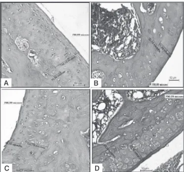
When cellularity was compared between the different groups, it was observed that only the negative control and IAA groups were not statistically different, with averages (41.6 and 40.1 cells/μm2)
showing significantly a trend of IAA group to normality. This is illustrated in Figure 1, respectively, by images 1A and 1B. ISS group averaged (63.1 μm2) with characteristics of hypercellula-rity grade 1, so that there is an increase in the number of cells (P <0.05). However, it is a disorderly growth, since it does not obey the peculiar columnar requirements of its layers as seen in seen in Figure 1C. However, the C+ group (positive control) showed results that are different from others with a lower sco-re (13.0 μm2) of the number of chondrocytes with thinning the
number of cells (P<0.05), showing a deteriorating state of the chondrocytes with general immaturity chondral characteristics, showing hypocellularity grade 3 found on Figure 1D.
When thickness of the cartilage were compared between the two groups it was seen that the positive control group showed the greatest mean thickness of calcified cartilage (104.8 microns), statistically equal to no other (P<0.05), which can be seen in Figure 1C. On the other hand, the negative control group, IAA group and ISS lower averages of calcified cartilage (p<0.05) were observed, respectively 39.6μm2, 21.6 μm2, and 28.5 μm2,
which can be seen in the in Figures 1C, 1B and 1D respectively, suggesting a maintenance of the physiological characteristics of these layers. Regarding the specific thickness of the non--calcified cartilage layer, what should be seen is a reversal in its distribution, noting that the positive control group has the lowest average non-calcified cartilage (53.3 μm2), therefore has
a lower thickness, as opposed to any other treatment (p<0.05), as illustrated in Figure 1C. The negative control group showed the highest average thickness (134.1 μm2), while ISS and IAA
groups obtained means of 91.8 μm2 and 75.5 μm2, respectively,
suggesting that the normal thickness compared with the positive control group (p <0.05) as demonstrated in Figure 1D and 1B.
Figure 1. Photomicrographs of the distal femoral region stained with he-matoxylin-eosin.
DISCUSSION
The results show that the cellular and morphological behavior usually varied with the treatments to articular cartilage chan-ged its behavior to electrical therapy. Regarding the variable cellularity, chondrocytes groups that were treated with galvanic current with or without L-ascorbate had increased cellularity suggesting comparative normality between IAA and negative control groups. This fact indicates that the treatment with direct current was effective regarding the secondary prevention of loss of chondrocytes, making this increase in cell number co-herent with the same arrangement of layers that form cartilage, with statistical significance. ISS group had the highest average cellularity, which means an increase in this variable, but not to converge to a physiological improvement, because there was isogenous group formation.
This induction feature in tissue proliferation is consistent and meets the study of Grace et al.9 and Aaron et al.12 which
exa-mined the effect of electric field in osteochondral defects in the trochlear groove of rats, where they could observe initial vascu-lar reaction, matrix synthesis, tissue repair and chondrogenesis with the application of continuous current.
The devices of therapeutic electrical current are designed to mimic the functions of the human body bioelectrical signals, generating an electric current to compensate for the bioelectri-city that was reduced in the injured tissue. This increases the body’s ability to transport nutrients to the cells in the injured tissue.13,14 According to the results obtained by comparing the
positive control group and the groups based on iontophoresis saline and ascorbic acid shows an improvement in the prolife-rative state denoted histological slides clearly show that a gain variable cellularity.
Comparing groups, treatment with ascorbic acid in this study also showed an improvement in cell allocation in the group that it was administered, and in accordance with the study proposed by Chowcat et al.,15 indicating that collagenase is synthesized
A
C
B
D
205 and controlled by a tissue inhibitor of metalloproteinase. The
authors add that supplementation with vitamin C, despite ha-ving no influence on these enzymes, provides greater flow of substrate, which could be responsible for chemical reactions that induce the synthesis of colagen.16,17
The thickness alteration of cartilage tissue was also a factor of investigation and is an important indicator of degeneration, since the articular cartilage depends on the composition and organization of the extracellular matrix beyond the relationship itself between the chondrocyte matrix and resistance to me-chanical load.
There is little research in the literature on the evaluation of the thickness of different regions of cartilage. Some theories pro-posed by Carter et al.18 show that the decrease in hydrostatic
load promotes an inhibition of physiological evolution in the subchondral bone and calcified cartilage layer, preventing it from moving toward the articular surface. According to a stu-dy by Del Carlo et al.,19 it has been observed an increase in
calcified cartilage in animals with degradation of the most su-perficial layer. It was also found that the subchondral bone of rats which cartilage had lost the ability to absorb mechanical stress and, thus, needed to receive higher load increased the layer of calcified cartilage.
Thus, the present study confirms what was described by these authors, showing that an increase of calcified cartilage in the positive control group, i.e. groups which were not treated with direct current alone and L-ascorbic acid, characterizing, thus, reduction and alteration in cellular distribution.
The limitations of this study, regarding the failure to use addi-tional histological analysis is justified by the possibility of tes-ting the digestion of protein from samples for quantification of proteoglycans using an agarose gel, and therefore, leaving the impregnation by safranin for further studies.
CONCLUSION
The study of the histomorphometric response of articular carti-lage in arthritis-induced rats, compared to treatment with ionto-phoresis alone and in the presence of L-ascorbic acid allowed to conclude that:
The electric current with l-ascorbic acid procedure used in this study was beneficial for articular cartilage with osteoarthritis previously induced by Zymosan, promoting changes in the car-tilage tissue leading to normality of the variable cellularity and maintaining the thickness of the non-calcified cartilage. Regar-ding the untreated group, it showed cellular and non-calcified cartilage layer reduction and increased of the calcified cartilage.
REFERENCES
1. Pizzorno J. Natural medicine approach to treating osteoarthritis. Altern Com-plement Ther. 1995;1:93-5.
2. Chubinskaya S, Hurtig M, Rueger DC. OP-1/BMP-7 in cartilage repair. Int Orthop. 2007;31(6):773-81.
3. Goodheart G. A presentation of a new approach to correction of disc lesions, ACA Journ. Chiro. 1954;36-7.
4. Nakayama J, Fujioka H, Nagura I, Kokubu T, Makino T, Kuroda R, et al. The effect of fibroblast growth factor-2 on autologous osteochondral transplanta-tion. Int Orthop. 2009;33(1):275-80.
5. Frasnelli ME, Tarussio D, Chobaz-Péclat V, Busso N, So A. TLR2 mo-dulates inflammation in zymosan-induced arthritis in mice. Arthritis Res Ther.2005;7(2):R370-9.
6. Fenske NA, Lober CW. Structural and functional changes of normal aging skin. J Am Acad Dermatol. 1986;15(4 Pt 1):571-85.
7. Ross ML, Reith EJ, Rownell LJ. Histologia texto e atlas. São Paulo: Panameri-cana;1993. p. 47-115, 347-76.
8. Rocha FA, Aragão AG Jr, Oliveira RC, Pompeu MM, Vale MR, Ribeiro RA. Periarthritis promotes gait disturbance in zymosan-induced arthritis in rats. Inflamm Res. 1999;48(9):485-90.
9. Grace KL, Revell WJ, Brookes M. The effects of pulsed electromagnetism on fresh fracture healing: osteochondral repair in the rat femoral groove. Ortho-pedics. 1998;21(3):297-302.
10. Leroux MA, Cheung HS, Bau JL, Wang JY, Howell DS, Setton LA. Altered
mechanics and histomorphometry of canine tibial cartilage following joint im-mobilization. Osteoarthritis Cartilage. 2001;9(7):633-40.
11. Vanwanseele B, Lucchinetti E, Stüssi E. The effects of immobilization on the characteristics of articular cartilage: current concepts and future directions. Osteoarthritis Cartilage. 2002;10(5):408-19.
12. Aaron RK, Ciombor DM. Acceleration of experimental endochondral ossifi-cation by biophysical stimulation of the progenitor cell pool. J Orthop Res. 1996;14(4):582-9.
13. Cheng N, Van Hoof H, Bockx E, Hoogmartens MJ, Mulier JC, De Dijcker FJ,et al. The effects of electric currents on ATP generation, protein synthesis, and mem-brane transport of rat skin. Clin Orthop Relat Res.1982;(171):264-72.
14. Green PG. Iontophoretic delivery of peptide drugs. J Control Release. 1996; 41(1-2):33-48.
15. Chowcat NL, Savage FJ, Hembry RM, Boulos PB. Role of collagenase in colonic anastomoses: a reappraisal. Br J Surg. 1988;75(4):330-4.
16. Arantes VN, Okawa RY, Silva AA, Barbosa AA, Petroianu A. Efeito da metilpredni-solona sobre a tensão anastomótica jejunal. Arq Gastroenterol. 1994; 31(3): 97-102. 17. Barbul A. Immune aspects of wound repair. Clin Plast Surg. 1990;17(3):433-42. 18. Carter DR, Wong M. The role of mechanical loading histories in the
develop-ment of diarthrodial joints. J Orthop Res. 1988;6(6):804-16.
19. Del Carlo RJ, Galvão MR, Viloria MIV, Natali AJ, Barbosa ALT, Monteiro BS, et al. Experimental immobilization and remobilization rat knee joints: clinical and microscopic study Arq Bras Med Vet Zootec. 2007;59(2):363-70.