Review
Emerging Functions for the
Staphylococcus aureus
RNome
Julien Guillet., Marc Hallier., Brice Felden*
Rennes University, Inserm U835-UpresEA2311, Pharmaceutical Biochemistry, Rennes, France
Abstract: Staphylococcus aureus is a leading pathogen for animals and humans, not only being one of the most frequently isolated bacteria in hospital-associated infec-tions but also causing diseases in the community. To coordinate the expression of its numerous virulence genes for growth and survival, S. aureus uses various signalling pathways that include two-component regula-tory systems, transcription factors, and also around 250 regulatory RNAs. Biological roles have only been deter-mined for a handful of these sRNAs, including cis,trans, andcis-transacting RNAs, some internally encoding small, functional peptides and others possessing dual or multiple functions. Here we put forward an inventory of these fascinating sRNAs; the proteins involved in their activities; and those involved in stress response, metab-olisms, and virulence.
Introduction
Staphylococcus aureusis a commonly isolated bacterial pathogen in humans and animals and a serious threat to health. It can live as a commensal but, provided suitable opportunity, can initiate severe infections at various body sites. S. aureus is one of the most frequently isolated pathogens in hospital-associated infections but can also cause diseases in the community [1]. Nosocomial and community-acquired S. aureus infections include superficial skin lesions such as boils, abscesses, and impetigo, while invasive infections include septic arthritis, pneumonia, osteomyelitis, and endocarditis. S. aureus is an aggressive pathogen due to the combination of elevated antibiotic resistance and prominent virulence. The virulence of S. aureus is defined by a series of determinants that are often redundant in their functions. This bacterium produces an array of cell surface and secreted factors, including proteins that promote adhesion to host cells and tissues and some that bind proteins in blood toevade triggered immune responses. The organism also secretes extracellular enzymes such as proteases, a hyaluronidase, a lipase, and a nuclease that facilitate host tissue destruction and spreading. It produces membrane-damaging toxins that lyse host cells, as well as superantigens that are immunostimulatory exotoxins [2].
To face and adapt to various environmental conditions, including host colonization and spreading, S. aureus possesses many signaling pathways, some that are redundant, to coordinate the expression of its numerous virulence genes. At least 12 two-component regulatory systems and several transcription factors control these regulatory circuits, with multiple and intricate interplays to specifically reprogram the expression of target genes for continuous adaptation. Dozens of regulatory RNAs (sRNAs) are also involved in such dedicated control of gene expression, but their direct mRNA targets are, for the most part, currently unknown. Additionally, translation control and decay of selected S. aureus mRNAs, in response to specific signals duringS. aureus
growth and adaptation, can be achieved by specific ribonucleases [3] organized into large multi-enzyme complexes [4]. Widespread mRNA antisense transcription all over theS. aureusgenome [5], as well as dedicatedcisandtranssRNAs (reviewed in [6,7]), actively participate in these gene expression controls.
More than 250 srna genes were discovered and detected as expressed transcripts in variousS. aureusstrains and experimental conditions [8–15]. The vast majority of these sRNAs are only expressed inS. aureus, a few are detected in Bacillaceae (e.g. RsaE), and several housekeeping sRNAs are detected in all eubacteria (e.g. tmRNA, RNase P RNA, 6S RNA). MostS. aureussRNAs are located within the core genome, with a few expressed from the pathogenicity islands and from plasmids. For the most part, their functional, structural, and mechanistic details are unknown. This review will focus on the current functional understanding of cis-andtrans- regulatory RNAs expressed in this organism, the unusual cases ofcissRNAs acting intrans, those expressing small peptides, and the sRNAs possessing multiple functions. We will exclude the S. aureus riboswitches that are cis-acting regulatory domains of mRNAs. The various proteins associated with S. aureus sRNA functions will be described, including the controversial roles of Hfq. The emphasis will be placed on sRNAs involved in stress response and metabolisms and on several sRNAs implicated inS. aureuspathogenesis.
A Multiplicity of sRNAs Expressed by
S. aureus
Cis-encoded antisense RNAs
Cis-encoded antisense sRNAs are transcribed on the strand opposite to their target mRNAs [16,17] and regulate gene expression by base-pairing with their complementary mRNAs (Figure 1A). Despite an extended complementarity with their primary target encoded on the opposite DNA strand, the initial interaction between the mRNA and the sRNA, ‘‘a kissing inter-action,’’ occurs by contact between a few nucleotides usually
Citation: Guillet J, Hallier M, Felden B (2013) Emerging Functions for the Staphylococcus aureus RNome. PLoS Pathog 9(12): e1003767. doi:10.1371/ journal.ppat.1003767
Editor:Chetan E. Chitnis, International Centre for Genetic Engineering and Biotechnology, India
PublishedDecember 12, 2013
Copyright:ß2013 Guillet et al. This is an open-access article distributed under the terms of the Creative Commons Attribution License, which permits unrestricted use, distribution, and reproduction in any medium, provided the original author and source are credited.
Funding:This work was supported by the Institut National de la Sante´ et de la Recherche Me´dicale (INSERM; BF) and by the Agence Nationale pour la Recherche (ANR-09-MIEN-030; BF). The funders had no role in study design, data collection and analysis, decision to publish, or preparation of the manuscript.
Competing Interests:The authors have declared that no competing interests exist.
* E-mail: [email protected]
located in accessible hairpins. This interaction is followed by additional pairings involving structural rearrangements of the two interacting RNAs [18]. InS. aureus, the firstcis-encoded sRNA identified controls the rolling-circle replication of plasmid pT181 by transcriptional attenuation [13]. pT181 regulates its replication by the expression of an antisense RNA (RNAI) that blocks the expression of the plasmid-encoded replication initiation protein RepC. This mechanism involves pairings between complementary loops in the mRNA leader and the antisense RNA, which results in the formation of a transcription-termination hairpin 59to the Rep initiation codon. Attenuation is very efficient, aborting .90% of the Rep transcripts under standard growth conditions. Several othercis-encoded sRNAs expressed byS. aureuswere detected in mobile genetic elements (PIs, plasmids, transposons) that are complementary to mRNAs expressing transposases involved in genome plasticity and integrity. RsaOX is complementary to the coding sequence of the SA0062 mRNA encoding a putative transposase [11]. Another transposase, IS1181, is probably also regulated by two additional sRNAs, RsaOW/Teg17 and Teg24as, which are complementary to its 59 UTR, including a portion of
the Shine Dalgarno (SD) sequence, and also to the 39 UTR [10,11]. In strain N315, the gene encoding the transposase is repeated eight times and these two sRNAs are systematically detected on the tnp locus [10]. Cis-encoded sRNAs can also interact with additional mRNA targets at distant genetic loci, in trans.
Trans-encoded sRNAs
In contrast to thecis, thetrans-encoded sRNAs are transcribed at distant genetic loci from their molecular targets and share only partial and often interrupted pairing complementarities, as for the eukaryotic microRNAs (Figure 1B). Although a seeding interaction of six to seven nucleotides is sufficient to initiate the ‘‘sRNA– mRNA’’ interaction in E. coli [19], pairings are usually much longer for the sRNAs expressed byS. aureus, probably due to its AT-rich genome. In most cases, the interaction involves the 59
domains of the sRNAs that encompass the translation initiation signals (TIS) of the mRNA targets [20]. Conserved and accessible motifs were detected in several S. aureus sRNAs containing consensus sequences involved in the initial pairing with their
Figure 1. A variety of mechanisms of actions for theS. aureussRNAs.(A)Cis-encoded sRNAs bind via perfect complementarities with mRNA targets at the translation initiation sequence, preventing ribosome binding and therefore translation. (B)Trans-acting sRNAs. Thetrans-encoded sRNAs bind and block the ribosome binding site by interrupted pairings, using one or two hairpin(s) to repress translation initiation. (C)Cis-encoded antisense sRNAs acting intrans. In the SprA1/A1ASTA module, SprA1ASprevents SprA1 translation to prevent toxic peptide expression. On the two
target mRNAs. Several sRNAs from the Rsa family contain unpaired and accessible UCCC motives located in conserved hairpin regions of the sRNAs predicted to interact with the target mRNA translation initiation signals [12]. The RNAIII also harbors several UCCC motifs in the three loops H7, H13, and H14, which interact with the SD sequences of several target mRNAs. These UCCC motives were also detected in sRNAs expressed from other gram-positive bacteria such asL. monocytogenes andB. subtilis[21–24]. Accessible C-rich boxes could be a general signature of regulatory RNAs controlling translation initiation of various target mRNAs.
Cis-encoded antisense sRNAs acting in trans
Type I Toxin/Antitoxin (TA) pairs are present on plasmids or chromosomes, or both simultaneously [25], and consist of stable toxins and labile antitoxins encoded within small genetic modules. InS. aureus, several candidates of type I TA were detected based on sequence homology [26], among which are SprA1/A1ASand
the SprG/F modules. SprA1-SprA1ASis an RNA pair transcribed
from a pathogenicity island, a genetic element acquired by horizontal transfers. SprA1 is a stable and structured 208 nt-long RNA that contains an internal open reading frame (ORF) encoding a cytolytic peptide, pepA1 [27]. PepA1 inserts within the biological membranes, alters their integrity, and induces cell death [28]. A smallcis-antisense RNA of SprA1, named SprA1AS
(Teg152, [10]), is transcribed from the complementary DNA strand. Although 35 nt at SprA1AS 39-end are perfectly
comple-mentary to a sequence located at SprA1 39-end, SprA1ASacts in
transby base pairing with the 59domain of SprA1 to repress pepA1 translation by occluding the TIS, preventing its toxicity for the bacteria (Figure 1C). By analogy, the SprA2/A2AS pair,
consid-ered as a second copy due to elevated sequence identity with SprA1/A1AS, may also act intrans. Another unconventional case
of a cis-trans RNA was detected in a plasmid from Enterococcus faecalis, another gram-positive bacterium, within the par stability determinant of a plasmid required for stable inheritance in its host [29]. In that case, overlapping RNAs I and II share a bidirectional transcription terminator but interact with one another by two non-overlapping direct repeats.
sRNAs with Multiple Functions
SsrA/tmRNA responsible fortrans-translation and acting as atrans-RNA
Transfer-messenger RNA (tmRNA or SsrA) is an sRNA expressed in all bacteria that displays both tRNA and mRNA properties. tmRNA, with the help of the SmpB protein, governs trans-translation, a process that rescues the ribosomes stalled during translation of defective mRNAs, such as those lacking in-frame termination codons [30,31]. tmRNA is recruited by the ribosomes through the smpB protein, an essential participant for ribosome rescue, and acts first as transfer RNA (tRNA) to add an alanine to the stalled polypeptide chain. Translation then switches from the problematic mRNA to a short tmRNA internal ORF that encodes a proteolytic tag [32,33]. The stalled ribosome is released at the tmRNA termination codon, and the problematic mRNA and the tagged protein are degraded by specific RNAses and proteases, respectively. Trans-translation allows ribosome recy-cling and degradation of potentially toxic truncated mRNAs and proteins. A recent study [34] showed that tmRNA activity inS. aureus was not restricted totrans-translation. Inactivation of ssra-expressing tmRNA leads to an increase of pigment synthesis that is counteracted by expressing a tmRNA harboring a mutated tag. Furthermore, this phenotype is not imputable to the alteration of
trans-translation since SmpB inactivation did not modify the quantity of pigments produced byS. aureus. The phenotype is due to the overexpression of the crtMN operon which encodes two enzymes involved in pigment synthesis. As the tmRNA sequence displays partial complementarity with the 59 UTR of thecrtMN mRNA, it could also act as an antisense sRNA acting intransto regulatecrtMNmRNA translation [34].
RNAIII, the Paradigm
influence of RNAIII on membranes, surface proteins, and cell wall turnovers contributes to virulence by controlling nutrients’ entries and host defences, struggle, and resistance by regulating hemoly-sins production and host immune evasion. We expect that RNAIII possesses additional targets involved inS. aureusvirulence control, which will be progressively identified by high throughput methods using Deep RNA sequencing technologies combined with target affinity purifications.
Proteins Involved in
S. aureus
sRNA FunctionsHfq: A controversial factor
Since trans-encoded sRNAs display short and imperfect com-plementarity to their target mRNAs, the effective sRNA-mRNA annealing requires an auxiliary factor in some bacterial species. Hfq, a member of the conserved RNA-binding Sm-like protein family, is needed for the efficient annealing of some sRNAs to target mRNAs and for the intracellular stability of these sRNAs. In the case of canonical sRNA, the Hfq protein enhances the binding of sRNA on the translational start site of their mRNA targets and prevents ribosome binding. Hfq is only active in its multimeric form. The Hfq ring formed by homohexameric Hfq proteins displays a characteristic doughnut-shaped structure containing two single-stranded RNA-binding faces located on opposite sides of the ring. The proximal face binds to AU-rich sequences and sRNAs, whereas the distal face interacts with poly(A) sequences [46,47]. The dual RNA-binding surface allows the simultaneous recruit-ment of an sRNA and its mRNA target on one Hfq molecule. Hfq facilitates sRNA–mRNA interaction by increasing the local concentration of the RNA species and/or by enhancing the base pairing interaction through a restructuring of these RNAs. InE. coli, the chaperon Hfq affects the turnover of some target mRNA by recruiting an RNase E in an activated state on the sRNA– mRNA duplex [48,49]. The requirement of Hfq for riboregula-tions by trans-encoded sRNAs depends on the small RNAs, mRNAs, and the bacterial species. Potential links between the free energy for sRNAs–mRNA pairing, the GC-content of the bacterial genome, and the involvement of Hfq protein have been proposed [50]. A highestDG value and a lowest GC-content correlate with a dispensability of Hfq protein for the sRNAs–mRNAs pairing. Accordingly, Hfq is required in sRNAs regulations, which are involved in the growth, the sensitivity to various environmental stresses, and the virulence of several gram-negative strains. Moreover, Hfq or Hfq-like proteins are absent in several ‘‘low GC’’ gram-positive strains such as Streptococcus pneumoniae and Lactococcus lactis. InS. aureus, the function of Hfq remains unclear. Inactivation of the hfq gene in three S.aureus strains (RN6390, COL, and NEWMAN) did not affect the phenotypes of these strains [51]. InS. aureus, Hfq was not involved in more than 2,000 phenotypes tested, including sensitivity to different stress condi-tions, antibiotic sensibility, and virulence [51]. In agreement with the Hfq dispensability in the riboregulation, the protein has no effect on the stability/turnover of several trans-acting sRNAs [12,27,43,45,52,53]. The S. aureus Hfq does not enhance the binding of RNAIII tospamRNA orSA1000mRNA, whereas an Hfq-RNAIII complex from RN6390 cells co-immunoprecipitates with an antibody against Hfq [43,52]. However, Hfq seems to be functional in S. aureus, since the overexpression of Hfq in an agr2 strain leads to the stimulation of the c-haemolysin translation. This discrepancy between the ability of Hfq to tightly bind RNA in vitro and its inability to affect the riboregulation in vivo could be explained by the very low expression levels of Hfq in the laboratory-adapted strains RN6390 and COL. Indeed, inactivation of thehfqgene in theS. aureus8325-4 strain expressing
a detectable Hfq level alters the expression profiles of 116 genes potentially involved in the decrease of the pathogenicity of the muted strain in a murine peritonitis infection model and in the increase of expression of the surface carotenoid pigment [54]. The relationship between Hfq and carotenoid production was also revealed in anotherS. aureusstrain. Low-fluid-shear culture of N315 cells, which promotes attachment-independent biofilm formation, leads to a decrease of carotenoid production associated with a down-regulation of the Hfq protein [55]. Hfq specifically binds to 49 of the 116 genes down-regulated in thehfqmutant of S. aureus 8325-4. In particular, some mRNA targets of sRNAs, such assbi,sucD, androtfor respectively SprD, RsaE, and RNAIII, were copurified with Hfq, suggesting that Hfq could be implied in the translational regulation of some S. aureus genes [54]. Altogether, these studies show that the modulation of virulence and stress response could be attributed to Hfq in some strains. However, the direct involvement of sRNAs in these Hfq-dependent phenotypes and the mechanisms of actions of potential ‘‘sRNA/Hfq’’ complexes remain unknown. AsS. aureusdoes not express RNase E, Hfq could recruit another endoribonuclease to affect the turnover of mRNA targets. Hfq proteins from different bacteria contain an evolutionarily conserved core of 65 amino acids and a divergent positively charged terminal end. The C-terminus extensions are short in gram-positive bacteria (like S. aureus,B. subtilis, andL. monocytogenes) and longer in gram-negative bacteria (102 amino acids inE. coliandSalmonella). Recently the C-terminal extension ofE. coliHfq protein was shown to be required for a non-canonical sRNA pathway, a translational regulation involving the binding of sRNA outside the canonical ribosome entry site, probably by recruiting additional RNAs or proteins on the mRNA target [56]. Thus, the short C-tail extension mainly present inS. aureuscould be involved in the recruitment of specific ligands during the Hfq-dependent riboregulation by non-canonical sRNAs. InS. aureusstrains that do not express the protein, the role of Hfq might be superseded by other RNA-binding molecules.
RNAse III: The major RNase involved in the sRNA-dependent mRNA turnover
improving the stability of RNAIII–mRNA duplexes. RNAse III only cleaves RNAIII when the sRNA is bound to its mRNA targets [52,62]. RNAIII binds to the ribosome binding site ofcoa androtmRNAs and recruits RNase III, which cleaves the mRNA target at an equivalent position of the loop–loop pairing. The RNase III cleavage site is independent of the sequence of the base pairs. The loop–loop interaction forms a unique hairpin motif creating a single binding site for the RNase III, which leads to a specific cleavage at single positions of the kissing interactions and irreversible repression of mRNA translation [62]. Several RNase III-binding sRNAs were identified by deep sequencing of RNA coimunoprecipitated with a wild-type RNase III and/or cleavage-defective mutants in vivo. Among the 58 sRNAs detected, many have been previously identified, such as the pathogenicity island-encoded small RNAs SprA, SprA3, SprB, SprC, SprF3/G3 [15], and RsaA, RsaE, RsaH, RsaI, RsaJ [11,12], as well as RNAIII. Some of these sRNAs were copurified with the cleavage-defective mutant and display hairpin motifs recognizable by the enzyme, suggesting that there are substrates of RNase III [60,62]. Thus, it appears that most of these known and unknown sRNAs are potential trans-acting factors which regulate gene expression by antisense mechanisms and recruit RNAse III to direct the mRNA decay. Also, RNase III could mediate specific cleavage of type I toxin/antitoxin pairs (SprA1/SprA1AS, SprG/SprF) to prevent
toxic peptide expression. RNase III is associated with a large number of antisense transcripts, covering 44% of the annotated genes [60,63]. These antisense (as) RNAs are issued from a genome-wide process of overlapping transcription and are perfectly complementary to the 59ends, 39ends, middle, or entire genes or operons [5,60,64]. RNase III-associatedcis-asRNAs are usually expressed at lower levels than their complementary mRNAs, and they direct the degradation of residual mRNAs. The RNase III-mediated digestion of sense/antisense transcripts generates a large collection of short, 22 nucleotide-long, double-stranded RNAs that could also have functions [64]. Pervasive transcriptions lead to the expression of RNase III-associated mRNAs, which overlap at their 59or 39UTRs. RNase III-induced cleavages of the 59overlapping regions of divergent mRNAs allow the fine regulation of the expression of genes which have to be expressed in a coordinated manner as the tagG/tagH teichoic acid biosynthetic genes encoding the TagGH ABC transporter complex [60,63]. These cleavages generate mRNAs with shorter 59ends which could be more sensitive to degradations and/or influence their translations. In some cases, the 59UTRs of mRNAs extend into the coding sequence of their neighbouring genes [64]. Similar large asRNAs are encoded at particular genomic loci called the ‘‘excludons’’ in the gram-positive Listeria monocytogenes [65]. These long asRNAs span divergent genes or operons with related or opposing functions, and allow meticulous regulatory switches in bacteria [66], probably also occurring inS. aureus. In S. aureus, the activity of RNase III, in association with large asRNAs, could be considered a general mechanism to regulate and coordinate the expression of neighbouring genes.
Other RNases: RNase J1-J2 and RNase Y
InE. coli, sRNA-mediated mRNA decay mainly involves the recruitment of RNase E on the mRNA target. The RNase E is the central component of the degradosome that is composed of a 39-exoribonuclease polynucleotide phosphorylase (PNPase), a RNA helicase (RhlB) and a glycolytic enzyme (enolase). RNase E catalyzes the initial endoribonucleolytic cleavage of mRNA targets which is followed by a directional, 39to 59, degradation by the PNPase with the help of RhlB [67]. Most gram-positive bacteria do not contain an RNase E and use RNase E functional
orthologs and other degradosome components to direct mRNA decay. BothS. aureus and B. subtilis contain a similar multicom-ponent ribonucleolytic degradosome complex formed around RNase Y, a functional homologue of RNase E [4,68]. The S. aureus degradosome includes both RNases J1/J2 originally proposed to act also as functional orthologs of RNase E [69], the PNPase, the enolase, the RNA helicase CshA, and RNase P [4]. The CshA Dead-box RNA helicase plays an essential role in the regulation of quorum sensing by controlling the agrBDCA mRNA turnover [70,71]. Both RNases Y and J1, which exhibit RNase-E–like 59 end-dependent endonucleolytic activity, play a central role in the degradation of mRNAs in B. subtilis. The endonucleolytic cleavage by the RNase Y initiates the mRNA decay, and the resulting RNA fragments are likely to be degraded by the 59-39exoribonuclease activity of RNase J1 and by the 39–59
activity of PNPase [72,73]. In contrast toB. subtilis, the membrane-associated RNase Y of S. aureus is not essential for growth but is required for virulence [74,75]. The enzyme is involved in the processing of the global virulence regulator sae and in the expression of various virulence genes by an indirect mechanism [75]. The RNase Y controls the stability of specific mRNAs and sRNAs. Interestingly, inactivation of the rnc gene encoding for RNase Y inS. aureusresults in an increase of the half-life of two sRNAs, RsaA and Sau63 [8,12], whereas the RNAIII steady-state level is unaffected. The specific activity of RNase Y represents a way to control both the expression of sRNAs and their mRNA targets in S. aureus. A similar regulation was detected in E.coli, where RNase E specifically affects the steady-state level of several sRNAs [59]. In contrast with the activity of RNase E inE. coli, an implication of RNase Y in the coupled degradation of sRNAs and their mRNA targets has not been revealed.
sRNAs Involved in Stress Response, Metabolisms, and Regulatory Networks
Sigma B-inducible small RNA encoding genes
The pathogenicity ofS. aureusdepends on its ability to respond quickly and specifically to a variety of environmental stresses and to control virulence genes expression. TheS. aureusgenome allows expression of the alternative sigma B transcriptional factor (sB
) that is an essential part of the complex regulatory network controlling the expression of around 200 genes involved in virulence, cell wall metabolism, and membrane transport processes [76–78]. sB
is involved in stress responses and contributes to pathogenesis in animal models of infections [79]. TheS. aureus sigma B operon resembles that of the homologous B. subtilis operon. It containssB, an anti-sBfactor RsbW, an anti-anti-sB
factor RsbV, and RsbU, a Mn2+-dependent phosphatase that positively controlssB
activity by dephosphorylating RsbV [80,81]. The sigma B regulon includes genes directly up-regulated bysB
and genes indirectly regulated via sB
-dependent expression of regulatory factors such as the SarA transcription factor [76,77]. In particular, the inactivation ofsBhas an indirect impact on theagr quorum-sensing system by enhancing RNAIII expression [82]. By computational approaches based on the search forsB
consensus binding sites (GWWT_N14–17GGGWWW) and transcriptional
terminator sequence within the intergenic regions ofS. aureusstrain N315, three sB
-regulated genes coding for new sRNAs were identified and validated [53]. Two of these sRNAs, SbrA and SbrB, are highly conserved amongStaphylococci(forsB
potential dual function of SbrB: a peptide-coding sRNA and activity as an sRNA regulator [53]. The third sRNA, SbrC, is a potential cis-acting antisense targeting the 39 end of themntABC operon encoding for an ABC transporter dedicated in the uptake of manganese. The manganese acquisition is crucial for defence systems against oxidative stress and contributes to the virulence of S. aureus[83]. InS. aureus,sB
-dependent transcription is induced by the presence of MnCl2, probably via the stimulation of the
Mn2+-dependent phosphatase activity of RsbU [77]. The sB
-dependent induction of SbrC could be a way for sB to autoregulate its own activity and to modulate manganese uptake in function of Mn availability. The transcription of other sRNAs like RsaA, RsaD, and RsaF is induced in S. aureus strains expressing an activesB
factor [12]. These sRNAs are differently transcribed in response to environmental stresses such as oxidative stress, heat stress, cold stress, osmotic stress, and acidic pH. A conserved sB
promoter sequence was found upstream rsaA, suggesting its direct regulation by the sB
. As RsaA is a trans-acting regulator that potentially targets three mRNAs repressed by sB [12,76,77], it could be an intermediate in the regulatory
network controlled bysB.
Metabolisms regulations
In S. aureus, all macromolecules are synthetized from 13 biosynthetic intermediates produced by glycolytic, pentose phosphate, and tricarboxylic acid cycle (TCA cycle) pathways. These three central metabolic pathways are closely linked to the expression of several virulence factors. The alteration of pentose phosphate and glycolytic pathways affects the
quorum-sensing–dependent regulation of RNAIII [84,85], and TCA cycle inactivation induces a reduction in the production of several secreted virulence factors and cell-associated adhesion factors [86,87], thus, slowing down central metabolism reduces bacterial virulence. Recently, RsaE, a sRNA conserved in allS. aureusstrains, and also in firmicutes, has been shown to regulate several metabolic pathways [11,12] (Figure 2). The overexpression of RsaE induces a growth defect that is partially alleviated by the addition of acetate, arguing for a role of RsaE in both catabolisms and anabolisms. Indeed, RsaE down-regulates the synthesis of enzymes from the TCA cycle (succinyl-Coa synthetase sucC and sucD, aconitase (citB), citrate synthase (citZ), and isocitrate dehydrogenase (citC)), and from the folate-dependent one-carbone metabolism (bi-functional protein fold and the formate-tetrahy-drofolate ligase (Fhs)), which is involved in the purine biosynthesis pathway. RsaE also affects the amino acid pool inS. aureus. It up-regulates the expression of valine, leucine, and isoleucine operons and potentially alters aspartate biosynthesis by inducing the expression of pyruvate carboxylase. Moreover, RsaE down-regulates theopp-3operon coding for an oligopeptide transporter involved in the uptake of specific peptides and in the regulation of extracellular protease production [88,89]. As the transcription of genes encoding for some enzymes of the TCA cycle is regulated by the availability of amino acids [90], RsaE could indirectly modulate the TCA cycle via the regulation of the pool of free intracellular amino acids. The distinction between metabolic pathways directly and/or indirectly regulated by RsaE is difficult to apprehend because these pathways are highly interconnected. RsaE can directly regulate the TCA cycle and amino acid uptake
by inhibiting the formation of ribosomal initiation complex on sucD and opp3A/opp3B mRNAs, respectively [11,12]. The TCA cycle is involved in the energetic transition which uses the acetate accumulated in the extracellular medium during the glycolysis and amino acids as an alternative carbon source. Although the expression profile of RsaE is a subject of controversy [11,12], in someS. aureusstrains, RsaE is expressed at late exponential phase and repressed at stationary phase, and it could facilitate the transition of energy metabolisms, the purine biosynthesis, and amino acid transport in response to the nutrients’ availability. Moreover, the RsaE expression seems to be dependent on theagr quorum-sensing system and sBactivity, suggesting that it could modulate the metabolism profile in function of stress responses and/or virulence [12].
sRNAs involved in global regulatory networks
S. aureusexpresses a large array of extracellular and cell-wall– associated virulence factors at different stages of the infectious
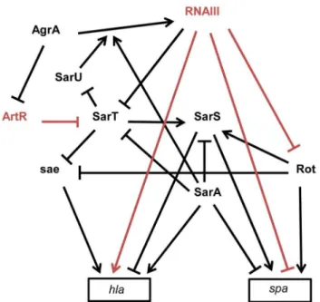
process. The exoproteins and cell-wall–associated adhesins, respectively involved in host immune evasion and host cell adhesion, are expressed early during the initial colonization, while the production of toxins that facilitateS. aureusgrowth and spread in the host tissues occurs late during infection. Their temporal expressions are controlled by two component regulatory systems (e.g. saeRS, arlRS, lytSR, srrAB) and global transcriptional regulatory factors (e.g. the sarA protein family, spX). The overlapping regulation between two component systems and global transcriptional factors constitutes a fine-tuning system for an efficient transcriptional control of virulence genes expression [91]. These factors affect the expression of virulence genes by directly binding to the promoters of target genes and/or indirectly through the regulation of the expression of global regulatory elements targeting the same set of virulence genes. RNAIII has character-istics similar to global regulatory factors that regulate directly and indirectly the expression of virulence genes, such as spa and hla genes [14]. The expression ofspaencoding an adhesin acting as an host immune evasion protein is directly controlled, at the translational level, by an ‘‘RNAIII-mRNA’’ direct pairing mechanism [14], as well as at the transcriptional level by three members (SarA, SarS, and Rot) of the SarA family of transcrip-tional regulators (Figure 3) [92].spais positively regulated by the transcriptional factors Rot (Repressor of toxins) and SarS and negatively regulated by SarA. RNAIII affects the mRNA level of spaby inhibitingrottranslation by a base pairing mechanism [45]. As Rot activates SarS transcription, RNAIII-mediated inhibition of Rot expression down-regulates the two transcriptional activators of spa. These transcriptional and translational controls avoid putative leakages in spamRNA expression. RNAIII uses similar double controls to up-regulate the expression ofhlaencoding the a-hemolysin [93]. RNAIII enhances a-hemolysin translation by a pairing interaction athlamRNA 59UTR [40] and up-regulates hla mRNA expression by down-regulating Rot, which acts as a repressor ofhlatranscription in SaeRS- and SarS-dependent ways [93,94]. In accordance with the antagonism between Rot and RNAIII, cellular amounts of Rot are inversely correlated to the RNAIII level in most S. aureus strains [95]. However, the transcriptomes ofS. aureusstrains deleted in RNAIII or in Rot only partially overlap, suggesting that RNAIII affects the expression levels of additional transcription factors [96]. One potential target of RNAIII could be the transcriptional factor SarT, which is down-regulated by agr at the post-exponential phase of growth [97] and contains a putative pairing interaction with RNAIII at its 59UTR [43]. RNAIII-mediated down-regulation of SarT, which acts as a positive and negative transcriptional regulator of sarSandhla, respectively, could be another way to controlspaand hlaexpression. The involvement ofS. aureussRNAs in the global gene regulatory network is not restricted to RNAIII. Recently, a new sRNA named ArtR (AgrA-repressed,toxin-regulating sRNA) was reported to activatea-hemolysin expression by binding to the sarTmRNA, promoting its degradation [98]. Although RNAIII and ArtR both similarly regulate hla expression, they display different expression patterns. In contrast to RNAIII, ArtR transcription is repressed by agrA, suggesting that ArtR-mediated hla up-regulation could be enhanced in agr-deficient strains. Multiple sRNAs controlling the expression of a similar compo-nent from a regulatory network allows the sharp regulation of virulence genes. Given the importance of multiple components from regulatory networks to express virulence genes and the elevated variability of their expression levels among theS. aureus strains, it is most likely thatS. aureusexpresses many other sRNAs that deeply interact with this network to influence bacterial virulence.
Figure 3. Schematic overview of the multiple interactions between sRNAs and transcriptional regulators involved inspa (protein A) andhla(a-hemolysin) expression inS. aureusstrain
8325-4. The arrows indicate the stimulations and the bars, the repressions. The direct effects of two sRNAs on gene expression are indicated in red. RNAIII repressesrotandspatranslation by direct pairing interactions [14,45]. Rot requires SarT to stimulate SarS in the presence of SarA [92,120]. In contrast to SarA, Rot and SarS are direct activators ofspa expression [92,121]. In the exponential phase of growth,spa transcrip-tion is stimulated by Rot and by SarS. In the post-exponential phase,spa transcription and translation are repressed by SarA and RNAIII, respectively, and the direct inactivation of Rot by RNAIII leads to the repression of the Rot and the SarS-dependent transcription activations ofspa.hlais up-regulated by SarA and down-regulated by SarS [93]. Rot and SarT represshlatranscription by asae-dependent way [94]. In the post-exponential phase of growth, RNAIII enhanceshlatranslation by direct pairings at thehlamRNA 59UTR and stimulateshlatranscription by down-regulating the expression of SarT and Rot. AgrA, the master transcriptional regulator of quorum sensing, stimulates RNAIII expres-sion [122] but also represses ArtR expresexpres-sion. ArtR indirectly activateshla transcription by repressing sarT translation [98]. SarA stimulates the AgrA-dependent expression of RNAIII [122]. SarT directly represses SarU which activatesagr(RNAIII) transcription [123].
sRNAs and Virulence Gene Regulations
The dual-function SCCmec-encoded psm-mec RNA suppresses agrA translation and attenuates MRSA virulence
SCCmec is a mobile genetic element that confers methicillin resistance to the methicillin-resistant S. aureus (MRSA) strains. SCCmec contains several genes, including the cytolysin psm-mec gene, whose transcription product suppresses colony spreading and the expression of phenol-soluble modulina, a cytolytic toxin [99]. The psm-mec RNA binds the agrA mRNA, encoding a virulence regulatory factor and inhibits its translation [100]. Deletion ofpsm-mecin MRSA clinical isolates increases virulence on mice skin infection models (Figure 4). The psm-mec RNA suppresses MRSA virulence by agrA translation inhibition, and the absence ofpsm-mecin community-acquired (CA) MRSA strains is responsible for their elevated virulence.
SprD, a pathogenicity island-encoded RNA regulating an immune-evasion molecule from the core genome
Small pathogenicity island rNA D9(SprD) is among the fewS. aureusRNAs with an identified function. SprD is expressed from the genome of a converting phage [15], a horizontally-acquired pathogenicity island (PI) being the repository of toxins, adherence, invasion factors, superantigens, and secretion systems [101]. SprD down-regulates, at the translational level, the expression of the Sbi immune evasion molecule located on the core genome [102]. One of its four hairpins binds the 59UTR of the sbimRNA by an antisense pairing mechanism. The initial binding involves the hairpin loop, and the interaction extends farther upstream and downstream from that initial binding site. The ‘‘SprD-sbimRNA’’ interaction sequesters the sbi mRNA TIS and, consequently, prevents translation initiation of the Sbi protein. Sbi is an immunoglobulin-binding protein expressed byS. aureus[103] that impairs the host immune response. Sbi acts as a complement inhibitor and forms a tripartite complex with host complement factors H and C3b [104]. SprD contains four hairpins, one of which interacts with the ribosome binding site of sbi mRNA to form a long imperfect duplex that prevents translation initiation in vivo. SprD contributes to causing disease in a mouse model of infection, although this effect is not only linked to the deregulation of Sbi production. It suggests that SprD regulates the expression of other targets playing important roles during host infection.
The implication of the 891 nucleotides-long small stable RNA42 inS. aureusvirulence
Small stable RNAs (SSRs) are RNAs specifically produced and/ or stabilized in response to various environmental conditions [104]. Among the SSRs, SSR42 is involved in host erythrocyte lysis, resistance to human polymorphonuclear leukocyte killing, and pathogenesis in a murine model of bacterial infection [3]. SSR42 is primarily expressed during the stationary phase of S. aureus growth, is a stable RNA with a,30 minute half-life, and appears to control the expression of a large set of target genes (,80) including virulence factors, which is the rationale of its involvement inS. aureuspathogenesis and virulence.
sRNAs Expressions during Infections
Staphylococcus aureus is a common resident of human skin and nasopharynx. It is also a cause of life-threatening illness, producing virulence factors that enable survival and spreading in various hosts. Its switch from commensalism to an infectious pathogen is poorly understood, whereas nasal carriage and clinical isolates
belong to the same genetic clusters [105]. The fewS. aureussRNAs with known targets regulate major biochemical pathways, some ultimately implicated in virulence [6]. TheS. aureussRNAs were detected and studied in various strains and their specific expression profiles during infection in humans are, for the vast majority of the ,250 sRNAs expressed by this bacterium, unknown. However, RNAIII expression in clinical samples, such as nasal secretions or cystic fibrosis sputa, has been monitored [106–108]. The majority of clinical isolates isolated from acute infections has functionalagr and produces RNAIII in vivo [109]. These data suggests that RNAIII influences the virulence phenotype. Agr-defective mu-tants, however, were detected in infected patients, and a mixture of agr positive and defective strains were detected in healthy humans [110]. Thus, agr is involved during acute infection, while agr mutants can be selected during chronic infections and dormant states. A recent study reported the expression profiles of the five sRNAs (RNAIII, RsaA, RsaE, RsaG, and RsaH) in strains isolated from patients with acute cutaneous infection, chronic cystic fibrosis, or nasal colonization [111]. The expression profiles of these five sRNAs are strain-specific and do not correlate to the type of infections or colonization, but the authors noticed that sRNA expression was more uniform among the strains from colonization compared to those responsible for infections. This observation might reflect the fact that S. aureus was primarily a commensal and then became an opportunistic pathogen [112,113]. Deep RNA sequencing technologies now allow global analyses of theS. aureusRNome in various clinical isolates to detect putative differences of expression of all sRNAs, with possible applications in the early diagnostic of strains that are susceptible to cause life-threatening infections.
Phenotypes Associated with sRNAs Expressions
Conclusion
Recent advances in the characterization of the plethora of regulatory RNAs expressed by S. aureus have provided novel
insights about how they monitor various cellular activities. Most of the few sRNAs whose physiological roles have been determined control the expression of genes involved in central metabolisms, in response to quorum sensing, and on virulence by pairing to target
Figure 4. sRNAs from theS. aureusRNome implicated in bacterial virulence.Multitasking RNAIII is the effector of quorum sensing to perceive bacterial population density and regulates multiple targets involved in peptidoglycan metabolism, adhesion, exotoxins production, and virulence. RNAIII internally encodes hemolysind(blue). RNAIII contains at least three repressor domains (red) containing accessible UCCC motifs that interact, by antisense pairings, with the ribosome binding sites of numerous target mRNAs for translational repression (Tr.R), some triggering endoribonuclease III (RNase III) cleavages to induce target mRNA degradations and irreversible gene expression decay. Translation of at least two exotoxins is activated by RNAIII, one encoded (hld), and another (hla) by translation activation (Tr.A). SprD is expressed from the genome of a converting phage and interacts, by antisense pairings, with the 59part of thesbimRNA encoding an immune evasion molecule. SprD possesses an important role inS. aureusvirulence, but the mechanism of its control is yet to be elucidated, with Sbi being only one player among others. The 891-nucleotide long SSR42 affects extracellular virulence expression, hemolysis, neutrophil virulence, and pathogenesis and contains a putative internal ORF. The mechanisms of target regulation remain to be elucidated. The SCCmec-encodedpsm-mecRNA suppressesagrAtranslation and attenuates MRSA virulence, acting as a dual-function RNA regulator.
mRNAs to modulate their translational activities and stabilities. Several sRNAs encode and express small peptides that may play important roles in virulence or in bacterial growth control. As is the case for some sRNAs expressed in other bacteria, it is likely that other mechanisms of action are used byS. aureussRNA such as molecular mimicry (e.g. the 6S RNA) or binding to regulatory proteins. The number of sRNAs identified in S. aureus has considerably increased in the past decade, up to 250 members, but the biological functions of most of them remain unknown. Some sRNAs that are expressed from mobile genetic elements can regulate target genes located on the core genome, as for SprD with Sbi, implying an efficient functional integration of the accessory genetic elements into the overall regulatory networks from theS. aureusgenome required for virulence. The sRNAs expressed from the core genome probably are involved in wider biological functions. Most of the well characterized sRNAs act as fine-tuning regulators by repressing the translational level of only one gene (Table 1), but it is likely that they target other genes and that one gene is regulated by different sRNAs. The identification of their molecular targets becomes a critical step to further understand their roles in bacterial homeostasis and pathogenesis. Bioinfor-matics approaches based on the prediction of sRNA base pairing within the TIS of mRNAs allowed identifying antisense targets of some sRNAs. However, these approaches often lead to false positive predictions and do not highlight the interactions outside the TIS that are not uncommon inS. aureus[44] and also in other bacterial species. In rare cases, quantitative proteomics and
microarray analyses of sRNA mutant strains have allowed the identification of target genes, but these genetic approaches are not well suited to detect the primary targets of the sRNAs involved in broad regulatory networks, such as RNAIII, which regulates the expression of the Rot transcription factor. Until now, few global regulatory sRNAs were identified. The identification of new sRNAs that have an impact on the regulatory network control and the characterization of mechanisms that allow them to connect environmental responses to other cellular processes are future challenges. InS. aureus, the characterization of sRNA functions is complicated by the elevated genetic variability between the strains. Such a high variance in the expression of virulence and transcription factors among theS. aureusstrains makes it difficult to generalize the functional impacts of a given sRNA to all the S. aureusstrains. The characterization of the input of these sRNAs in global gene expression will provide a better understanding of the processes allowing the extraordinary adaptation ofS. aureusin its various environments and its elevated pathogenicity in humans and animals. It should provide fundamental insights for potential therapeutic applications in using some of these sRNAs as early diagnostic markers and putative drug targets. Other future challenges will be to comprehend the contribution of S. aureus sRNAs during the various steps of the infectious process, host– pathogen interactions, colonization, spread, and antibiotic resis-tance. To tackle these ambitious goals, it will require developing elegant technologies in living animals to analyze the implications of theS. aureusRNome during infection.
References
1. David MZ, Daum RS (2010) Community-associated methicillin-resistant Staphylococcus aureus: epidemiology and clinical consequences of an emerging epidemic. Clin Microbiol Rev 23: 616–687.
2. Xu SX, McCormick JK (2012) Staphylococcal superantigens in colonization and disease. Front Cell Infect Microbiol 2: 52.
3. Morrison JM, Miller EW, Benson MA, Alonzo F, 3rd, Yoong P, et al. (2012) Characterization of SSR42, a novel virulence factor regulatory RNA that contributes to the pathogenesis of a Staphylococcus aureus USA300 representative. J Bacteriol 194: 2924–2938.
4. Roux CM, DeMuth JP, Dunman PM (2011) Characterization of components of the Staphylococcus aureus mRNA degradosome holoenzyme-like complex. J Bacteriol 193: 5520–5526.
5. Lasa I, Toledo-Arana A, Dobin A, Villanueva M, de los Mozos IR, et al. (2011) Genome-wide antisense transcription drives mRNA processing in bacteria. Proc Natl Acad Sci U S A 108: 20172–20177.
6. Felden B, Vandenesch F, Bouloc P, Romby P (2011) The Staphylococcus aureus RNome and its commitment to virulence. PLoS Pathog 7: e1002006. doi: 10.1371/journal.ppat.1002006.
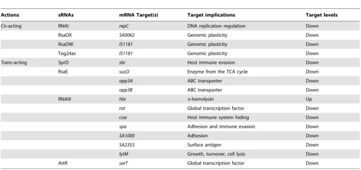
Table 1.Cis- andtrans-acting sRNAs, their corresponding mRNA targets, and physiological consequences.
Actions sRNAs mRNA Target(s) Target implications Target levels
Cis-acting RNAI repC DNA replication regulation Down
RsaOX SA0062 Genomic plasticity Down
RsaOW IS1181 Genomic plasticity Down
Teg24as IS1181 Genomic plasticity Down
Trans-acting SprD sbi Host immune evasion Down
RsaE sucD Enzyme from the TCA cycle Down
opp3A ABC transporter Down
opp3B ABC transporter Down
RNAIII hla a-hemolysin Up
rot Global transcription factor Down
coa Host immune system hiding Down
spa Adhesion and immune evasion Down
SA1000 Adhesion Down
SA2353 Surface antigen Down
lytM Growth, turnover, cell lysis Down
ArtR sarT Global transcription factor Down
7. Romilly C, Caldelari I, Parmentier D, Lioliou E, Romby P, et al. (2012) Current knowledge on regulatory RNAs and their machineries in Staphylo-coccus aureus. RNA Biol 9: 402–413.
8. Abu-Qatouseh LF, Chinni SV, Seggewiss J, Proctor RA, Brosius J, et al. (2010) Identification of differentially expressed small non-protein-coding RNAs in Staphylococcus aureus displaying both the normal and the small-colony variant phenotype. J Mol Med (Berl) 88: 565–575.
9. Anderson KL, Roberts C, Disz T, Vonstein V, Hwang K, et al. (2006) Characterization of the Staphylococcus aureus heat shock, cold shock, stringent, and SOS responses and their effects on log-phase mRNA turnover. J Bacteriol 188: 6739–6756.
10. Beaume M, Hernandez D, Farinelli L, Deluen C, Linder P, et al. (2010) Cartography of methicillin-resistant S. aureus transcripts: detection, orientation and temporal expression during growth phase and stress conditions. PLoS One 5: e10725. doi: 10.1371/journal.pone.0010725.
11. Bohn C, Rigoulay C, Chabelskaya S, Sharma CM, Marchais A, et al. (2010) Experimental discovery of small RNAs in Staphylococcus aureus reveals a riboregulator of central metabolism. Nucleic Acids Res 38: 6620–6636. 12. Geissmann T, Chevalier C, Cros MJ, Boisset S, Fechter P, et al. (2009) A
search for small noncoding RNAs in Staphylococcus aureus reveals a conserved sequence motif for regulation. Nucleic Acids Res 37: 7239–7257.
13. Novick RP, Iordanescu S, Projan SJ, Kornblum J, Edelman I (1989) pT181 plasmid replication is regulated by a countertranscript-driven transcriptional attenuator. Cell 59: 395–404.
14. Novick RP, Ross HF, Projan SJ, Kornblum J, Kreiswirth B, et al. (1993) Synthesis of staphylococcal virulence factors is controlled by a regulatory RNA molecule. Embo J 12: 3967–3975.
15. Pichon C, Felden B (2005) Small RNA genes expressed from Staphylococcus aureus genomic and pathogenicity islands with specific expression among pathogenic strains. Proc Natl Acad Sci U S A 102: 14249–14254. 16. Gottesman S, Storz G (2011) Bacterial small RNA regulators: versatile roles
and rapidly evolving variations. Cold Spring Harb Perspect Biol 3: a003798. 17. Waters LS, Storz G (2009) Regulatory RNAs in bacteria. Cell 136: 615–628. 18. Storz G, Vogel J, Wassarman KM (2011) Regulation by small RNAs in
bacteria: expanding frontiers. Mol Cell 43: 880–891.
19. Papenfort K, Vogel J (2010) Regulatory RNA in bacterial pathogens. Cell Host Microbe 8: 116–127.
20. Romby P, Vandenesch F, Wagner EG (2006) The role of RNAs in the regulation of virulence-gene expression. Curr Opin Microbiol 9: 229–236. 21. Gaballa A, Antelmann H, Aguilar C, Khakh SK, Song KB, et al. (2008) The
Bacillus subtilis iron-sparing response is mediated by a Fur-regulated small RNA and three small, basic proteins. Proc Natl Acad Sci U S A 105: 11927– 11932.
22. Mandin P, Repoila F, Vergassola M, Geissmann T, Cossart P (2007) Identification of new noncoding RNAs in Listeria monocytogenes and prediction of mRNA targets. Nucleic Acids Res 35: 962–974.
23. Nielsen JS, Olsen AS, Bonde M, Valentin-Hansen P, Kallipolitis BH (2008) Identification of a sigma B-dependent small noncoding RNA in Listeria monocytogenes. J Bacteriol 190: 6264–6270.
24. Toledo-Arana A, Dussurget O, Nikitas G, Sesto N, Guet-Revillet H, et al. (2009) The Listeria transcriptional landscape from saprophytism to virulence. Nature 459: 950–956.
25. Fozo EM, Hemm MR, Storz G (2008) Small toxic proteins and the antisense RNAs that repress them. Microbiol Mol Biol Rev 72: 579–589.
26. Fozo EM, Makarova KS, Shabalina SA, Yutin N, Koonin EV, et al. (2010) Abundance of type I toxin-antitoxin systems in bacteria: searches for new candidates and discovery of novel families. Nucleic Acids Res 38: 3743–3759. 27. Sayed N, Jousselin A, Felden B (2011) A cis-antisense RNA acts in trans in Staphylococcus aureus to control translation of a human cytolytic peptide. Nat Struct Mol Biol 19: 105–112.
28. Sayed N, Nonin-Lecomte S, Rety S, Felden B (2012) Functional and structural insights of a Staphylococcus aureus apoptotic-like membrane peptide from a toxin-antitoxin module. J Biol Chem 287: 43454–43463.
29. Greenfield TJ, Ehli E, Kirshenmann T, Franch T, Gerdes K, et al. (2000) The antisense RNA of the par locus of pAD1 regulates the expression of a 33-amino-acid toxic peptide by an unusual mechanism. Mol Microbiol 37: 652– 660.
30. Dulebohn D, Choy J, Sundermeier T, Okan N, Karzai AW (2007) Trans-translation: the tmRNA-mediated surveillance mechanism for ribosome rescue, directed protein degradation, and nonstop mRNA decay. Biochemistry 46: 4681–4693.
31. Gillet R, Felden B (2001) Emerging views on tmRNA-mediated protein tagging and ribosome rescue. Mol Microbiol 42: 879–885.
32. Haebel PW, Gutmann S, Ban N (2004) Dial tm for rescue: tmRNA engages ribosomes stalled on defective mRNAs. Curr Opin Struct Biol 14: 58–65. 33. Keiler KC (2008) Biology of trans-translation. Annu Rev Microbiol 62: 133–
151.
34. Liu Y, Wu N, Dong J, Gao Y, Zhang X, et al. (2010) SsrA (tmRNA) acts as an antisense RNA to regulate Staphylococcus aureus pigment synthesis by base pairing with crtMN mRNA. FEBS Lett 584: 4325–4329.
35. Novick RP, Geisinger E (2008) Quorum sensing in staphylococci. Annu Rev Genet 42: 541–564.
36. Benito Y, Kolb FA, Romby P, Lina G, Etienne J, et al. (2000) Probing the structure of RNAIII, the Staphylococcus aureus agr regulatory RNA, and
identification of the RNA domain involved in repression of protein A expression. RNA 6: 668–679.
37. Kreger AS, Kim KS, Zaboretzky F, Bernheimer AW (1971) Purification and properties of staphylococcal delta hemolysin. Infect Immun 3: 449–465. 38. Mellor IR, Thomas DH, Sansom MS (1988) Properties of ion channels formed
by Staphylococcus aureus delta-toxin. Biochim Biophys Acta 942: 280–294. 39. Verdon J, Berjeaud JM, Lacombe C, Hechard Y (2008) Characterization of
anti-Legionella activity of warnericin RK and delta-lysin I from Staphylococcus warneri. Peptides 29: 978–984.
40. Morfeldt E, Taylor D, von Gabain A, Arvidson S (1995) Activation of alpha-toxin translation in Staphylococcus aureus by the trans-encoded antisense RNA, RNAIII. Embo J 14: 4569–4577.
41. Liu Y, Mu C, Ying X, Li W, Wu N, et al. (2011) RNAIII activates map expression by forming an RNA-RNA complex in Staphylococcus aureus. FEBS Lett 585: 899–905.
42. Chavakis T, Hussain M, Kanse SM, Peters G, Bretzel RG, et al. (2002) Staphylococcus aureus extracellular adherence protein serves as anti-inflam-matory factor by inhibiting the recruitment of host leukocytes. Nat Med 8: 687– 693.
43. Boisset S, Geissmann T, Huntzinger E, Fechter P, Bendridi N, et al. (2007) Staphylococcus aureus RNAIII coordinately represses the synthesis of virulence factors and the transcription regulator Rot by an antisense mechanism. Genes Dev 21: 1353–1366.
44. Chevalier C, Boisset S, Romilly C, Masquida B, Fechter P, et al. (2010) Staphylococcus aureus RNAIII binds to two distant regions of coa mRNA to arrest translation and promote mRNA degradation. PLoS Pathog 6: e1000809. doi: 10.1371/journal.ppat.1000809.
45. Geisinger E, Adhikari RP, Jin R, Ross HF, Novick RP (2006) Inhibition of rot translation by RNAIII, a key feature of agr function. Mol Microbiol 61: 1038– 1048.
46. Link TM, Valentin-Hansen P, Brennan RG (2009) Structure of Escherichia coli Hfq bound to polyriboadenylate RNA. Proc Natl Acad Sci U S A 106: 19292–19297.
47. Schumacher MA, Pearson RF, Moller T, Valentin-Hansen P, Brennan RG (2002) Structures of the pleiotropic translational regulator Hfq and an Hfq-RNA complex: a bacterial Sm-like protein. Embo J 21: 3546–3556. 48. Bandyra KJ, Said N, Pfeiffer V, Gorna MW, Vogel J, et al. (2012) The seed
region of a small RNA drives the controlled destruction of the target mRNA by the endoribonuclease RNase E. Mol Cell 47: 943–953.
49. Urban JH, Vogel J (2007) Translational control and target recognition by Escherichia coli small RNAs in vivo. Nucleic Acids Res 35: 1018–1037. 50. Jousselin A, Metzinger L, Felden B (2009) On the facultative requirement of the
bacterial RNA chaperone, Hfq. Trends Microbiol 17: 399–405.
51. Bohn C, Rigoulay C, Bouloc P (2007) No detectable effect of RNA-binding protein Hfq absence in Staphylococcus aureus. BMC Microbiol 7: 10. 52. Huntzinger E, Boisset S, Saveanu C, Benito Y, Geissmann T, et al. (2005)
Staphylococcus aureus RNAIII and the endoribonuclease III coordinately regulate spa gene expression. Embo J 24: 824–835.
53. Nielsen JS, Christiansen MH, Bonde M, Gottschalk S, Frees D, et al. (2011) Searching for small sigmaB-regulated genes in Staphylococcus aureus. Arch Microbiol 193: 23–34.
54. Liu Y, Wu N, Dong J, Gao Y, Zhang X, et al. (2010) Hfq is a global regulator that controls the pathogenicity of Staphylococcus aureus. PLoS One 5: e13069. doi: 10.1371/journal.pone.0013069.
55. Castro SL, Nelman-Gonzalez M, Nickerson CA, Ott CM (2011) Induction of attachment-independent biofilm formation and repression of Hfq expression by low-fluid-shear culture of Staphylococcus aureus. Appl Environ Microbiol 77: 6368–6378.
56. Salim NN, Faner MA, Philip JA, Feig AL (2012) Requirement of upstream Hfq-binding (ARN)x elements in glmS and the Hfq C-terminal region for GlmS upregulation by sRNAs GlmZ and GlmY. Nucleic Acids Res 40: 8021– 8032.
57. Chevalier C, Huntzinger E, Fechter P, Boisset S, Vandenesch F, et al. (2008) Staphylococcus aureus endoribonuclease III purification and properties. Methods Enzymol 447: 309–327.
58. Herskovitz MA, Bechhofer DH (2000) Endoribonuclease RNase III is essential in Bacillus subtilis. Mol Microbiol 38: 1027–1033.
59. Stead MB, Marshburn S, Mohanty BK, Mitra J, Pena Castillo L, et al. (2011) Analysis of Escherichia coli RNase E and RNase III activity in vivo using tiling microarrays. Nucleic Acids Res 39: 3188–3203.
60. Lioliou E, Sharma CM, Caldelari I, Helfer AC, Fechter P, et al. (2012) Global regulatory functions of the Staphylococcus aureus endoribonuclease III in gene expression. PLoS Genet 8: e1002782. doi: 10.1371/journal.pgen.1002782. 61. Liu Y, Dong J, Wu N, Gao Y, Zhang X, et al. (2011) The production of
extracellular proteins is regulated by ribonuclease III via two different pathways in Staphylococcus aureus. PLoS One 6: e20554. doi: 10.1371/journal. pone.0020554.
62. Romilly C, Chevalier C, Marzi S, Masquida B, Geissmann T, et al. (2012) Loop-loop interactions involved in antisense regulation are processed by the endoribonuclease III in Staphylococcus aureus. RNA Biol 9: 1461–1472. 63. Lioliou E, Sharma CM, Altuvia Y, Caldelari I, Romilly C, et al. (2013) In vivo
64. Lasa I, Toledo-Arana A, Gingeras TR (2012) An effort to make sense of antisense transcription in bacteria. RNA Biol 9: 1039–1044.
65. Wurtzel O, Sesto N, Mellin JR, Karunker I, Edelheit S, et al. (2012) Comparative transcriptomics of pathogenic and non-pathogenic Listeria species. Mol Syst Biol 8: 583.
66. Sesto N, Wurtzel O, Archambaud C, Sorek R, Cossart P (2013) The excludon: a new concept in bacterial antisense RNA-mediated gene regulation. Nat Rev Microbiol 11: 75–82.
67. Carpousis AJ (2007) The RNA degradosome of Escherichia coli: an mRNA-degrading machine assembled on RNase E. Annu Rev Microbiol 61: 71–87. 68. Lehnik-Habrink M, Newman J, Rothe FM, Solovyova AS, Rodrigues C, et al.
(2011) RNase Y in Bacillus subtilis: a Natively disordered protein that is the functional equivalent of RNase E from Escherichia coli. J Bacteriol 193: 5431– 5441.
69. Even S, Pellegrini O, Zig L, Labas V, Vinh J, et al. (2005) Ribonucleases J1 and J2: two novel endoribonucleases in B.subtilis with functional homology to E.coli RNase E. Nucleic Acids Res 33: 2141–2152.
70. Oun S, Redder P, Didier JP, Francois P, Corvaglia AR, et al. (2013) The CshA DEAD-box RNA helicase is important for quorum sensing control in Staphylococcus aureus. RNA Biol 10: 157–165.
71. Redder P, Linder P (2012) DEAD-box RNA helicases in gram-positive RNA decay. Methods Enzymol 511: 369–383.
72. Durand S, Gilet L, Bessieres P, Nicolas P, Condon C (2012) Three essential ribonucleases-RNase Y, J1, and III-control the abundance of a majority of Bacillus subtilis mRNAs. PLoS Genet 8: e1002520. doi: 10.1371/journal. pgen.1002520.
73. Lehnik-Habrink M, Lewis RJ, Mader U, Stulke J (2012) RNA degradation in Bacillus subtilis: an interplay of essential endo- and exoribonucleases. Mol Microbiol 84: 1005–1017.
74. Kaito C, Kurokawa K, Matsumoto Y, Terao Y, Kawabata S, et al. (2005) Silkworm pathogenic bacteria infection model for identification of novel virulence genes. Mol Microbiol 56: 934–944.
75. Marincola G, Schafer T, Behler J, Bernhardt J, Ohlsen K, et al. (2012) RNase Y of Staphylococcus aureus and its role in the activation of virulence genes. Mol Microbiol 85: 817–832.
76. Bischoff M, Dunman P, Kormanec J, Macapagal D, Murphy E, et al. (2004) Microarray-based analysis of the Staphylococcus aureus sigmaB regulon. J Bacteriol 186: 4085–4099.
77. Pane-Farre J, Jonas B, Forstner K, Engelmann S, Hecker M (2006) The sigmaB regulon in Staphylococcus aureus and its regulation. Int J Med Microbiol 296: 237–258.
78. Shaw LN, Aish J, Davenport JE, Brown MC, Lithgow JK, et al. (2006) Investigations into sigmaB-modulated regulatory pathways governing extracel-lular virulence determinant production in Staphylococcus aureus. J Bacteriol 188: 6070–6080.
79. Jonsson IM, Arvidson S, Foster S, Tarkowski A (2004) Sigma factor B and RsbU are required for virulence in Staphylococcus aureus-induced arthritis and sepsis. Infect Immun 72: 6106–6111.
80. Pane-Farre J, Jonas B, Hardwick SW, Gronau K, Lewis RJ, et al. (2009) Role of RsbU in controlling SigB activity in Staphylococcus aureus following alkaline stress. J Bacteriol 191: 2561–2573.
81. Senn MM, Giachino P, Homerova D, Steinhuber A, Strassner J, et al. (2005) Molecular analysis and organization of the sigmaB operon in Staphylococcus aureus. J Bacteriol 187: 8006–8019.
82. Lauderdale KJ, Boles BR, Cheung AL, Horswill AR (2009) Interconnections between Sigma B, agr, and proteolytic activity in Staphylococcus aureus biofilm maturation. Infect Immun 77: 1623–1635.
83. Horsburgh MJ, Wharton SJ, Cox AG, Ingham E, Peacock S, et al. (2002) MntR modulates expression of the PerR regulon and superoxide resistance in Staphylococcus aureus through control of manganese uptake. Mol Microbiol 44: 1269–1286.
84. Seidl K, Stucki M, Ruegg M, Goerke C, Wolz C, et al. (2006) Staphylococcus aureus CcpA affects virulence determinant production and antibiotic resistance. Antimicrob Agents Chemother 50: 1183–1194.
85. Zhu Y, Nandakumar R, Sadykov MR, Madayiputhiya N, Luong TT, et al. (2011) RpiR homologues may link Staphylococcus aureus RNAIII synthesis and pentose phosphate pathway regulation. J Bacteriol 193: 6187–6196. 86. Somerville GA, Chaussee MS, Morgan CI, Fitzgerald JR, Dorward DW, et al.
(2002) Staphylococcus aureus aconitase inactivation unexpectedly inhibits post-exponential-phase growth and enhances stationary-phase survival. Infect Immun 70: 6373–6382.
87. Somerville GA, Said-Salim B, Wickman JM, Raffel SJ, Kreiswirth BN, et al. (2003) Correlation of acetate catabolism and growth yield in Staphylococcus aureus: implications for host-pathogen interactions. Infect Immun 71: 4724– 4732.
88. Borezee-Durant E, Hiron A, Piard JC, Juillard V (2009) Dual role of the oligopeptide permease Opp3 during growth of Staphylococcus aureus in milk. Appl Environ Microbiol 75: 3355–3357.
89. Hiron A, Borezee-Durant E, Piard JC, Juillard V (2007) Only one of four oligopeptide transport systems mediates nitrogen nutrition in Staphylococcus aureus. J Bacteriol 189: 5119–5129.
90. Zhu Y, Xiong YQ , Sadykov MR, Fey PD, Lei MG, et al. (2009) Tricarboxylic acid cycle-dependent attenuation of Staphylococcus aureus in vivo virulence by selective inhibition of amino acid transport. Infect Immun 77: 4256–4264.
91. Priest NK, Rudkin JK, Feil EJ, van den Elsen JM, Cheung A, et al. (2012) From genotype to phenotype: can systems biology be used to predict Staphylococcus aureus virulence? Nat Rev Microbiol 10: 791–797. 92. Oscarsson J, Harlos C, Arvidson S (2005) Regulatory role of proteins binding to
the spa (protein A) and sarS (staphylococcal accessory regulator) promoter regions in Staphylococcus aureus NTCC 8325-4. Int J Med Microbiol 295: 253–266.
93. Oscarsson J, Kanth A, Tegmark-Wisell K, Arvidson S (2006) SarA is a repressor of hla (alpha-hemolysin) transcription in Staphylococcus aureus: its apparent role as an activator of hla in the prototype strain NCTC 8325 depends on reduced expression of sarS. J Bacteriol 188: 8526–8533. 94. Li D, Cheung A (2008) Repression of hla by rot is dependent on sae in
Staphylococcus aureus. Infect Immun 76: 1068–1075.
95. Jelsbak L, Hemmingsen L, Donat S, Ohlsen K, Boye K, et al. (2010) Growth phase-dependent regulation of the global virulence regulator Rot in clinical isolates of Staphylococcus aureus. Int J Med Microbiol 300: 229–236. 96. Said-Salim B, Dunman PM, McAleese FM, Macapagal D, Murphy E, et al.
(2003) Global regulation of Staphylococcus aureus genes by Rot. J Bacteriol 185: 610–619.
97. Schmidt KA, Manna AC, Gill S, Cheung AL (2001) SarT, a repressor of alpha-hemolysin in Staphylococcus aureus. Infect Immun 69: 4749–4758. 98. Xue T, Zhang X, Sun H, Sun B (2013) ArtR, a novel sRNA of Staphylococcus
aureus, regulates alpha-toxin expression by targeting the 59 UTR of sarT mRNA. Med Microbiol Immunol. E-pub ahead of print. doi: 10.1007/s00430-013-0307-0.
99. Kaito C, Saito Y, Nagano G, Ikuo M, Omae Y, et al. (2011) Transcription and translation products of the cytolysin gene psm-mec on the mobile genetic element SCCmec regulate Staphylococcus aureus virulence. PLoS Pathog 7: e1001267. doi: 10.1371/journal.ppat.1001267.
100. Kaito C, Saito Y, Ikuo M, Omae Y, Mao H, et al. (2013) Mobile Genetic Element SCCmec-encoded psm-mec RNA Suppresses Translation of agrA and Attenuates MRSA Virulence. PLoS Pathog 9: e1003269. doi: 10.1371/ journal.ppat.1003269.
101. Novick RP, Christie GE, Penades JR (2010) The phage-related chromosomal islands of Gram-positive bacteria. Nat Rev Microbiol 8: 541–551.
102. Chabelskaya S, Gaillot O, Felden B (2010) A Staphylococcus aureus small RNA is required for bacterial virulence and regulates the expression of an immune-evasion molecule. PLoS Pathog 6: e1000927. doi: 10.1371/journal. ppat.1000927.
103. Zhang L, Jacobsson K, Vasi J, Lindberg M, Frykberg L (1998) A second IgG-binding protein in Staphylococcus aureus. Microbiology 144: 985–991. 104. Haupt K, Reuter M, van den Elsen J, Burman J, Halbich S, et al. (2008) The
Staphylococcus aureus protein Sbi acts as a complement inhibitor and forms a tripartite complex with host complement Factor H and C3b. PLoS Pathog 4: e1000250. doi: 10.1371/journal.ppat.1000250.
105. Lamers RP, Stinnett JW, Muthukrishnan G, Parkinson CL, Cole AM (2011) Evolutionary analyses of Staphylococcus aureus identify genetic relationships between nasal carriage and clinical isolates. PLoS One 6: e16426. doi: 10.1371/journal.pone.0016426.
106. Burian M, Wolz C, Goerke C (2010) Regulatory adaptation of Staphylococcus aureus during nasal colonization of humans. PLoS One 5: e10040. doi: 10.1371/journal.pone.0010040.
107. Cheung AL, Bayer AS, Zhang G, Gresham H, Xiong YQ (2004) Regulation of virulence determinants in vitro and in vivo in Staphylococcus aureus. FEMS Immunol Med Microbiol 40: 1–9.
108. Goerke C, Campana S, Bayer MG, Doring G, Botzenhart K, et al. (2000) Direct quantitative transcript analysis of the agr regulon of Staphylococcus aureus during human infection in comparison to the expression profile in vitro. Infect Immun 68: 1304–1311.
109. Traber KE, Lee E, Benson S, Corrigan R, Cantera M, et al. (2008) agr function in clinical Staphylococcus aureus isolates. Microbiology 154: 2265–2274. 110. Shopsin B, Drlica-Wagner A, Mathema B, Adhikari RP, Kreiswirth BN, et al.
(2008) Prevalence of agr dysfunction among colonizing Staphylococcus aureus strains. J Infect Dis 198: 1171–1174.
111. Song J, Lays C, Vandenesch F, Benito Y, Bes M, et al. (2012) The expression of small regulatory RNAs in clinical samples reflects the different life styles of Staphylococcus aureus in colonization vs. infection. PLoS One 7: e37294. doi: 10.1371/journal.pone.0037294.
112. von Eiff C, Becker K, Machka K, Stammer H, Peters G (2001) Nasal carriage as a source of Staphylococcus aureus bacteremia. Study Group. N Engl J Med 344: 11–16.
113. Wertheim HF, Vos MC, Ott A, van Belkum A, Voss A, et al. (2004) Risk and outcome of nosocomial Staphylococcus aureus bacteraemia in nasal carriers versus non-carriers. Lancet 364: 703–705.
114. von Eiff C, Heilmann C, Proctor RA, Woltz C, Peters G, et al. (1997) A site-directed Staphylococcus aureus hemB mutant is a small-colony variant which persists intracellularly. J Bacteriol 179: 4706–4712.
115. Proctor RA, von Eiff C, Kahl BC, Becker K, McNamara P, et al. (2006) Small colony variants: a pathogenic form of bacteria that facilitates persistent and recurrent infections. Nat Rev Microbiol 4: 295–305.
keratinocytes: a cause for antibiotic treatment failure in a patient with darier’s disease. Clin Infect Dis 32: 1643–1647.
118. Lannergard J, von Eiff C, Sander G, Cordes T, Seggewiss J, et al. (2008) Identification of the genetic basis for clinical menadione-auxotrophic small-colony variant isolates of Staphylococcus aureus. Antimicrob Agents Che-mother 52: 4017–4022.
119. Chatterjee I, Kriegeskorte A, Fischer A, Deiwick S, Theimann N, et al. (2008) In vivo mutations of thymidylate synthase (encoded by thyA) are responsible for thymidine dependency in clinical small-colony variants of Staphylococcus aureus. J Bacteriol 190: 834–842.
120. Schmidt KA, Manna AC, Cheung AL (2003) SarT influences sarS expression in Staphylococcus aureus. Infect Immun 71: 5139–5148.
121. Gao J, Stewart GC (2004) Regulatory elements of the Staphylococcus aureus protein A (Spa) promoter. J Bacteriol 186: 3738–3748.
122. Reyes D, Andrey DO, Monod A, Kelley WL, Zhang G, et al. (2011) Coordinated regulation by AgrA, SarA, and SarR to control agr expression in Staphylococcus aureus. J Bacteriol 193: 6020–6031.