Andrea Maria Campagnolo
Estudo histológico da ação do corticosteróide
injetável no processo agudo de cicatrização das
pregas vocais de coelhos
Tese apresentada à Faculdade de Medicina da
Universidade de São Paulo para obtenção do título de Doutor em CiênciasÁrea de concentração: Otorrinolaringologia Orientador: Prof. Dr. Domingos Hiroshi Tsuji
Dados Internacionais de Catalogação na Publicação (CIP) Preparada pela Biblioteca da
Faculdade de Medicina da Universidade de São Paulo
©reprodução autorizada pelo autor
Campagnolo, Andrea Maria
Estudo histológico da ação do corticosteróide injetável no processo agudo de cicatrização das pregas vocais de coelhos / Andrea Maria Campagnolo. -- São Paulo, 2009.
Tese(doutorado)--Faculdade de Medicina da Universidade de São Paulo.
Departamento de Oftalmologia e Otorrinolaringologia. Área de concentração: Oftalmologia e Otorrinolaringologia. Orientador: Domingos Hiroshi Tsuji.
Descritores: 1.Corticosteróides 2.Cicatriz 3.Cordas vocais 4.Colágeno 5.Inflamação
“Se você não mudar a direção,
terminará exatamente onde
partiu.”
Antigo provérbio chinês
DEDICATÓRIA
Aos meus pais Ivo e Umbelina pela dedicação,
exemplo e amor incondicional.
Aos meus irmãos Marcelo e Paulo pelo amor e
amizade eterna.
Ao meu querido Marcelino pelo amor, paciência,
AGRADECIMENTOS
Ao Prof. Dr. Domingos Tsuji, orientador desta tese, meu amigo e mestre que desde o início me apoiou na realização deste trabalho, com seus conhecimentos e paciência, tornou possível a concretização deste sonho. É meu exemplo profissional e pessoal que admiro profundamente. Seus conselhos me ajudaram muito e sempre serão bem-vindos.
Ao Prof. Dr. Luiz Ubirajara Sennes, Coordenador do programa de Pós-graduação em Otorrinolaringologia da Faculdade de Medicina da USP, com grande respeito e admiração, pelas contribuições indispensáveis a esse projeto, pelo incentivo e principalmente pelo carinho e amizade que você dispensa aos pós-graduandos.
Ao Prof. Dr. Paulo Hilário Nascimento Saldiva, Professor Titular do Departamento de Patologia da FMUSP, pela ajuda fundamental no desenvolvimento deste projeto, pelo altruísmo e amor à pesquisa.
Ao Dr. Bruno Ctenas, médico do Departamento de Patologia da FMUSP, pela realização das análises histológicas deste estudo.
Ao Dr. Rui Imamura, pelas críticas e sugestões valiosas que auxiliaram muito na elaboração desta tese.
Aos Dr. Ronaldo Frizzarini e Prof. Dr. Ivan Dieb Mizziara pelas observações pertinentes e fundamentais por ocasião do exame de qualificação.
Ao Dr. Flávio Sakae pela ajuda solidária e amizade.
Aos colegas da Pós-Graduação pelo privilégio do convívio, em especial Adriana Hachya e Roberta Garcia pela amizade e ajuda.
Às secretárias do Departamento de Otorrinolaringologia: Marilede, Márcia e Luci pelo auxílio sempre que necessário.
À funcionária do LIM, Melissa, pela ajuda durante a pesquisa.
Às funcionárias da patologia da FMUSP pela elaboração das lâminas histológicas.
SUMÁRIO
Lista de abreviaturas, símbolos e siglas Lista de figuras
Lista de gráficos Lista de tabelas Resumo
Summary
Instructions to authors
Artigo ix x xi xii xiii xiv xv xix 1 INTRODUÇÃO... 2 OBJETIVO... 3 REVISÃO DA LITERATURA ...
3.1 Histologia da prega vocal, inflamação aguda e cicatrização ... 3.2 Corticosteróide ... 3.3 Uso dos corticosteróides em fonocirurgia ... 3.4 Processo cicatricial em fonocirurgia ...
4 MATERIAL E MÉTODOS ...
4.1 Aspecto ético ... 4.2 Material ... 4.3 Métodos ... 4.3.1 Técnica cirúrgica ... 4.3.2 Preparo da laringe ... 4.4 Estudo histológico da prega vocal ... 4.4.1 Análise microscópica... 4.4.2 Método da Picrossirius-Polarização ... 4.5 Análise morfométrica ...
4.5.1 Células inflamatórias e fibroblastos ... 4.5.2 Fibras colágenas ... 4.6 Análise estatística ...
5 RESULTADOS ...
5.1 Reação inflamatória ... 5.2 Fibras colágenas ...
6 DISCUSSÃO ...
6.1 Considerações finais ...
7 CONCLUSÃO ... 8 ANEXOS ... Anexo 1 - Média da densidade numérica de cada célula (MRI2)
(linfócitos, neutrófilos, eosinófilos, macrófagos, fibroblastos e plasmócitos) em relação à intervenção (corticóide x controle) ...
Anexo 2 - Média geral da densidade numérica de todas as células (MRI3)
em função do tempo (3 dias) e da intervenção (corticóide x controle) ...
Anexo 3 - Média geral da densidade numérica de todas as células (MRI3)
em função do tempo (7 dias) e da intervenção (corticóide x controle) ...
Anexo 4 - Média da área do colágeno do controle e da intervenção em 3 e
7 dias. (Área de colágeno dividida pela área total X 100) ...
LISTA DE ABREVIATURAS, SÍMBOLOS E SIGLAS
CAPPesq Comissão para Análise de Projetos de Pesquisa
ºC Grau Celsius
ed edição
et al. e outros
EUA Estados Unidos da América
FMUSP Faculdade de Medicina da Universidade de São Paulo
HC Hospital das Clínicas
HE Hematoxilina e Eosina
IL Estado de Illinois, Estados Unidos da América Inc do inglês Incorporation, empresa
LIM Laboratório de Investigação Médica
µm micrometro
µm2 micrometro quadrado mg miligrama
mL mililitro
MD Estado de Maryland, Estados Unidos da América p significância estatística
p. página
% porcentagem, por cento
® marca registrada
SPSS Statistical Package for the Social Sciences
LISTA DE FIGURAS
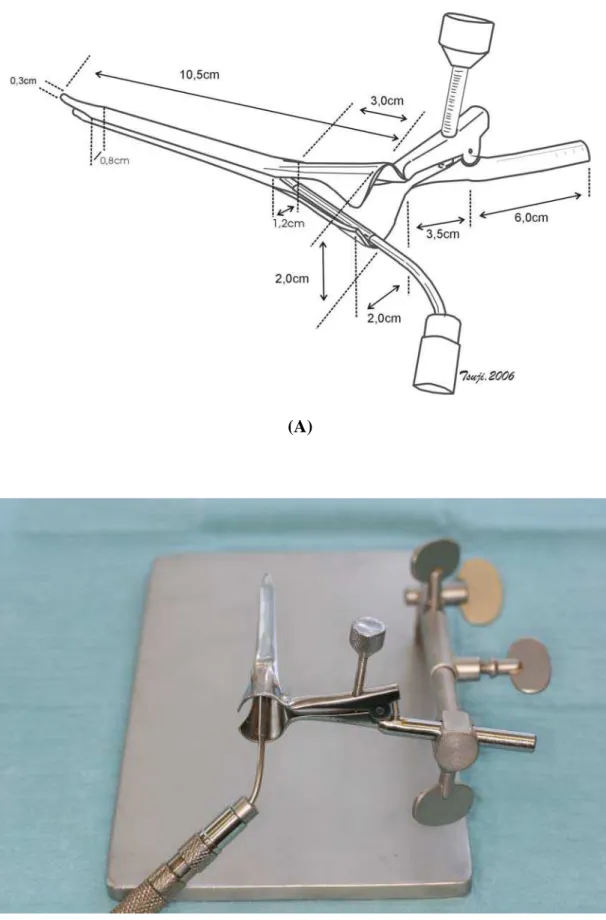
Figura 1 Laringoscópio: (A) esquema; (B) foto ... 24
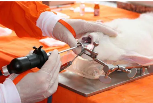
Figura 2 Fonocirurgia com uso do telescópio ... 25
Figura 3 Imagem visualizada no monitor de vídeo das pregas vocais do coelho ... 25
Figura 4 Bisturi utilizado para realização da lesão ... 26
Figura 5 Agulha para infiltração ... 27
Figura 6 Prega vocal após coloração com picrossírius após 7 dias com corticóide: (A) luz não-polarizada (100X); (B) luz polarizada (100X) ... 31
Figura 7 Imagem do retículo utilizado para quantificação das células inflamatórias ... 33
Figura 8 Reação inflamatória na lesão da prega vocal após o 30 dia: (A) controle. HE (400x); (B) corticosteróide. HE (400x) (HE = hematoxilina-eosina) ... 36
Figura 9 Reação inflamatória na lesão da prega vocal após o 70 dia: (A) controle, HE (400x); (B) corticosteróide, HE (400x). (HE = hematoxilina-eosina) ... 37
LISTA DE GRÁFICOS
Gráfico 1 Média da densidade numérica de cada célula (MRI2) (linfócitos, neutrófilos, eosinófilos, macrófagos, fibroblastos e plasmócitos) em relação à intervenção (corticóide x
controle) ... 39
Gráfico 2 Média geral da densidade numérica de todas (MRI3) as células em função do tempo (3 e 7 dias) e da intervenção
(corticóide x controle) ... 40
Gráfico 3 Média da área do colágeno, em valores porcentuais, medido
do controle e da intervenção em 3 e 7 dias ... 42
Gráfico 4 Média da área do colágeno aos 3 e 7 dias em relação à
intervenção (p = significância) ... 45
LISTA DE TABELAS
Tabela 1 Média da densidade numérica de todas as células (MRI1) em
relação à intervenção (corticóide x controle) ... 38
Tabela 2 Média da área total analisada versus intervenção ... 41
Tabela 3 Média da medida do colágeno em função do tempo ... 42
RESUMO
Campagnolo AM. Estudo histológico da ação do corticosteróide injetável no processo agudo de cicatrização das pregas vocais de coelhos [tese]. São Paulo: Faculdade de Medicina, Universidade de São Paulo; 2009. 119p.
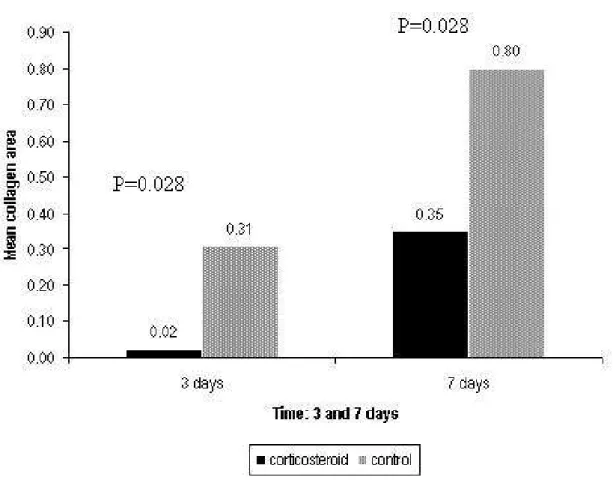
Corticosteróides injetáveis têm sido usados em fonocirurgia com o intuito de se prevenir fibrose da prega vocal pelos seus efeitos na cicatrização e assegurar uma melhor qualidade vocal. O objetivo deste estudo é avaliar histologicamente os efeitos da infiltração de dexametasona no processo agudo de cicatrização, 3 e 7 dias após uma lesão induzida cirurgicamente na prega vocal de coelhos, através da análise quantitativa da reação inflamatória e da deposição de colágeno. Uma incisão cirúrgica foi realizada nas pregas vocais de 12 coelhos, seguido da injeção de 0,1ml de dexametasona na prega vocal esquerda. A prega vocal direita não recebeu nenhuma injeção e serviu como grupo controle. As laringes foram coletadas 3 e 7 dias após a cirurgia. As pregas vocais foram histologicamente analisadas com hematoxilina-eosina para quantificar a resposta inflamatória e picrossírius-red para quantificar a deposição de colágeno. Quantitativamente, não houve diferença significativa na resposta inflamatória das pregas vocais com corticosteróide em relação ao controle. A deposição de colágeno no grupo com corticóide foi estatisticamente menor em 3 dias e permaneceu menor em 7 dias após a lesão cirúrgica (p=0,002). Esses resultados sugerem que os corticosteróides diminuem a deposição do colágeno no processo agudo da cicatrização.
Descritores: Corticosteróides injetáveis, cicatriz pregas vocais, colágeno pregas
vocais, reação inflamatória pregas vocais.
SUMMARY
Campagnolo AM. Histologic study of corticosteroid injection in the acute vocal fold wound healing in a rabbit model. [thesis]. São Paulo: “Faculdade de Medicina, Universidade de São Paulo”; 2009. 119p.
Injectable corticosteroids have been used in phonosurgery to prevent scarring of the vocal fold due to their effects on wound healing, and to assure better voice quality. The aim of this study is to evaluate histologically the effects of dexamethasone infiltration on acute vocal fold wound healing in rabbits 3 and 7 days after surgically-induced injury, by quantification of the inflammatory reaction and collagen deposition. A standardized surgical incision was made in the vocal folds of 12 rabbits, and 0.1 ml dexamethasone (4 mg/ml) was injected into the left vocal fold. The right vocal fold was not injected and served as the control. The larynges were collected 3 and 7 days after surgery. For histological analysis, the vocal folds were stained with hematoxylin-eosin for quantification of the inflammatory response and with picrosirius red for quantification of collagen deposition. There was no quantitative difference in the inflammatory response between vocal folds injected with the corticosteroid and the control folds. However, collagen deposition was significantly lower in the corticosteroid-treated group at 3 and 7 days after the injury (p = 0.002). The present results suggest that dexamethasone reduces collagen deposition during acute vocal fold wound healing.
Descriptors: Injectable corticosteroids, vocal fold scar, collagen, inflammatory
INSTRUCTIONS TO AUTHORS
SUBMISSION
Send manuscripts to Richard J. H. Smith, MD, Editor, Department of Otolaryngology– Head and Neck Surgery, The University of Iowa Hospitals and Clinics, 200 Hawkins Dr, Iowa City, IA 52242. Original manuscripts dealing with clinical or scientific aspects of otolaryngology, bronchoesophagology, head and neck medicine and surgery, maxillofacial and plastic surgery, audiology, speech pathology, or related specialties are considered for publication. All materials submitted for publication undergo peer review. Submit 3 complete copies (including figures) and an electronic version with a signed copyright transfer statement (see “Copyright,” below). Include an e-mail address for notification of receipt of manuscript. All submitted manuscripts must be accompanied by a cover letter. This letter should disclose any financial interests the authors have in relation to the work, or any financial support provided by companies toward the completion of the work. The letter should also indicate whether the manuscript has been presented before any professional otolaryngological association and the place and year of presentation. Manuscripts
submitted without this letter will not be reviewed and will be returned to the author.
Written permission from both senior author and publisher must be provided to the Annals in order to republish tables or illustrations copyrighted elsewhere. Submit this permission with the manuscript.
Papers are scheduled for publication in chronological order of acceptance, but manuscripts received in im-proper form require longer production time. Manuscripts are edited in accordance with the AMA Manual of Style , 10th edition (2007), and with the
Uniform Requirements for Manuscripts Submitted to Biomedical Journals : Writing and Editing for Biomedical Publication (updated Oct 2007; http://www.icmje.org).
Manuscripts not accepted for publication are not Returned.
MANUSCRIPT PREPARATION
Limit papers to a size that will make up to no more than six printed pages, figuring three double-spaced typewritten pages of text to one journal page; see the journal for estimating space required for references, illustrations, and tables. If a manuscript of greater length is accepted for publication by the Editor, all journal pages over six are charged to the author at the publisher’s cost of $175 per page. Submit an original and two copies of the manuscript on white bond paper with margins of at least 25 mm (1 inch), double-spaced throughout, including abstract, references, tables, and legends. Use a type size no smaller than 10 points, preferably 12. Begin each com-ponent on a new page in the following sequence: title page, abstract, text, acknowledgments, references, tables, and figure legends. Number pages consecutively in the upper right corner, beginning with the title page. Avoid the use of staples.
The author is responsible for all statements in the paper, as approved on the copyedited galley proofs. Alterations made by the author after the paper has been typeset are charged to the author.
Use standard abbreviations given in the Uniform Requirements. Express all measurements in metric terms; if original measurements were made in another system, include these parenthetically. Plot audiograms according to ISO standards. Use generic names whenever possible.
Title page must include 1) a concise but informative title, worded to facilitate indexing; 2) an abbreviated form of the title to be used as a running head; 3) authors’ full names (inclusion of more than five authors requires written explanation to the Editor at time
of submission ) and no more than two academic degrees per author; 4) department(s) and
institution(s) to which the work is attributed, with authors’ present affiliations and addresses, if different, separately noted; 5) statement of grant or other support; 6) name and address of author to whom reprint requests should be sent; and 7) name, address, telephone and fax numbers, and e-mail address of corresponding author. Manuscripts that report animal research performed in the United States must carry the following statement on the title page: “This study was performed in accordance with the PHS Policy on Humane Care and Use of Laboratory Animals, the NIH Guide for the Care and Use of Laboratory Animals, and the Animal Welfare Act (7 U.S.C. et seq.); the animal use protocol was approved by the Institutional Animal Care and Use Committee (IACUC) of __________ University (or institution).”
Abstracts must be less than 200 words and structured to include objectives, methods,
results, and conclusions.
Key Words, chosen as far as possible from the National Library of Medicine medical subject headings, are listed after the abstract. A maximum of 6 are permitted. References, double-spaced, are numbered consecutively in the order in which they are cited in the text. Primary references should be used whenever possible. The author is charged $2.00 for each reference over 30. Use the style of references given in the Uniform
Requirements or a current issue of the Annals. Include the names of all authors and the
inclusive page numbers of an article. If a manuscript accepted but not yet published is included in the reference list, give the accepting journal’s name, followed by “in press.” Manuscripts still in review or not yet accepted formally should be cited within the text as “unpublished observations.” A reference to a personal communication is also placed in the text, accompanied by a date (year). Papers presented at scholarly meetings but not published are considered “unpublished bservations.” Papers published only in abstract form are listed as references with “[Abstract]” after the title.
Tables should be on separate sheets, numbered consecutively and headed by a concise title. Put explanatory matter in footnotes. Tables are adjuncts to the text and should not repeat material presented therein. The cost of preparing tables is billed to the author. Illustrations must be submitted in three complete sets, unmounted. Only professional-quality glossy photographs and black-and-white line drawings are acceptable. Multi-part illustrations should be labeled (A, B, etc) on the reverse side, not on the illustration itself. Put legends (detailed explanations) to the photographs on a separate page in the manuscript. Affix a label to the reverse side of each illustration, indicating figure number, first author’s name, and top of the figure. Cite each figure in the text in consecutive order. Written permission from identifiable subjects is required. The cost of preparing illustrations for publication
(scanning if necessary, sizing, lettering, etc) is charged to the author without exception. Color illustrations are accepted; cost estimates for color separations and printing
are provided on an individual basis. Illustrations should enhance, not repeat, material presented in the manuscript and should be kept to a minimum.
ELECTRONIC SUBMISSION
Software and format must be Microsoft Word. Do not use complex formatting or desktop publishing software. Do not deliver files that contain hidden text. For example, do not use your word processor’s automated features to create footnotes and reference lists. Submit text, tables, and figure legends as a single file. Do not include illustrations in this file. Illustrations should be in TIFF, EPS, JPEG, or PSD formats. Do not submit illustrations in Powerpoint. Do not submit native application formats. Journalquality reproduction will require grayscale and color files at resolutions of at least 300 dpi. Bitmapped line art should be submitted at resolutions of at least 600 dpi. (These resolutions refer to the output size of the file.)
Send files to: annals-orl@uiowa.edu.
REPRINTS
COPYRIGHT
The following statements must accompany the manuscript, signed by all authors (only original signatures are acceptable: 1) “I warrant that my contribution to the work is original
and that I have full power to enter into this agreement. The content of this paper, all or in part, has not been published, has not been submitted for publication elsewhere, and is not in press elsewhere.” 2) “I verify that I have met all of the following criteria for authorship and am qualified to be listed as an author of this work by my substantive contribution to the conception and design of the project or analysis of the data, my drafting or critical revision of the content of this manuscript, and my approval of the final version to be published.” 3) “In consideration of the Annals of Otology, Rhinology & Laryngology taking action in reviewing and editing my (our) submission, I hereby transfer, assign, or otherwise convey all copyright ownership to Annals Publishing Company in the event such work is published in the Annals of Otology, Rhinology & Laryngology.” After acceptance, no author may be added to or removed from a paper.
SUPPLEMENTS
A manuscript too long for inclusion in the Annals (over 12 typeset pages) may be
published as a supplement if approved by the Editor. All costs are borne by the author, and estimates are provided upon request. Supplements have the advantages of separate identification and rapid publication, but undergo the same peer review as journal articles.
REVIEWS OF SOFTWARE AND COMPUTER APPLICATIONS
Programs submitted for review must be compatible with one of the following operating systems: Windows 2000 and above; or Macintosh OS 9.0 and above. The author must specify hardware and system requirements. Submit the software on CD. If the program is a template, submit also a runtime version of the source program. Software considered for review includes educational software, artificial intelligence software to aid in the diagnosis of otolaryngic disorders, software for logging operations, software to aid in research, and software that addresses specific problems in otolaryngology. Also, manuscripts reviewing software and computer applications relating directly to otolaryngology are considered for publication.
February 2008
September 2, 2009
MS: 09-4357; Histological study of acute vocal fold wound healing after corticosteroid injection in a rabbit model
Dear Dr. Campagnolo:
I am pleased to inform you that the Editorial Board of the Annals of Otology, Rhinology & Laryngology has found your manuscript titled “Histological study of acute vocal fold wound healing after corticosteroid injection in a rabbit model” suitable for publication. Editorial preparations will now begin in the Managing Editor’s office in St. Louis.
Only a small fraction of the manuscripts submitted to the Annals are accepted for publication, and I congratulate you on your accomplishment.
With kind regards,
Histological study of acute vocal fold wound healing after corticosteroid injection in a rabbit model
Acute vocal fold wound healing after corticosteroid injection
Andréa M Campagnolo1, Domingos Hiroshi Tsuji2, Luiz Ubirajara Sennes3, Rui Imamura4,Paulo H. N. Saldiva5
(1) Otorhinolaryngologist, PhD Student, São Paulo University School of Medicine. (2) Associate Professor, Discipline of Otorhinolaryngology, São Paulo University
School of Medicine.
(3) Associate Professor, Discipline of Otorhinolaryngology, São Paulo University School of Medicine.
(4) Collaborating Professor, Discipline of Otorhinolaryngology, São Paulo University School of Medicine.
(5) Full Professor, Department of Pathology, São Paulo University School of Medicine.
From the Department of Otolaryngology (a.m.c., d.h.t., l.u.s., r.i.,) and Department of Pathology (p.h.n.s.), University of São Paulo School of Medicine, São Paulo, Brazil.
Send correspondence to Andrea M Campagnolo. R. Alceu Amoroso Lima 105/1802, Barra da Tijuca, 22631010, Rio de Janeiro/RJ, Brasil. E-mail: amcampag@ig.com.br. Phone: +552194214122, fax: +552124955044.
Abstract
Introduction: Injectable corticosteroids have been used in phonosurgery to prevent scarring of the vocal fold due to their effects on wound healing, and to assure better voice quality.
Objective: To histologically evaluate the effects of dexamethasone infiltration on acute vocal fold wound healing in rabbits 3 and 7 days after surgically-induced injury by quantification of the inflammatory reaction and collagen deposition.
Study design: Experimental study in rabbits.
Material and Methods: A standardized surgical incision was made in the vocal folds of 12 rabbits, and 0.1 ml dexamethasone (4 mg/ml) was injected into the left vocal fold. The right vocal fold was not injected and served as the control. The larynges were collected 3 and 7 days after surgery. For histological analysis, the vocal folds were stained with hematoxylin-eosin for quantification of the inflammatory response and with picrosirius red for quantification of collagen deposition.
Results: There was no quantitative difference in the inflammatory response between vocal folds injected with the corticosteroid and control folds. However, collagen deposition was significantly lower in the corticosteroid-treated group at 3 and 7 days after injury (p = 0.002).
Conclusion: The present results suggest that dexamethasone reduces collagen deposition during acute vocal fold wound healing.
Introduction
Phonosurgery techniques and instruments have been improved over the years and now permit the removal of benign vocal fold lesions with maximum preservation of adjacent normal tissue. Understanding of the body-cover theory of the vocal fold is fundamental for phonomicrosurgery. The mucosal wave is generated by sliding the mucosal cover over the vocal ligament and muscle. This cover can be divided into different layers with distinct mechanical properties that can be distinguished by their concentration of collagen and elastin fibers.1 Most benign vocal fold lesions occur in the superficial layer of the lamina propria, and surgery should therefore be confined to this layer. According to Schweinfurth and Ossoff,2 damage to the deep layers of the lamina propria and to the vocal ligament is associated with a more intense formation of scar tissue. This scarring causes the adhesion of the mucosal cover to deep tissues, impairing the mucosal wave and consequently causing dysphonia. Vocal fold scarring is the most common cause of dysphonia (35%) after phonosurgery.3
Studies have shown that an increase in collagen provides the basis of fibrosis and scar resistance, which mainly depends on collagen content.4-7 Scar formation resulting from the replacement of healthy tissue by fibrous tissue may permanently alter vocal fold function and may lead to a decrease or loss of the mucosal wave.8 In an attempt to obtain data that would contribute to the development of a treatment method that minimizes fibrosis in the vocal fold after injury, many studies are aimed at a better understanding and manipulation of the vocal fold wound healing process.9,10,11-15
Systemic corticosteroids, especially injectable corticosteroids, are frequently used in laryngeal microsurgery of the vocal folds in order to prevent scar formation and, consequently, to assure better voice quality.2,16,17 Corticosteroids delay the healing process, permitting a better organization of scar tissue. However, the effects of these drugs on the vocal folds have not been well established.18,19
The objective of the present study was to histologically evaluate the effects of dexamethasone infiltration on acute vocal fold wound healing in rabbits 3 and 7 days after surgically-induced injury by quantitative analysis of the inflammatory reaction and collagen deposition. The results were compared to control vocal folds that were submitted to similar surgery.
Materials and Methods
Animals and Surgery
The study was approved by the Ethics Committee for the Analysis of Research Projects (CAPPesq) of the Clinical Board of the University Hospital and São Paulo University School of Medicine. The study was conducted according to guidelines for the care and use of laboratory animals following the norms of Federal Law No. 6.638 (enacted May 8, 1979), and the ethical principles of the Brazilian College on Animal Experimentation (COBEA).
manufacturer and consisted of a spear-shaped knife (2 mm wide, 2 mm deep at its central end and 1 mm deep at its borders) with a transverse shield, which restricted the depth of penetration into the vocal fold to 2 mm in the center of the lesion. The needle used for dexamethasone infiltration was also fabricated for this study and contained a pin that permitted the standardization of injection at a depth of 2 mm. The caliber of the needle corresponded to the diameter of the needle used for insulin injection (0.4 x 12 mm), and the needle was coupled to an insulin syringe to permit standardization of the injection volume.
Laryngeal microsurgery was performed on 12 rabbits. The animals were anesthetized by intramuscular injection of xylazine (5 mg/kg) and ketamine (50 mg/kg) and maintained under spontaneous ventilation. Adequate exposure of the glottis was obtained for all animals.
Surgery was performed under endoscopic vision using a 4-mm diameter telescope with an angle of 30 degrees (Karl Storz®). A puncture wound was made with the knife in the upper surface of the vocal folds in the midpoint of its membranous portion (both in the anteroposterior and lateromedial direction), resulting in a longitudinal lesion parallel to the free border and in the center of the vocal folds. The dimensions of the injury were standardized and were similar to that of the tip of the knife. Next, 0.1 ml (0.4 mg) dexamethasone was injected into the left vocal fold of each rabbit lateral to the incision. The right vocal fold was not injected and served as control.
Half of the animals were sacrificed 3 days after injury; the remaining rabbits were sacrificed at 7 days post-injury. Larynges were removed en bloc from the base of the tongue to the fourth tracheal ring by cervical incision. An anterior longitudinal
incision was made in the larynx and the right and left wings of the thyroid cartilage were separated. Each hemi-larynx was identified for subsequent blind assessment.
Histological study of the vocal fold
The excised larynges were fixed in 10% formaldehyde and processed at the Pathological Anatomy Laboratory of the institution (University Hospital and São Paulo University School of Medicine). The vocal folds were cut into three parts (anterior, middle and posterior) and the middle third containing the injury was selected for the study. Specimens were gradually dehydrated in progressive alcohol concentrations and embedded in paraffin melted in an oven at 60°C. The specimens were then oriented, cut into 4-µm coronal sections with a microtome, deparaffinized, and stained. For standardization, specimens were prepared by a single experienced technician who carried out the whole staining procedure.
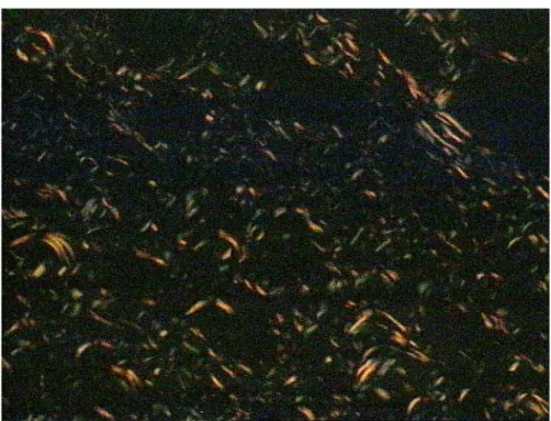
Hematoxylin-eosin staining was used for the identification of cell nuclei of the inflammatory infiltrate surrounding the injury. Picrosirius staining was used for the visualization and analysis of collagen fibers. This method consists of staining with Sirius red dissolved in saturated aqueous picric acid. The slides were analyzed by polarized light microscopy.20 The picrosirius polarization method permits the differentiation between collagen and non-collagen tissue. A non-collagen substance is stained black and collagen is stained red (thicker), orange, yellow or green (thinner) according to its thickness and maturity (Figure 1 A and B).
an inflammatory reaction and signs of wound healing. A single blinded pathologist determined the number of inflammatory cells and collagen in all vocal folds, examining three to five fields per histological slide containing the injured vocal folds. An eyepiece grid with 50 horizontal lines connecting 100 points was used for the counting of inflammatory cells.21 Cells associated with inflammation and mucosa remodeling were counted (lymphocytes, neutrophils, eosinophils, macrophages, fibroblasts, and plasma cells). The results are expressed as numerical density, which corresponds to the relative frequency of these cells (number of cells in 62,500 µm2) in each field at 400X magnification. Collagen fibers were quantified with a digital image analysis system using the Image Pro Plus software, version 4.5.0.29 (Media Cybernetics, Bethesda, MD). In the image capture system used, the camera was set to capture images of higher birefringence and to calculate the results in µm2. The concentration of collagen fibers was determined by the number of collagen fibers of higher birefringence per area unit measured (area of collagen fibers divided by total area x 100).
The measurements were transferred to the SPSS® program (version 14.0) for statistical analysis.
Statistical analysis
The nonparametric Mann-Whitney test was used for statistical analysis of the inflammatory response and collagen deposition to determine possible differences between study times. Possible differences between the intervention and control groups were evaluated by the nonparametric Wilcoxon test. The level of significance was set at p < 0.05.
Results
Inflammatory reaction
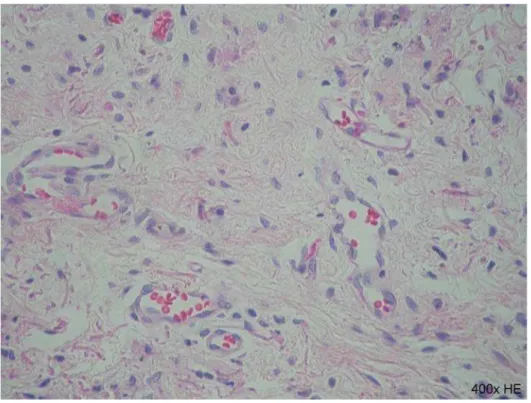
Figures 2 (A and B) and 3 (A and B) show the inflammatory reaction in control and corticosteroid-treated vocal folds, respectively, at 3 and 7 days after injury.
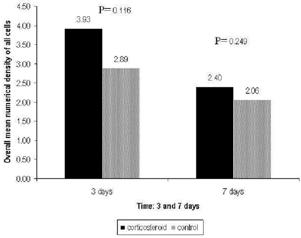
The mean overall numerical density of all cells was 3.16 (± 1.12) in the corticosteroid-treated group and 2.48 (± 0.98) in the control group, with no significant difference between groups (p = 0.099). The mean numerical density of each cell type (lymphocytes, neutrophils, eosinophils, macrophages, fibroblasts, and plasma cells) did not differ between the corticosteroid-treated and control groups (Figure 4). The mean overall numerical density of all cells as a function of time (3 and 7 days after injury) was similar in the corticosteroid-treated and control groups (Figure 5).
Collagen
The mean total area per vocal fold analyzed was 48,478.42 µm2 in the corticosteroid-treated group and 47,608.82 µm2 in the control group, with no significant difference between groups (p = 0.386).
The concentration of collagen fibers in each vocal fold was calculated per µm2 (Figure 6).
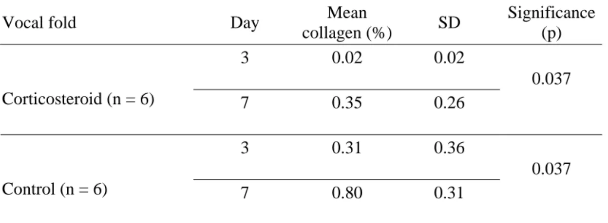
The collagen fiber concentration showed a significant increase at 7 days in both corticosteroid-treated and control vocal folds (p = 0.037) (Table 1).
(273.6686 µm2) in the control group. This difference was statistically significant (p = 0.002), demonstrating that corticosteroid treatment promoted a reduction in the quantity of collagen during wound healing .
Collagen deposition was significantly lower in the corticosteroid-treated group at 3 and 7 days after injury (Figure 7).
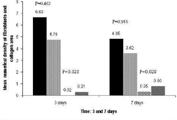
Figure 8 shows the number of fibroblasts and quantity of collagen in the corticosteroid-treated and control groups at 3 and 7 days. The number of fibroblasts was higher in the corticosteroid-injected group compared to control, but the difference was not significant (p = 0.463 in 3 days and p = 0.916 in 7 days), whereas the quantity of collagen was significantly lower in the corticosteroid-treated group (p = 0.028 in 3 days and p = 0.028 in 7 days).
Discussion
No currently available therapeutic method is effective in the treatment of vocal fold scarring.22 Injection of corticosteroids is widely used for vocal fold wound healing and has been associated with a reduction of stiffness and improvement of glottic closure and voice quality.16,23 However, according to these authors, some degree of fibrosis always persists.
Local injection of corticosteroids is an effective method to achieve a high concentration only in the target organ, such as the larynx, and has a low frequency of adverse effects, since these agents are rapidly inactivated after absorption (with the result that only inactive metabolites enter the bloodstream).24 Bouchayer and Cornut16 used injectable hydrocortisone to reduce the post-traumatic inflammatory
response. We chose dexamethasone because of its high long-term efficacy and low cost.
surgically-induced vocal fold wound during this period. Evaluation of vocal fold wound healing on day 7 was based on the fact that new collagen is deposited by day 5, with fibroblasts being the predominant cells at that time.15 More mature collagen is detected by day 7.27 However, it should be emphasized that the results of the present study and of other investigations using rabbit and rat models13,15 should be evaluated with caution since these animals present a shorter healing period than humans.
In the present study, the number of inflammatory cells was not affected by corticosteroid injection (p > 0.05). Coleman et al.18 evaluated the effects of triamcinolone injected into the vocal folds of dogs after the introduction of a lateral microflap (surgical injury). The contralateral vocal fold, which was submitted to the same type of surgery and not injected with the corticosteroid, served as the control. Paired analysis revealed an increase in the inflammatory infiltrate around the microflap in corticosteroid-treated vocal folds at 2, 4 and 6 weeks. In that study, triamcinolone caused a delay in the corticosteroid-treated vocal fold tissue response of 12 days for the inflammatory infiltrate. No delay in the inflammatory response was observed in the present study. The difference between our results and the findings of Coleman et al.18 might be due to methodological differences, such as the type of injury and period studied.
The finding that corticosteroid treatment apparently did not affect the number of inflammatory cells should be interpreted with caution and some considerations can be made. In contrast to what the result shows, the presence of the corticosteroid inside the tissue might have had an effectively inhibitory action on the migration of inflammatory cells. However, the fact that the test vocal fold suffered traumas caused by the incision, introduction of the needle and tissue expansion due to the injected
volume may have resulted in a greater migration of inflammatory cells than observed in the control fold, apparently abolishing the inhibitory effect of the corticosteroid. Another consideration is that, since the volume of the injected drug is relatively large when compared to the small dimensions of the rabbit vocal fold, the possibility that drug diffusion by contiguity to the control fold might have exerted an anti-inflammatory effect cannot be completely ruled out. However, this hypothesis does not seem to be very likely since, if this would have really occurred, no significant differences in collagen fiber concentration can be expected. Finally, another possibility is that the injected corticosteroid acts on the control fold through its systemic effect, i.e., bloodstream diffusion. However, this hypothesis seems unlikely since, according to Wannmacher and Ferreira,24 local injection of corticosteroids results in a high concentration of the drug in the target tissue, which is rapidly inactivated after its absorption so that only inactivated metabolites enter the bloodstream.
investigation collagen deposition was analyzed on the third and seventh day, a fact impairing technically precise comparison between studies.
We observed a larger number of fibroblasts in the corticosteroid-treated group compared to the control vocal fold, but the difference was not significant. One explanation for this apparently unexpected result is the fact mentioned earlier that injection of the corticosteroid into the vocal fold might have caused greater damage and tissue distension. On the other hand, despite the lack of a significant difference in the number of fibroblasts, the quantity of collagen was significantly lower in the corticosteroid-treated group. This finding might be explained by the fact that fibroblasts produced less collagen in the corticosteroid-treated fold. According to Kloth and McCulloch,25 corticosteroids may interfere with the mechanisms of collagen production by these cells.
We believe that a better understanding of the physiopathological mechanisms underlying collagen deposition during wound healing is important for the treatment and prevention of vocal fold scars after phonosurgery. The present results suggest that corticosteroids, frequently used in phonosurgery to prevent fibrosis, influence collagen deposition during acute wound healing. However, further studies are necessary to determine whether these alterations persist into the late phase of collagen remodeling.
Conclusion
In the present study, corticosteroid injection into the vocal fold of rabbits after a surgical procedure reduced collagen deposition at the site of injury during the acute inflammatory response. The number of inflammatory cells at the site of injury was not affected by corticosteroid treatment.
References
1. Gray SD, Hirano M, Sato K. Molecular and cellular structure of vocal fold tissue. In: Titze IR, eds. Vocal Fold Physiology: Frontiers of Basic Science. San Diego: Singular Publishing; 1993:1-34.
2. Schweinfurth JM, Ossoff RH. Surgery for dysphonia. Curr Opin Otolaryngol Head Neck Surg 1999; 7(6): 357-64.
3. Woo P, Casper J, Colton R, Brewer D. Diagnosis and treatment of persistent dysphonia after laryngeal surgery: a retrospective analysis of 62 patients. Laryngoscope 1994; 104: 1084-91.
4. Ehrlich HP. Collagen considerations in scarring and regenerative repair. In Garg HG, Longaker MT eds. Scarless Wound Healing. New York, NY: Marcel Dekker Inc.; 2000, p. 99-113.
5. Hirano M, Phonosurgery. Basic and clinical investigation. Otologia (Fukuoka) 1975; 21(suppl 1): 299-303.
6. Rogerson AR, Clark KF, Bandi SR, et al. Voice and healing after vocal fold epithelium removal by CO2 laser vs. microlaryngeal stripping. Otolaryngol
Head Neck Surg 1996; 115: 352-9.
7. Blakeslee DB, Banks RE, Eusterman V, et al. Analysis of vocal fold function in the miniswine model. J Invest Surg 1995; 8: 409-24.
8. Benninger MS, Alessi D, Archer S. Vocal fold scarring in a canine model. Laryngoscope 2003; 113: 620-7.
10. Thibeault SL, Gray SD, Bless DM, Chan RW, Ford CN. Histologic and rheologic characterization of vocal fold scarring. J Voice 2002; 16:96-104. 11. Rousseau B, Hirano S, Chan RW, et al. Characterization of chronic vocal fold
scarring in a rabbit model. J Voice 2004; 18: 116-24.
12. Tateya T, Tateya I, Sohn JH, Bless D. Histologic characterization of rat vocal fold scarring. Ann Otol Rhinol Laryngol 2005; 114(3): 183-91.
13. Tateya T, Tateya I, Sohn JH, Bless D. Histological study of acute vocal fold injury in a rat model. Ann Otol Rhinol Laryngol 2006; 115(4): 285-92.
14. Lim X, Tateya I, Tateya T, Muñoz-Del-Rio A, Bless D. Immediate inflammatory response and scar formation in wounded vocal folds. Ann Otol Rhinol Laryngol 2006; 115(12): 921-9.
15. Branski RC, Verdolini K, Rosen CA, Hebda PA. Acute vocal fold wound healing in a rabbit model. Ann Otol Rhinol Laryngol 2005; 114: 19-24.
16. Bouchayer M, Cornut G. Microsurgery for benign lesions of the vocal fold. Ear Nose Throat J 1998; 67: 446-66.
17. Courey MS, Gardner GM, Stone RE, Ossoff RH. Endoscopic vocal fold microflap: a three-year experience. Ann Otol Rhinol Laryngol 1995; 104: 267-73.
18. Coleman JR, Smith S, Reinisch L, Billante CR, Ossof J, Deriso W, Garret CG. Histomorphometric and laryngeal videostroboscopic analysis of the effects of corticosteroids on microflap healing in the dog larynx. Ann Otol Rhinol Laryngol 1999; 108: 119-27.
19. Alencar BLF, Baptistella E, Malafaia O, Czeczko NG, Scopel TF, Trotta F, Costa AFCB, Watanahe SS, Silva TP. Estudo experimental de corticóide
injetável em microcirurgia laríngea. Arq Int Otorrinolaringol 2007; 11(4): 438-43.
20. Mello ECM, Lemos M, Ximenes Filho JA, Sennes UL, Saldiva PHN, Tsuji DH. Distribution of collagen in the lamina propria of the human vocal fold. Laryngoscope 2003, 113: 2187-91.
21. Mallik MK, Kapila K, Das DK, Haji BE, Anim JT. Cytomorphology of hyaline-vascular Castleman’s disease: a diagnostic challenge. Cytopathology 2007; 18: 168-174.
22. Hirano S. Current treatment of vocal fold scarring. Curr Opin Otolaryngol Head Neck Surg 2005; 13(3): 143-7.
23. Mortensen M, Woo P. Office steroid injections of the larynx. Laryngoscope 2006; 116(10): 1735-39.
24. Wannmacher L, Ferreira MBC. Antiinflamatórios esteróides. In Fuchs D, Wannmacher L, eds. Farmacologia Clínica. 2nd ed. Rio de Janeiro: Guanabara Koogan; 1998, p. 194-202.
25. Kloth LC, McCulloch JM. The inflammatory response to wounding. In McCulloch JC, Kloth LC, Feedar JA eds. Wound healing alternatives in management. Philadelphia: CPR F. A. Company; 1998, p. 3-15.
26. Piccinato CE, Netto JC, Cherri J. Cicatrização. In Campos CAH, Costa HOO, eds. Tratado de Otorrinolaringologia. São Paulo: Roca 2002, p. 189-200.
Table 1: Mean collagen content as a function of time.
Vocal fold Day Mean
collagen (%) SD
Significance (p)
3 0.02 0.02
Corticosteroid (n = 6) 7 0.35 0.26
0.037
3 0.31 0.36
Control (n = 6) 7 0.80 0.31
0.037
n = number of rabbits; SD: standard deviation.
Figure 1A
Figure 1B
Figure 1: Coronal sections of a surgically-injured vocal fold 7 days after
Figure 2A
Figure 2B
Figure 2: Coronal sections of a control (A) and corticosteroid-treated vocal fold (B)
showing the inflammatory reaction on day 3 after injury (400X, HE).
Figure 3A
Figure 3B
Figure 4: Comparison of mean numerical density of each cell type (lymphocytes,
neutrophils, eosinophils, macrophages, fibroblasts, and plasma cells) between corticosteroid-treated and control vocal folds, combining the results obtained at 3 and 7 days. SD = Standard deviation. (SD corticosteroid-treated group: lymphocytes = 2.02; neutrophils = 2.40; eosinophils = 1.50; macrophages = 1.68; fibroblasts = 4.51; plasma cells = 2.11; SD control group: lymphocytes = 2.86; neutrophils = 1.42; eosinophils = 1.30; macrophages = 2.37; fibroblasts = 2.87; plasma cells = 1.77).
Figure 5: Comparison of overall mean numerical density of all cells 3 and 7 days
Figure 6: Mean collagen area (µm2 x 100) in the control and corticosteroid-treated groups 3 and 7 days after injury.
Figure 8: Comparison of the number of fibroblasts and collagen area (µm2 x 100) between corticosteroid-treated and control vocal folds 3 and 7 days after injury. SD = Standard deviation. (SD 3 days corticosteroid-treated group: fibroblasts = 5.2, collagen= 0.02; SD 3 days control group: fibroblasts = 3.34, collagen = 0.36; SD 7 days corticosteroid-treated group: fibroblasts = 3.97, collagen = 0.26; SD 7 days control group fibroblasts = 2.49, collagen = 0.31).
Introdução 2
Com o passar dos anos as técnicas e os instrumentos em fonocirurgia têm sido aprimoradas no intuito de remover as lesões benignas das pregas vocais com o máximo possível de preservação dos tecidos normais adjacentes. O entendimento do princípio de corpo-cobertura da prega vocal é fundamental em uma abordagem cirúrgica. A onda mucosa é gerada pelo deslizamento da cobertura mucosa sobre o ligamento vocal e o músculo vocal. Essa cobertura pode ser dividida em camadas com propriedades mecânicas distintas e podem ser distinguidas pela concentração de fibras colágenas e elastina (Gray et al., 1993). A maioria das lesões benignas ocorre na camada superficial da lâmina própria, portanto as abordagens cirúrgicas devem idealmente ser confinadas nessa camada. A violação das camadas profundas da lâmina própria está associada com formação de tecido cicatricial. Essa cicatriz causa fixação da cobertura mucosa aos tecidos profundos, prejudicando a onda mucosa e causando disfonia (Schweinfurth, Ossoff, 1999). Uma cicatriz na prega vocal é considerada a causa mais comum de disfonia (35%) após fonocirurgia (Woo et al., 1994).
Em geral, uma cicatriz está relacionada com o tecido fibroso. Estudos têm demonstrado que um aumento do colágeno é a base da fibrose e que a resistência da cicatriz depende principalmente do conteúdo de colágeno (Hirano, 1975; Blakeslee et al., 1995; Rogerson et al., 1996; Ehrlich, 2000).
Introdução 3
da onda mucosa da prega vocal (Benninger et al., 2003). Em busca de conhecimentos que contribuam para o desenvolvimento de um método de tratamento que minimize a formação de fibrose na prega vocal após uma lesão, muitas pesquisas estão sendo direcionadas para o entendimento e manipulação do processo cicatricial na prega vocal (Thibeault et al., 2002; Rosseau et al., 2003; Rosseau et al., 2004; Branski et al., 2005; Tateya et al., 2005; Tateya et al., 2006; Lim et al., 2006).
Os corticosteróides sistêmicos, principalmente os injetáveis, são usados com freqüência em microcirurgia de laringe nas pregas vocais, operadas com intuito de prevenir a formação de cicatriz e assim assegurar uma melhor qualidade vocal. (Courey et al., 1995; Bouchayer; Cornut, 1998; Schweinfurth, Ossoff, 1999).
Objetivo 5
Revisão da literatura 7
3.1 Histologia da prega vocal, inflamação aguda e cicatrização
Uma cicatriz iatrogênica na prega vocal pode resultar em disfonia pela
diminuição de suas propriedades vibratórias (Rousseau et al., 2003). Alguns estudos
têm demonstrado que essas alterações são decorrentes da desorganização do sistema
de colágeno (Thibeault et al., 2002).
Na histologia, a prega vocal é composta por cinco camadas: o epitélio de
revestimento; a lâmina própria com suas camadas superficial, intermediária e
profunda; e, o músculo vocal (Hirano, 1974). A lâmina própria é composta
principalmente pela matriz extracelular, que tem propriedades biomecânicas
determinantes na fonação e é composta por dois grupos de macromoléculas: as
proteínas fibrosas (fibras elásticas e colágenas) e as proteínas intersticiais
(glicosaminoglicanas, proteoglicanas e glicoproteínas). O colágeno e as fibras
elásticas conferem, respectivamente, resistência e elasticidade à lâmina própria,
enquanto que as glicosaminoglicanas e os proteoglicanos formam géis que
preenchem os espaços, retêm água e controlam a viscosidade (Gray et al., 2000;
Mello et al., 2003).
O processo de cicatrização de uma lesão das pregas vocais é similar àquele
classicamente descrito para derme, embora algumas diferenças tenham sido citadas,
como uma reepitalização mais prolongada na subglote (Goldstein et al., 1998) e na
estabilização do colágeno, que se processa em 21 dias na derme (Lawrence, 1998) e
pode levar até seis meses na prega vocal (Rousseau et al., 2003).
A seqüência de eventos durante os quais um tecido lesado é substituído por
Revisão da literatura 8
superpostas: inflamatória, proliferativa e de formação e remodelamento da matriz, e
essa última caracterizará a resistência da cicatriz (Piccinato et al., 2002).
A resposta inflamatória começa pelo sangue extravasado pela lesão que forma
um coágulo que ocupa o espaço entre as margens da ferida. A partir do coágulo e do
tecido lesado, surgem fatores quimiotáticos e vasoativos que promovem o exudato de
neutrófilos, macrófagos e monócitos do sangue para as margens da lesão. Por volta
de 24 horas o coágulo é invadido por essas células, em 48 horas os macrófagos
passam a predominar e passam a liberar citoquinas adicionais, tais como: fator de
crescimento derivado das plaquetas (PDGF), fator de crescimento dos fibroblastos,
fator β transformador de crescimento (TGF-β) e fator ativador das plaquetas (PAF),
que ativam e estimulam a proliferação de fibroblastos que migram em direção ao
coágulo e atuam na sua reabsorção e começam a sintetizar os componentes da matriz
extracelular (Piccinato et al., 2002). Nesse intervalo, as células da camada basal da
epiderme entram em mitose e migram sobre a superfície do coágulo, recompondo o
epitélio. Desse modo, após a exsudação dos macrófagos, um tecido conjuntivo
vascularizado cresce, preenchendo o espaço antes ocupado pelo coágulo. Esse tecido
conjuntivo frouxo, rico em capilares sangüíneos, leucócitos e matriz extracelular
formada por fibras colágenas finas (colágeno tipo III), ácido hialurônico e
proteinoglicanos recebe o nome de tecido de granulação (Pereira et al., 1994). Cerca
de cinco dias após, o tecido de granulação preenche todo o espaço da ferida e o
epitélio da epiderme já adquire sua espessura normal iniciando a queratinização
(Lawrence, 1998).
O remodelamento é o estágio final no processo de cicatrização. A quantidade
Revisão da literatura 9
na matriz extracelular. Ao mesmo tempo, diminui a síntese de glicosaminoglicano,
especialmente do ácido hialurônico. O colágeno tipo I, com fibras mais grossas e
compactas passa a predominar em relação ao tipo III. Nas semanas seguintes, o
colágeno vai sendo remodelado, com aumento de ligações transversais, tornando-se
mais resistente e estável (Pereira et al., 1994).
3.2 Corticosteróides
Os corticóides são amplamente empregados por suas propriedades:
antiinflamatória e imunomoduladora. Eles têm um mecanismo de ação muito original
que é essencialmente genômico (transcriptacional) e caracterizado pela ativação
(transativação) ou inibição (transrepressão) de numerosos genes alvo. Essas
moléculas agem em muitas células, envolvendo a imunidade inata (macrófagos,
granulócitos, mastócitos) e a imunidade adaptativa (linfócitos), também em outras
células (fibroblastos, células epiteliais e endoteliais). A eficácia antiinflamatória dos
corticóides relaciona-se com a inibição da síntese de numerosas citoquinas, enzimas
e mediadores da inflamação, e pela indução de citoquinas e moléculas
antiinflamatórias como a lipocortina que inibe a liberação de substâncias vasoativas e
fatores quimiotáticos. (Wannmacher, Ferreira, 1998; Sibila, 2003; Campagnolo et al.,
2008)
Enzimas lipo e proteolíticas são também diminuídas por estabilização dos
lisossomos, assim como o extravasamento de leucócitos para zona de lesão. Há
Revisão da literatura 10
afetam elementos e etapas da reação inflamatória (Quadro 1). (Wannmacher,
Ferreira, 1998; Sibila, 2003)
Quadro 1 - Sítios e mecanismos de atuação de glicocorticóides em reações inflamatórias e imunitárias.
Células Elemento afetado Resposta induzida
ácido araquidônico e seus
produtos (prostaglandinas e
leucotrienos)
Diminuição de vasodilatação,
permeabilidade vascular,
hipersensibilização de
nociceptor, resposta imune Macrófagos e
monócitos
Citocinas (interleucina-1 e -6,
TNF-α)
Diminuição de ativação de
linfócitos T e fibroblastos
ELAM-1 e ICAM-1 Diminuição da adesão de
leucócitos
Citocinas (interleucina-1) Mesmo efeito acima Células
endoteliais
Derivados do ácido
araquidônico
Mesmo efeitos acima
Basófilos Histamina; leucotrieno C4 inibição de IgE
Fibroblastos Derivados do ácido
araquidônico
Inibição de proliferação
Linfócitos Citocinas (1, 2, 3,
IL-6, TNF-α, GM-CSF, interferon
gama)
Inibição de proliferação
Fonte: Adaptado de Wannmacher e Ferreira (1998)
Os corticosteróides inibem múltiplos sítios do sistema imunitário. Interferem
tanto na imunidade humoral quanto celular. Pesquisas sugerem que seus efeitos em
doenças da imunidade devam-se mais ao bloqueio da resposta inflamatória do que à
Revisão da literatura 11
antiinflamatórios potentes que podem atuar sub-regulando a expressão de genes
alvos específicos, incluindo o COX2 (resulta na geração de prostaglandinas), genes
que codificam citocinas pró-inflamatórias (como a IL-1 e FNT-α: induzem a síntese e
expressão superficial das moléculas das moléculas de aderência endotelial, como a
E-selectina que não está presente no endotélio normal e medeiam a aderência de
neutrófilos, monócitos e certos linfócitos por ligação a seus receptores) e a óxido
nítrico sintetase (iNOS). Os corticosteróides também supra-regulam genes que
codificam proteínas antiinflamatórias potentes, como a lipocortina 1. A lipocortina 1
inibe a liberação de ácido araquidônico (seus metabólitos, os eucosanóides podem
mediar praticamente todas as etapas da inflamação) dos fosfolipídeos de membrana
(Collins, 2000).
No processo cicatricial, a ação dos corticosteróides ocorre nas duas primeiras
fases do processo cicatricial. Inicialmente a ação ocorre sobre a fase inflamatória,
inibindo a ação dos macrófagos e dos fibroblastos, bem como sua proliferação pela
diminuição da atividade mitótica. Em seguida ocorre uma diminuição na síntese do
colágeno. O efeito resultante é uma cicatrização com menos depósito de colágeno,
menor quantidade de tecido de granulação e menor quantidade de células
inflamatórias (Carlson et al., 1997; Mulder et al., 1998).
A ação dos corticosteróides sobre a cicatrização ocorre pela ação
antiinflamatória e por uma depressão geral na síntese de proteínas e inibição da
síntese de colágeno. No entanto, seus efeitos podem ser causados mais por sua
regulação da condição metabólica geral do que à modificação específica do processo
de cicatrização (Martinez-Hernandez, 1990; Collins, 2000).
Revisão da literatura 12
isso são geralmente dosificados em doses eqüipotentes. Apresentam atividade
mineralocorticóide diversificada. Classificam-se em função da sua duração de efeito.
(Quadro 2)
Quadro 2 - Equivalência dos glicocorticóides para administração sistêmica
Fármaco Potência anti- inflamatória1 Dose equivalente (mg) Retenção relativa de sódio2
Meia-vida plasmática (min) Meia-vida biológica (h)
Hidrocortisona 1 20 1 90 8 a 12
Cortisona 0,8 25 0,8 30 8 a 12
Prednisona 4 5 0,2 60 12 a 36
Prednisolona 5 4 0 180 12 a 36
Metilprednisolona 5 4 0 180 12 a 36
Triancinolona 5 4 0 300 12 a 36
Betametasona 25 0,5 0 100 a 300 24 a 72
Dexametasona 25 0,75 0 100 a 300 24 a 72
Fonte: Adaptado de Louzada e Sarti (2002); 1
Efeito glicocorticóide; 2 Efeito ineralocorticóide; mg = miligramas; min = minutos; h = horas
O Quadro 2 se refere à administração por via sistêmica (intramuscular,
endovenosa ou subcutânea). É importante enfatizar a diferença entre meia-vida
plasmática e a meia vida biológica. A primeira corresponde aos níveis hormonais no
sangue, a segunda refere-se ao tempo de disponibilidade do fármaco nos tecidos, e a
presença tecidual do glicocorticóide é que determina a duração de seu efeito
Revisão da literatura 13
com o qual o glicocorticóide é formulado. Por exemplo, se o corticosteróide estiver
preparado com sais que formam ésteres livremente solúveis (fosfato, mono e
dissódico, succinato sódico) ele será rapidamente absorvido. Por outro lado, se o
glicocorticóide estiver complexado com outros sais que formam derivados pouco
solúveis (acetato, dipropionato, acetonido, diacetato, hexcetonido) a absorção será
mais lenta (Louzada, Sarti, 2002; Campagnolo et al., 2008).
Os glicorticóides são os mais eficazes antiinflamatórios disponíveis,
promovendo melhora sintomática de uma série de manifestações clínicas. No
entanto, apresentam o risco de potenciais efeitos adversos, que afetam diversos
órgãos e estão relacionados às doses empregadas e principalmente à duração do
tratamento. Por curtos períodos (até duas semanas), mesmo em altas doses, tem baixa
probabilidade de ocasionar efeitos adversos. Em tratamento prolongado, surgem
efeitos adversos graves, limitantes da efetividade nas doenças crônicas
(Wannmacher, Ferreira, 1998; Campagnolo et al., 2008).
Injeção local de corticosteróides tem sido utilizada como uma maneira de
racionalizar seu uso. Essa via de administração permite uma alta concentração local
do fármaco com menor risco de efeitos adversos sistêmicos. (Wannmacher, Ferreira,
1998; Louzada, Sarti, 2002; Campagnolo et al., 2008)
3.3 Usos dos corticosteróides em fonocirurgia
Os esteróides têm sido usados clinicamente em fonocirurgia com o objetivo
Revisão da literatura 14
Bouchayer e Cornut (1998)relatam o uso de hidrocortisona injetável na prega
vocal ao final das microcirurgias de lesões benignas como nódulos, cistos, sulcos e
pontes, principalmente, se houver sinais inflamatórios presentes. No tratamento de
cicatriz iatrogênica os autores injetaram hidrocortisona na prega vocal, fizeram uma
incisão na superfície superior da prega vocal e elevaram o microflap separando-o do
ligamento vocal. Os autores relatam melhora da maleabilidade da prega vocal, do
fechamento glótico e da qualidade vocal. Courey et al. (1995) também descrevem o
uso de esteróide sob o microflap após excisão de lesões benignas no intuito de
reduzir a cicatriz decorrente do microflap.
Mortensen e Woo (2006) injetaram metilprednisolona, na prega vocal com
fibrose iatrogênica pós-fonocirúrgica de 12 pacientes, por laringoscopia indireta, sob
anestesia local. Descrevem melhora significativa da voz medida de acordo com a
escala GRABS (p<0,01). Na estroboscopia houve melhora da amplitude de vibração
(p<0,05) e da onda mucosa (p<0,05). Esse estudo também avaliou 18 pacientes com
nódulos ou pólipos nas pregas vocais e 4 pacientes com sarcoidose ou granuloma.
Dos 34 pacientes estudados, 28 (82%) apresentaram melhora. Os autores descrevem
que a injeção de corticosteróide nas lesões apresenta três indicações principais: 1)
redução do tecido de granulação e promoção da cicatrização primária; 2) redução da
formação de cicatriz hipertrófica; e, 3) redução da inflamação para evitar cirurgia.
Um estudo, que analisa histológica e funcionalmente o efeito do
corticosteróide injetado nas pregas vocais de cães, foi realizado por Coleman et al.
(1999). Nesse estudo, foi realizado um microflap lateral nas pregas vocais de 15 cães
e através dele foi injetada triancinolona em uma das pregas vocais, a outra serviu
Revisão da literatura 15
sacrificados e foram realizados cortes histológicos das pregas vocais para análise do
infiltrado inflamatório e neovascularização. Análises pareadas demonstraram
aumento do infiltrado inflamatório em volta do microflap na prega vocal tratada com
esteróide em 2, 4 e 6 semanas (p<0,02). A resposta neovascular na prega vocal
tratada com esteróide foi menor em 2 semanas (p<0,005), mas maior em 4 e 6
semanas (p<0,005). Para a avaliação funcional qualitativa e quantitativa foi realizada
videoestroboscopia pré-operatória e na data do sacrifício. Não foram identificadas
diferenças significativas na aparência, amplitude, onda mucosa ou maleabilidade
entre as duas pregas vocais. Nesse estudo, embora o corticosteróide tenha causado
um atraso no processo cicatricial, os parâmetros avaliados na videostrosboscopia não
demostraram diferenças entre a prega vocal tratada com corticosteróide e a prega
vocal controle em 2, 4 e 6 semanas.
Em um estudo recente, Alencar et al. (2007) avaliaram a presença de sinéquia
e a quantificação da deposição de fibras colágenas em pregas vocais de porcos, 30
dias após a exérese de um fragmento de mucosa. Doze porcos foram divididos em
dois grupos: controle e experimento. No grupo controle (6 porcos), em uma das
pregas vocais nada foi feito, a outra foi submetida ao procedimento cirúrgico com
retirada de um fragmento da mucosa e sem injeção de corticóide. No grupo do
experimento (6 porcos), uma prega vocal foi submetida ao mesmo procedimento
cirúrgico, porém com injeção pré-procedimento de dexametasona na prega vocal
operada. Na outra prega vocal, a lesão não foi realizada, sendo efetuada apenas a
injeção do corticóide. Após 30 dias, na comparação dos dois grupos, não foram
observadas sinéquias em ambos os grupos e não foi constatada diminuição
Revisão da literatura 16
corticóides (p=0,1320).
3.4 Processo cicatricial em fonocirurgia
Em geral, uma cicatriz está relacionada a tecido fibroso. Estudos têm
demonstrado que um aumento do colágeno, especialmente do colágeno tipo I, é a
base da fibrose e que a resistência da cicatriz depende do conteúdo do colágeno
(Ehrlich, 2000). No caso de fibrose da prega vocal, também tem sido encontrado
depósito de colágeno denso na lâmina própria lesada da prega vocal de animais
(Hirano, 1975; Blakeslee et al., 1995; Rogerson et al., 1996). Entretanto, pesquisas
recentes têm sugerido que outros componentes da matriz extracelular como o ácido
hialurônico, fibronectina e decorina também podem afetar o tecido fibroso (Hirano et
al., 2003b) O colágeno, apesar de ser a principal molécula, não é a única que
determina as propriedades viscoelásticas do tecido (Hirano, 2005).
A organização e a composição desses componentes são importantes para
assegurar a integridade das propriedades biomecânicas necessárias para uma boa
vibração das pregas vocais (Gray et al., 1999; Gray et al., 2000). A cicatriz resultante
da substituição do tecido normal por tecido fibroso pode alterar permanentemente a
função da prega vocal e levar a uma diminuição ou perda da onda mucosa da prega
vocal (Benninger et al., 2003). Em busca de conhecimentos que contribuam para o
desenvolvimento de um método de tratamento que minimize a formação de fibrose
na prega vocal após uma lesão, muitas pesquisas estão sendo direcionadas para o
Revisão da literatura 17
2002; Rousseau et al., 2003; Rousseau et al., 2004a; Branski et al., 2005; Tateya et
al., 2005; Tateya et al., 2006; Lim et al., 2006).
No tecido normal da prega vocal, o colágeno é organizado em feixes,
formando uma rede, que corre paralela à mucosa epitelial citada acima (Mello et al.,
2003).
Segundo Thibeault et al. (2002), o aumento da rigidez e da viscosidade do
tecido cicatricial da prega vocal aparentemente não resulta de um aumento do
colágeno e sim da presença de um colágeno novo e com estrutura desorganizada. Os
autores avaliaram histologicamente o colágeno, procolágeno (precursor do
colágeno), elastina e ácido hialurônico da lâmina própria de pregas vocais normais e
com tecido cicatricial de coelhos, dois meses após o procedimento cirúrgico. A
lâmina própria com cicatriz apresentou redução significante de colágeno e elastina,
mas um aumento no pró-colágeno. Outro estudo, também sobre a fase madura do
remodelamento em pregas vocais de coelhos, porém seis meses após uma lesão da
lâmina própria, demonstrou que o colágeno apresentava uma densidade
significativamente maior que nas pregas vocais normais (Rousseau et al., 2004).
Em um modelo canino de cicatrização das pregas vocais, os níveis de
procolágeno, colágeno e elastina foram similares aos do modelo com coelhos. Os
níveis de colágeno não foram significantemente diferentes em dois meses, porém, em
seis meses eles estavam acima do encontrado em pregas vocais normais e ainda
mostraram marcada desorganização e feixes densos. Também aqui o tecido
cicatricial é caracterizado por elevados níveis de procolágeno nos primeiros dois
meses de cicatrização, e é substituído por feixes de colágeno denso e desorganizado