the 5
9
UTR and the NS2/NS3 Proteins of Hepatitis C Virus:
Bioinformatic and Functional Analyses
Hung-Yu Sun1, Nai-Ying Ou2, Shainn-Wei Wang3, Wen-Chun Liu4, Ting-Fang Cheng3, Shiou-Jiuan Shr5, Koun-Tem Sun5, Ting-Tsung Chang6,7, Kung-Chia Young1,2,7*
1Institute of Basic Medical Sciences, College of Medicine, National Cheng Kung University, Tainan, Taiwan, 2Department of Medical Laboratory Science and Biotechnology, College of Medicine, National Cheng Kung University, Tainan, Taiwan,3Institute of Molecular Medicine, College of Medicine, National Cheng Kung University, Tainan, Taiwan,4Department of Biotechnology, College of Applied Science, MingDao University, ChangHua, Taiwan,5Department of Information and Learning Technology, Science and Engineering College, National University of Tainan, Tainan, Taiwan,6Department of Internal Medicine, College of Medicine, National Cheng Kung University, Tainan, Taiwan,7Center of Infectious Disease and Signaling Research, National Cheng Kung University, Tainan, Taiwan
Abstract
Molecular covariation of highly polymorphic viruses is thought to have crucial effects on viral replication and fitness. This study employs association rule data mining of hepatitis C virus (HCV) sequences to search for specific evolutionary covariation and then tests functional relevance on HCV replication. Data mining is performed between nucleotides in the untranslated regions 59and 39UTR, and the amino acid residues in the non-structural proteins NS2, NS3 and NS5B. Results indicate covariance of the 243rdnucleotide of the 5
9UTR with the 14th, 41st, 76th, 110th, 211thand 212thresidues
of NS2 and with the 71st, 175thand 621stresidues of NS3. Real-time experiments using an HCV subgenomic system to
quantify viral replication confirm replication regulation for each covariant pair between 59UTR243and NS2-41, -76, -110,
-211, and NS3-71, -175. The HCV subgenomic system with/without the NS2 region shows that regulatory effects vanish without NS2, so replicative modulation mediated by HCV 59UTR243depends on NS2. Strong binding of the NS2 variants
to HCV RNA correlates with reduced HCV replication whereas weak binding correlates with restoration of HCV replication efficiency, as determined by RNA-protein immunoprecipitation assay band intensity. The dominant haplotype 59UTR243-NS2-41-76-110-211-NS3-71-175 differs according to the HCV genotype: G-Ile-Ile-Ile-Gly-Ile-Met for
genotype 1b and A-Leu-Val-Leu-Ser-Val-Leu for genotypes 1a, 2a and 2b. In conclusion, 59UTR243co-varies with specific
NS2/3 protein amino acid residues, which may have significant structural and functional consequences for HCV replication. This unreported mechanism involving HCV replication possibly can be exploited in the development of advanced anti-HCV medication.
Citation:Sun H-Y, Ou N-Y, Wang S-W, Liu W-C, Cheng T-F, et al. (2011) Novel Nucleotide and Amino Acid Covariation between the 59UTR and the NS2/NS3 Proteins of Hepatitis C Virus: Bioinformatic and Functional Analyses. PLoS ONE 6(9): e25530. doi:10.1371/journal.pone.0025530
Editor:Indra Neil Sarkar, University of Vermont, United States of America
ReceivedApril 14, 2011;AcceptedSeptember 6, 2011;PublishedSeptember 28, 2011
Copyright:ß2011 Sun et al. This is an open-access article distributed under the terms of the Creative Commons Attribution License, which permits unrestricted use, distribution, and reproduction in any medium, provided the original author and source are credited.
Funding:This study was supported by the National Science Council of Taiwan under grant numbers NSC 96-2628-B-006-007-MY3 and NSC 99-2320-B-006-015-MY3. The funders had no role in study design, data collection and analysis, decision to publish, or preparation of the manuscript.
Competing Interests:The authors have declared that no competing interests exist. * E-mail: [email protected]
Introduction
Co-evolution was initially defined as covarying genetic adapta-tion between species in an environment. More recently, the concept of covariation has been extended to covarying amino acids at the molecular level of proteins, mostly involving the coordinated change of certain amino acid residues in response to the change of other amino acid residues to maintain biologically relevant structures and functions [1]. Amino acid covariation is commonly observed in polymorphic viruses. Such behavior may result in compensatory mutations by which an evolving mutation with reduced fitness can be rescued. It is well known that triple mutations of Ile63Met, Val189Ile and Glu396Gly partially restore the enzymatic activity of a Trp229Tyr reverse transcriptase mutant of the human immunodeficiency virus type 1 [2]. As an alternative example, Leu180Met and Val173Leu mutations may enhance the replicative efficiency of a reverse transcriptase Tyr-Met-Asp-Asp motif mutant of the hepatitis B virus [3].
Chronic hepatitis C virus (HCV) infection is a primary factor leading to liver cirrhosis and hepatocellular carcinoma worldwide [4]. Genomic HCV RNA consists of an open reading frame encoding a polypeptide precursor of the sequence NH2
-core-envelope 1--core-envelope 2-p7- non-structural (NS) 2-NS3-NS4A-NS4B-NS5A-NS5B-COOH, flanked by the 59and 39untranslated regions (UTR). An internal ribosome entry site (IRES) within the 59UTR is essential for translational initiation of the viral RNA [5]. The cis-acting elements in the 39UTR and IRES(-) are
RNA polymerase responsible for replication of the HCV RNA genome [15].
The highly variable HCV genome has been classified into 6 genotypes [16] which affect pathogenesis and therapeutic outcome [17]. Covarying amino acid residues are believed to evolve for persistent viral replication or egression, to be functionally conserved and constrained within certain viral components. Certain compensatory mutations at the protein level have been reported recently [18-22]. Coordinated substitutions in NS3 and NS4A affect HCV replicationviamodulation of NS5A
phosphor-ylation [19]. Compensatory mutations in p7 and NS2 restore assembly-defective core protein mutants, whereas chimeric HCV with coordinated mutations in envelope 1, p7, NS2, and NS3 increase the intergenotypic compatibilities for virus assembly and release [20,22]. More importantly, amino acid covariance networks have been identified to predict the response in HCV patients receiving anti-viral therapy [18,21]. Such studies underscore the significance of the functional linkage of certain proteins and their covariant amino acid residues for HCV persistency, raising the possibility that molecular covariation can be computationally predicted during persistent infection for diagnosis, prognosis and optimal drug selection.
It is suspected that covariation might involve motifs in the UTRs which regulate HCV genome replication at transcriptional or translational levels and may be essential for persistent HCV. However, no studies have yet addressed covariation between the HCV UTRs and the NS proteins. In the present study, the authors explore the possibility that conserved covariation spots exist between functionally essential nucleotides in the UTRs and the amino acid residues in the 3 enzymatic NS proteins. The association data mining algorithm in the Weka software [23] was used to extract previously unknown and potentially meaning-ful covariation within the HCV sequences retrieved from the Los Alamos HCV database at the full-length genome level [24]. The functional relevance of the observed covariation sites was then tested in a cell-based HCV replicon system [25], analyzing the effects of either the individual or simultaneous substitutions of those sites with regard to replication efficiency and RNA-protein interactive ability.
Results
Bioinformatic analysis - Preparation of 217 full-length HCV genome sequences for association rule data mining
One of the most common applications of association rule mining is ‘market basket’ analysis, i.e. a search is performed from supermarket checkout data for groups of items that occur together in transactions. A similar technique is used in this study, whereby the nucleotides and amino acid positions are considered as attributes in an individual instance. Association rule mining searches for covariation rules between single nucleotides of the UTRs and the amino acid residues of the NS proteins. To this end, 217 full-length HCV genome sequences were downloaded from the Los Alamos HCV sequence database on Nov. 30, 2006. Analysis of the phylogenetic relationships of the HCV sequences indicated that most were clustered into 4 major genotypes, 1a, 1b, 2a and 2b, while the others sporadically presented as 14 minor genotypes (Table S1 and Fig. S1). The individual UTR RNA segments (59UTR and 39UTR) and the NS protein segments (NS2, NS3, NS5B) from each full-length genome sequence were retrieved and then connected to create new sequence components (Fig. 1) for covariation analysis. These 6 binary sequence components were input to the Weka software to determine the covariation association between each of the nucleotide sites and the amino acid residues. The unique association rules of these binary sequence datasets are summarized in Table S2. Thirty-nine unique association rules (12 for all genotypes, 11, 2, 8 and 6 for genotypes 1a, 1b, 2a and 2b, respectively) were identified. Results in the set for all genotypes indicate covariance of the 204th nucleotide of the 59UTR with 3 amino acid residues of the NS3 protein (71, 175 and 621) and the 243rdnucleotide of the 59UTR with 6 amino acid residues of the NS2 protein (14, 41, 76, 110, 211 and 212) and 3 amino acid residues of the NS3 protein (71, 175 and 621). Since the covariance between 59UTR243 and NS2-14,
-41, -76, -110, -211, -212 and NS3-71, -175 and -621 consists of associations involving the largest number of multiple sites, the functional relevance of 59UTR243in co-variation with the residues
in the NS2 and NS3 proteins but not the other pairings was examined in our cell-based experiments.
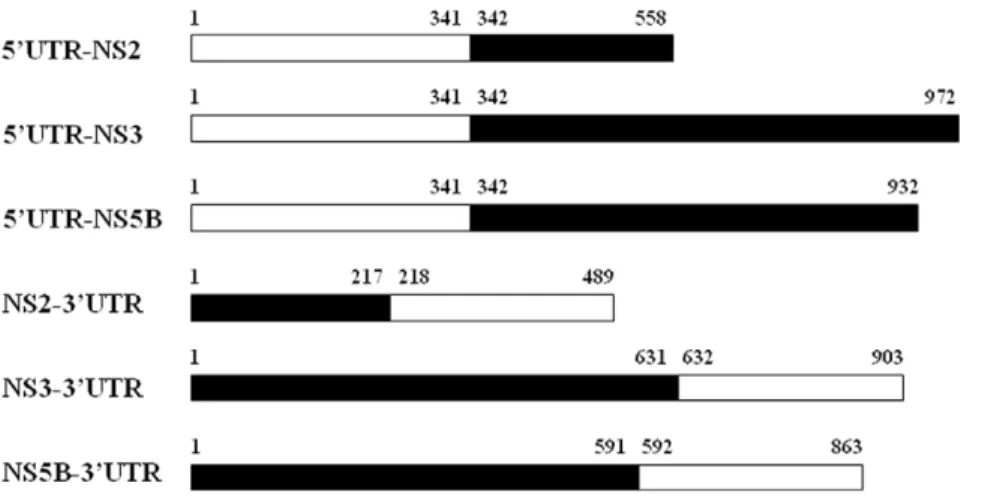
Figure 1. Binary HCV sequence components used for prediction of site-specific covariation between UTRs and NS proteins.Six sequence components for data mining were created by connecting UTR nucleotide segments and amino acid segments of the enzymatic NS proteins, including 59UTR-NS2, 59UTR-NS3, 59UTR-NS5B, NS2-39UTR, NS3-39UTR and NS5B-39UTR. The 59UTR and 39UTR contained 341 and 272 nucleotides, respectively; the NS2, NS3 and NS5B contained 217, 631 and 591 amino acid residues, respectively. White bars indicate nucleic acid segments; black bars indicate amino acid segments.
Discovery of covariation pairs between 59UTR243
nucleotide and NS2/3 amino acid residues
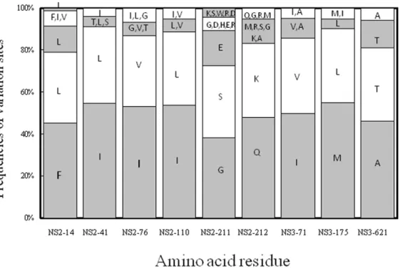
The data mining results showed a strong covariation relation-ship of the 243rdnucleotide of 59UTR to the 14th, 41st, 76th, 110th, 211thand 212thresidues of NS2 and to the 71st, 175thand 621st residues of NS3 (Fig. 2). Notably, the 59UTR243G was frequently
associated with NS2-14F, -41I, -76I, -110I, -211G, -212Q, NS3-71I, -175M and 621A, while the 59UTR243A was associated with
NS2-14L, -41L, -76V, -110L, -211S, -212K, NS3-71V, -175L and 621T. Neither C nor T was present at the 243rdnucleotide of the 59UTR. Simultaneous change of both sites of a covariant pair, as opposed to the change of a single site of a covariant pair, has been hypothesized to be better for HCV replication. With this in mind, the HCV replication consequences of the observed covariations were tested in a cell-based system, as detailed in the following.
Cell-based functional analysis - Evaluating HCV replication efficiency by mutation of coordinated variations between 59UTR243nucleotide and NS2/NS3
amino acid residues by using NS2-39replicon
Site-specific mutations matching various of the observed covariations were introduced in order to analyze their effects on the replication efficiency using a transient-replication assay. We constructed 9 pairs of variants in the context of the wild-type NS2-39
replicon (59UTR243G), each consisting of a single amino acid
substitution at the NS2 or NS3 region and double substitutions in combination with 59UTR-G243A and the corresponding amino acid
(Figs. 3A and B). Based on the normalized luciferase activities at 3 consecutive time points, the transient luciferase assays indicated that the 9 single amino acid variants decreased replication efficiency in the presence of 59UTR243G, but replication efficiency could be
rescued when any single variant of I41L, I76V, NS2-I110L, NS2-G211S, NS3-I71V and NS3-M175L was combined with 59UTR-G243A. On the contrary, the 59UTR-G243A could not
compensate the NS2-F14L, NS2-Q212K and NS3-A621T variants. Furthermore, different types of codon usage were introduced for NS2-I110L (CTT and TTG) and NS2-G211S (AGC and TCA), yielding comparable compensatory effects and indicating that differences of codon usage at the nucleotide level may not be a concern (Fig. 4). These results together suggest that the covariation of 59UTR-G243A with the NS2 and NS3 proteins was most likely
due to amino acid substitution, but this was not the case for the specific nucleotide sequences.
Dependence of NS2 protein in functional coordinated variations between 59UTR243and NS3
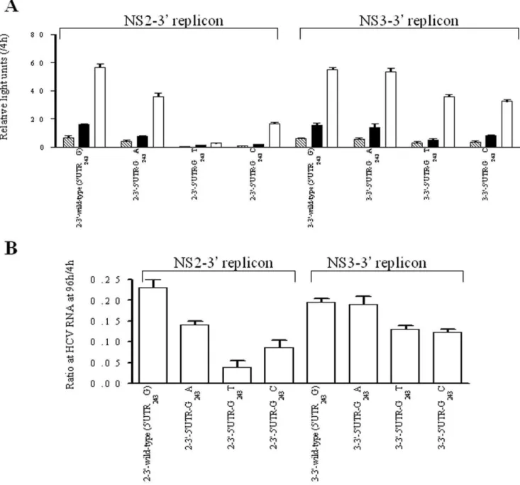
The effects of nucleotide substitution at the 59UTR243site with
regard to HCV replication were compared for the NS2-39or NS3-39 replicon backbone contexts. Compared to 59UTR243G in the
NS2-39replicon context, 59UTR-G243A showed a moderate decline
in replication efficiency, whereas 59UTR-G243T and 59UTR-G243C
showed a profound decline (Figs. 5A and B). However, 59 UTR-G243A, 59UTR-G243T and 59UTR-G243C showed no or little
influence on replication efficiency in the context of the NS3-39
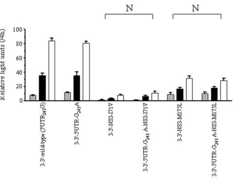
replicon (Figs. 5A and B). These results indicate that the NS2 protein may be of great importance in replicative modulation mediated by HCV 59UTR243. Further, the I71V and
NS3-M175L variants impaired HCV replication to similar levels in the NS3-39 replicons carrying either 59UTR243G or 59UTR-G243A
(Fig. 6). Because 59UTR-G243A could only compensate NS3-I71V
and NS3-M175L in the presence of NS2 (Fig. 3), the results suggest that these functional coordinated variations between 59UTR243and
NS3 depend on the NS2 protein.
Figure 2. Frequencies of variation sites assessed from Weka.The covariation sites were predicted at the 59UTR243nucleotide position and
their corresponding amino acid sites in HCV NS2 or NS3. Co-variation sites between the 59UTR243G (grey) and 59UTR243A (white) with the
corresponding amino acid residues (single-letter code) are shown in accumulative percentage. A frequency.10% for an amino acid residue is presented individually. Possible NS2 co-evolving sites include the 14th, 41st, 76th, 110th, 211thand 212thresidues; possible NS3 sites include the 71st
175thand 621stresidues.
Modulation of HCV replication efficiency by exogenously expressed NS2 proteins using NS3-39replicon
Next, we addressed the question of whether the NS2 protein variants expressed exogenously could modulate HCV replication activity of the HCV NS3-39replicon in the presence of 59UTR243G or 59UTR243A. Wild-type and variant forms of the
NS2-flag fusion protein were transfected into stable NS3-39 -Feo-59UTR243G or NS3-39-Feo-59UTR243A replicon cells, after which
the lysate luciferase activities were analyzed (Fig. 7). The results showed that the wild-type NS2 reduced by,10% the replication
activity of the NS3-39-Feo-59UTR243G replicon and by,40% the
replication activity of the NS3-39-Feo-59UTR243A replicon. As
compared to the wild-type NS2, the 6 individual variant NS2 proteins substantially reduced the replication activity of the NS3-39-Feo-59UTR243G replicon. However, the replication efficiencies
reduced by NS2-I41L, NS2-I76V, NS2-I110L and NS2-G211S
could be rescued in the NS3-39-Feo-59UTR243A replicon, but not
those reduced by NS2-F14L, NS2-Q212K (Fig. 7A). The NS2-flag proteins were immunostained by western blot analysis, exhibiting at levels comparable to the 7 proteins, i.e. the wild-type and the six variants (Fig. 7B). Thus, the results of the exogenously expressed wild-type and the variant NS2 agreed with the experiments based on NS2 expressed as replicon NS proteins, indicating that the compensatory effects did not depend on whether the NS2 proteins were expressed as a separate protein or in a polyprotein.
Regulation of HCV replication by 59UTR-mediated NS2 binding to HCV RNA
To study the RNA/protein interactions, stable NS3-39 -Feo-59UTR243G or NS3-39-Feo-59UTR243A replicon cells transfected
with each of the wild-type and variant NS2-flag expression plasmids were crosslinked at 48 hr after transfection.
Immuno-Figure 3. Transient-replication assay of NS2-39replicons.The replicons carried either wild-type (59UTR243G) or 59UTR-G243A and 9 pairs of
variants carrying specific amino acid residues as indicated at the NS2 or NS3 regions. Huh-7 cells were transfected with 2.5mg of replicon RNA. (A) Luciferase activity was determined in cell lysates at 48h (stripe), 72 h (black) and 96 h (white) posttransfection. Data were normalized by the values at 4 h posttransfection and expressed as mean6SD (n = 4). (B) HCV-RNA was quantified by real-time PCR at 96 h posttranfection. Data were normalized by the values at 4 h posttransfection and expressed as mean6SD (n = 3). Note that replication efficiency of replicons carrying amino acid variants was compensated by the presence of 59UTR-G243A, indicated as compensatory pair (C) and otherwise as non-compensatory pair (N).
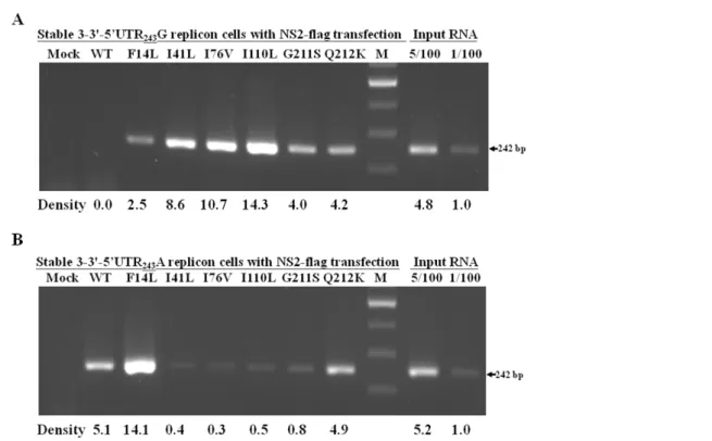
precipitation of the RNA-protein complexes was performed using anti-flag antibody to specifically recognize HCV NS2-flag protein. Bound RNA samples were then detected by reverse transcription-PCR using HCV-specific primers at the 59UTR region. The results showed that HCV-specific 242-bp product could be detected in the NS2-F14L, NS2-I41L, NS2-I76V, NS2-I110L, NS2-G211S and NS2-Q212K replicon cells that expressed NS3-39-Feo-59UTR243G, but not in the wild-type NS2 transfectant
(Fig. 8A). On the other hand, the HCV-specific 242-bp product was detected in the wild-type NS2, NS2-F14L and NS2-Q212K replicon cells that expressed NS3-39-Feo-59UTRG243A, but could
only barely be detected in those transfected with I41L, NS2-I76V, NS2-I110L and NS2-G211S (Fig. 8B). The stronger ability of NS2 to bind to the HCV-RNA of the NS2 variants relative to the wild-type HCV-RNA correlated with decreasing HCV replication in the 59UTR243G replicon cells, as shown in Figs. 3
and 7. As the NS2/HCV-RNA binding abilities of the NS2-I41L, NS2-I76V, NS2-I110L and NS2-G211S variants weakened in the 59UTR243A replicon cells, HCV replication levels were restored.
Together, these data indicate that 59UTR-mediated NS2 binding to HCV RNA can regulate HCV replication.
Genotype-specific covariation patterns between 59UTR243nucleotide and specific NS2/NS3 amino acid
residues
Analysis of the frequencies of the co-evolutionary sites in the 217 sampled HCV genome found distinct dominant sequences between the 1b and the non-1b genotype groups (Table S3). To confirm the genotype-specific covariation patterns, 381 additional full-length HCV genome sequences in an updated Los Alamos HCV sequence database were downloaded on May 19, 2009, making a total of 598 (217 in the Nov. 30, 2006 download+381 in the May 19, 2009 download = 598 total) analyzed nucleotide sequences. The combined results showed that G-Ile-Ile-Ile-Gly-Ile-Met was predominant (appearing in 53.8% of genotype 1b but
0.0% of genotype non-1b) for genotype-1b, while A-Leu-Val-Leu-Ser-Val-Leu was predominant (appearing in 79.1–100.0% of genotype non-1b but 0.0% of genotype 1b) in genotypes 1a, 2a and 2b (Table 1). These results confirmed that the initial data mining dataset for the nucleotide and amino acid residues found in 59UTR, NS2 and NS3 co-varied in a genotype-specific manner.
Discussion
Data mining involves finding patterns or rules in large data sets. Such patterns can be used to make predictions or form the basis of hypotheses for future experiments [26-28]. Data mining is being integrated into bioinformatic research [29]. In the present study, data mining methodology found previously unnoticed nonrandom covariance between HCV 59UTR with NS2 and NS3 proteins from a large HCV genomic database built from patient samples. This nonrandom association was experimentally verified to be of functional significance to viral replication by use of a cell-based HCV replicon system.
Protein residue covariation may suggest physical and/or functional constraints of paired amino acid positions [30]. As shown in previous studies, the covarying amino acid residues in the 10 HCV proteins display a scale-free network where central amino acid substitutions connect to many other sites [18,31]. Data mining analysis in the present study has revealed that coordinated variations occur between the untranslated 59UTR-RNA elements and the amino acid residues of the NS2 and NS3 proteins. UTRs are traditionally thought to have no influence on protein coding sequences. Accordingly, the data mining results of this study indicating coordinated variations between the 59UTR-RNA element and the NS2/NS3 proteins were surprising. Importantly, the computational results were confirmed by cell-based experi-ments using replicon replication and RNA-protein interaction assay to have significant effect on viral replication. Therefore, this study demonstrates a functionally significant pattern of linkage disequilibrium involving a non-coding nucleotide (59UTR243) and Figure 4. Transient-replication assay of NS2-39replicons.The replicons carried different types of codon usage for NS2-I110L (CTT and TTG) and NS2-G211S (AGC and TCA). Huh-7 cells were transfected with 2.5mg of replicon RNA. Resulting luciferase activities were determined from equal amounts of cell lysate harvested at 48 h (stripe), 72 h (black) and 96 h (white) posttransfection. Data were normalized by luciferase activity measured at 4 h posttransfection and expressed as mean6SD (n = 4). Compensatory pairs (C) are indicated as described in Fig. 3.
the amino acid residues (4 NS2 sites and 2 NS3 sites) in the HCV genome. The results suggest mutual communication in trans
between HCV 59UTR-RNA and individual NS2 proteins, or a combination of NS2 and NS3 proteins, by a mechanism that possibly involves direct binding or interaction with a common partner from either the HCV or host factors such as cellular protein or RNA. Strong binding of the NS2 proteins to the HCV 59UTR-RNA appears to diminish HCV replication, whereas weak binding correlates with restoration of HCV replicative efficiency. In cell-based systems, HCV NS2 is not an indispensable component for replication because HCV subgenomic replicon RNA allows replication in the absence of NS2 [32,33]. However, the NS2 protein may modulate IRES-dependent translation, NS3
kinetics and/or NS5B replication, thus affecting HCV synthesis of both viral RNA and proteins [34,35], and also may mediate HCV assembly and release [36,37]. It has been reported that NS2 sequences differ between nonresponder and relapser groups in HCV patients receiving antiviral therapy, with clinical relevance [38]. According to NS2 topology [12], the 14th, 41st and 76th residues are located at the first, the second and the third transmembrane domains, respectively. The present study suggests a novel regulatory mechanism involving NS2, whereby NS2 with a high binding affinity for 59UTR sequences may result in reduced HCV RNA flexibility, which in turn may compromise HCV RNA conformational rearrangement and/or the joining of other essential factors, resulting in less efficient HCV replication.
Figure 5. Transient-replication assay of the NS2-39and NS3-39replicons.The replicons each carried wild-type (59UTR243G), 59UTR-G243A,
59UTR-G243T and 59UTR-G243C. Huh-7 cells were transfected with 2.5mg of replicon RNA. (A) Luciferase activity was determined in cell lysate at 48 h
(stripe), 72 h (black) and 96h (white) posttransfection. Data were normalized by the values at 4 h posttransfection, expressed as mean6SD (n = 4). (B) HCV-RNA was quantified by real-time PCR at 96 h posttranfection. Data were normalized by the values at 4 h posttransfection, expressed as mean6 SD (n = 3).
HCV 59UTR243 is located at a non-Watson-Crick base pair
position between the IRES IIIc and IIId domains of the positive strand [5,39,40] and at the IIIc’domain of the replicative strand [41]. Both the positive- and negative-stranded domains of this non-coding region function as host protein binding sites which regulate translation and replication [42,43]. In previous studies using either rabbit reticulocyte or hepatic lysates in vitro,
59UTR243A and 59UTR243G had the same IRES translation
activities [40,44,45]. Furthermore, a G-to-A change at 59UTR243 exhibited preferential translation functions in
lym-phoblastoid cell lines and primary dentritic cells [45–47], suggesting that 59UTR243 might be a cell-specific determinant
in viral RNA translation. The present study revealed that changes in 59UTR243alter HCV replication, with G, A, C and T
displayed in order of decreasing activity with the NS2-39replicon assay. These results agree with G and A, but not with C and T at 59UTR243 in the HCV from patient samples, i.e. the HCV
sequences in patient samples show G and A, but no C and T at the 59UTR243 site. In addition, our results indicate that HCV
59UTR243may mutate in a genotype-specific covariant manner.
The dominant haplotype of 59UTR243
-NS2-41-76-110-211-NS3-71-175 differed among HCV 1b and non-1b genotypes. This variance relationship could only be seen in a population consisting of 1b and 1b genotypes, but not in the 1b or non-1b subpopulations alone. The distinct haplotype patterns suggest that the genotype-1b may split from the non-1b genotypes where fitness epistasis causes fixation of beneficial polymorphisms within a genotypic subpopulation. The genotype-1b haplotype was G-Ile-Ile-Ile-Gly-Ile-Met for 59UTR243, NS2-41, -76, -110,
-211, NS3-71 and -175; that of the non-1b haplotype was A-Leu-Val-Leu-Ser-Val-Leu. It should be noted that genotype-1b had 4 Ile residues but that genotype-non-1b had none. Genotype-non-1b had 3 Leu residues and 2 Val residues while genotype-Genotype-non-1b had none, suggesting that covarying substitutions differ but that the
Figure 6. Transient-replication assay of NS3-39replicons.The replicons carried either wild-type (59UTR243G) or 59UTR-G243A and 2 pairs of
variants carrying NS3-I71V and NS3-M175L. Huh-7 cells were transfected with 2.5mg of replicon RNA. Luciferase activity was determined in cell lysate at 48 h (strip), 72 h (black) and 96 h (white) posttransfection. Data were normalized by the values at 4 h posttransfection, expressed as mean6SD (n = 4). Non-compensatory pairs (N) are indicated as described in Fig. 3.
doi:10.1371/journal.pone.0025530.g006
Figure 7. Modulation of HCV replication by exogenously expressed NS2 protein.(A) Three micrograms of each wild-type or variant NS2-flag expression plasmids were transfected into stable NS3-39-Feo-59UTR243G (black bar) or NS3-39-Feo-59UTR243A (white bar)
replicon cells. Cell lysate was harvested at 48 h posttransfection and luciferase activity was determined. Data were calculated as the ratio to the corresponding untransfected control, expressed as mean 6 SD (n = 4). (B) Fifty micrograms of total protein from each cell lysate were separated by 12% sodium dodecyl sulfate-polyacrylamide gel electro-phoresis and immunostained with antibodies recognizing the flag epitope. Compensatory pairs (C) and non-compensatory pairs (N) are indicated as in Fig. 3.
physicochemical properties in these hydrophobic residues may be conserved between genotypes 1b and non-1b. These sites may be of significance in determining HCV functional changes during genome evolution.
Adaptive mutations at the NS regions in cell-culture based systems have been shown in prior work [25,48]. A recent study further identified an adapted Gly-to-Arg mutation at the 28th residue of NS2 in a chimpanzee-infected JFH-1 strain [49]. This present study reports coevoluationary sites of the NS2 and NS3 proteins in humans. Covariance of these sites during divergent genome evolution is assumed to be advantageous to HCV replication in vivo. It is most likely that the primary mutations
appear at residues of the NS2 and NS3 proteins, subsequently exerting structural-dynamic pressure that induces a conformation-al change of 59UTR243, which is located in the most conserved
region of the HCV genome.
In conclusion, the presented data mining analysis of HCV genome sequences has indicated by both computational method-ology and by cell-based HCV replicon assay that 59UTR243and
specific residues of the NS2 and NS3 proteins are involved in a previously unnoticed nucleotide and amino acid covariation, which may be associated with genome evolution which contributes to functional regulation of HCV replication. These results further support the premise that data mining methodology is an effective tool for finding useful patterns in the increasingly large database of contemporary virus research.
Materials and Methods
Data mining analysis
The employed data mining analysis involved the following steps: i) full-length HCV genome sequences were downloaded from the
Figure 8. Immunoprecipitation of RNA-protein assay using anti-flag antibody recognized crosslinked HCV NS2-flag protein and replicon RNA.Stable NS3-39-Feo-59UTR243G (A) or NS3-39-Feo-59UTR243A (B) replicon cells transfected with each of the wild-type and variant
NS2-flag expression plasmids were harvested for formaldehyde-mediated crosslinking. After immunoprecipitation with anti-NS2-flag antibody, purified RNA samples were analyzed by reverse transcription-PCR using a primer pair spanning 242 bases from the HCV 59UTR region. Total RNA samples from mock transfected replicon cells were analyzed by reverse transcription-PCR in parallel. Band intensity quantification was performed by AlphaImage 2200 software and a representative experimental run is shown (Lab Recyclers, MD).
doi:10.1371/journal.pone.0025530.g008
Table 1.Comparison of the predominant haplotypes of 59UTR243, NS2-41, -76, -110, -211, NS3-71 and -175 among the HCV
genotypes.
HCV genotype Number of full-length HCV sequences (n = 598) Haplotype of 59UTR243, NS2-41, -76, -110, -211, NS3-71 and -175 G-Ile-Ile-Ile-Gly-Ile-Met A-Leu-Val-Leu-Ser-Val-Leu
1a 218 (100%)a 0 (0.0%) 218 (100.0%)
1b 327 (100%) 176 (53.8%) 0 (0.0%)
2a 29 (100%) 0 (0.0%) 29 (100.0%)
2b 24 (100%) 0 (0.0%) 19 (79.1%)
aNumber (percentage).
Los Alamos HCV database [24]; ii) the nucleotide segments of the UTRs and the amino acid segments of the NS proteins were retrieved and combined, creating 6 new binary sequence components including 59UTR-NS2, 59UTR-NS3, 59 UTR-NS5B, NS2-39UTR, NS3-39UTR and NS5B-39UTR (Fig. 1); iii) multiple sequence alignments were constructed by the CLUSTAL W software program [50] and confirmed by visual inspection; iv) 100% conserved columns in multiple sequence alignment were eliminated by GeneDoc to avoid false covariation signals due to site conservation; v) the covariation relationships of the remaining individual sites were identified by association rule mining based on the Apriori algorithm using Weka software [23]. A format transformation system was used to transform the output of GeneDoc into ARFF format which is readable as Weka input. The Apriori algorithm satisfies 2 parameters: support (also known as coverage, proportion of instances that contain a particular code) and confidence (also known as accuracy, proportion of instances that it predicts correctly) to find the best association rules. In order to capture novel covariations from highly polymorphic sites, the support threshold was set at 0.33. In order to identify strong associations, the minimum confidence threshold was set at 1.0, which indicates that the identified rule is present in 100% of the sequences.
Cell cultures
Cell monolayers of cloned human Huh7 hepatoma cell line were grown in Dulbecco’s modified Eagle medium (HyClone, USA) supplemented with 10% heat-inactivated fetal bovine serum and 1% penicillin/streptomycin at 37uC in a 5% CO2
atmosphere.
Plasmid construction
The HCV replicon constructs pFK-i341-PI-Luc/NS2-39/ET (pNS2-39 replicon) and pFK-i341-PI-Luc/NS3-39/ET (pNS3-39
replicon) for transient replication were kindly provided by Professor R. Bartenschlager [25]. These constructs possessed the luciferase reporter gene and the NS gene frame starting from either NS2 or NS3 to NS5B and were used as backbone constructs to generate co-variation mutants. G-to-A mutation in the 59UTR and its paired NS2 or NS3 covariant mutations were introduced into the backbone constructs by site-directed mutagenesis using the QuickChange XL Site-Directed Mutagenesis kit (Stratagene, USA). The mutation sites generated in the NS2 region were F14L, I41L, I76V, I110L, G211S or Q212K; the mutation sites generated in the NS3 region were I71V, M175L or A621T. The oligonucleotides used for construction of replicon variants are listed in Table S4. To facilitate stable selection of covariant mutants, a gene fragment composed of fused firefly luciferase and neomycin phosphotransferase genes (Feo) was used to replace the firefly luciferase gene. The coding regions of the wild-type and the variant NS2 were PCR amplified from the corresponding replicon templates and subcloned into a p3XFLAG-CMV-14 expression vector (Sigma-Aldrich, Germany). DNA sequencing was used to verify the exact site-specific substitutions. No non-target sequence changes were introduced.
In vitrotranscription, electroporation and transient
replication assay
In vitrotranscripts were prepared and transfection by
electropo-ration was carried out as described previously [25], with slight modifications. Briefly, the replicon plasmids were prepared with a midi-plasmid extraction kit (Qiagen, USA), linearized with AseI
andScaI (New England Biolabs, USA), extracted with phenol and
chloroform, precipitated with ethanol and dissolved with nuclease-free water. T7 promoter-driven in vitro transcription was
performed with purified linearized replicon DNA using MEGA-script T7 kit (Ambion, USA) at 37uC for 2 hours. The DNA templates were digested by adding RNase-free DNase. After purification by RNeasy MinElute Cleanup kit (Qiagen), the replicon RNA was precipitated with alcohol and dissolved with nuclease-free water. RNA quantity and purity was determined by 260nm/280nm optical density measurements and agarose gel electrophoresis.
For electroporation, monolayered Huh7 cells were trypsinized and resuspended at a concentration of 56106 cells per mL in cytomix buffer [51] containing 2 mM of ATP and 5 mM of glutathione. Four-hundred microliters of the suspended cells were mixed with 2.5mg of replicon RNA and 5mg of total RNA from the Huh7 cells as a carrier. The cell mixture was transferred to a 4-mm cuvette and electroporated at 1300 V for 99msec using
electroporation equipment (ECM 830, BTX Harvard Apparatus, USA). After incubation at room temperature for 10 minutes, the cells were seeded into a 6-well and harvested at given time points after transfection.
For transient replication assay, the cells were washed with 1x PBS and lysed with 1x passive lysis buffer (Promega, USA), 120mL per well. Then, 20mL of supernatant was mixed with 100mL of Luciferase assay reagent (QuantiLum Recombinant Luciferase kit, Promega) and measured in a luminometer (Lumat LB9506, Berthold Technologies, Germany). The values of luciferase activity in the cell lysates harvested at 4 h posttransfection were used to normalize the transfection efficiency.
Quantitation of HCV RNA by real-time PCR
Total RNA was isolated from cells with a single-step method modified from the acid guanidinium–thiocyanate–phenol–chloro-form extraction procedure with REzolTM C&T reagent (Protech Technology, Taiwan). Intracellular HCV-RNA titers were measured quantitatively by reverse transcription coupled to real-time PCR assay using a LightCycler instrument (Roche, Germany) yielding a dynamic range from 800 to 100 million copies/mL [52].
Stable HCV replicon cells
At 48 h posttransfection, the Huh7 cells transfected with plasmids carrying Feo gene were selected by 500mg/mL G418 for approximately 1 month to obtain stable NS3-39-Feo-59UTR243G
and NS3-39-Feo-59UTR243A replicon cells. Antibodies and Western blot analysis
Monoclonal antibodies specific to flag tag (clone F1804) and actin (clone MAB1501) were purchased from Sigma-Aldrich (USA) and Chemicon (USA), respectively. Cell lysates were electrophoresed on sodium dodecyl sulfate-polyacrylamide gel electrophoresis and transferred to a polyvinylidene fluoride membrane. After blocking, the membrane was incubated with specific primary antibody, washed with 0.05% phosphate buffer saline-Tween 20, reacted with horseradish peroxidase-conjugated secondary antibody and developed with Western Lighting (Perkin-Elmer, USA).
Immunoprecipitation of RNA-protein assay
by 1% formaldehyde in phosphate buffer saline at room temperature for 30 min, followed by a quench solution (0.25 M glycine in phosphate buffer saline). The cells were then resuspended in RIPA buffer (50 mM Tris-HCl, pH 7.5, 1% Nonidet P-40, 0.5% sodium deoxycholate, 0.05% sodium dodecyl sulfate, 1 mM EDTA, 150 mM NaCl) containing a cocktail of protease inhibitors (Roche) and RNase inhibitor (Takara, Japan) and lysed by 3 freeze–thaw cycles. After centrifugation at 12000 rpm for 15 min to remove any insoluble materials, the clarified supernatant was collected, pre-cleared with protein G beads not coupled with ligand but accompanied by yeast tRNA as a nonspecific competitor, and then incubated with protein G beads coupled with anti-flag monoclonal antibody at room temperature for 90 min. The complex was washed with RIPA buffer, resuspended in a solution of 50 mM Tris-HCl, pH 7.0, 5 mM EDTA, 10 mM dithiothreitol and 1% sodium dodecyl sulfate, then incubated at 70uC for 45 min to reverse the crosslinking. The immunoprecipitated RNA was analyzed by reverse transcription-PCR. Briefly, the RNA was extracted with Rezol C&T (Protech) reagent, reversely transcribed into cDNA with Moloney murine leukemia virus RT (Promega) and PCR amplified for 35 cycles with a primer pair (sense primer: 59 -ACTCCACCATAGAT-CACTCC-39 and antisense primer: 59 -AACACTACTCGGC-TAGCAGT-39) spanning 242 bases from the HCV 59UTR region.
Supporting Information
Figure S1 Neighbor-joining phylogenetic tree of the
HCV sequences. The sequences were downloaded from the
Los Alamos HCV database on Nov. 30, 2006. 217 full-length HCV genome sequences were aligned using CLUSTAL software and phylogenetically analyzed by the neighbor-joining method
using the molecular evolutionary genetics analysis (MEGA) program. The constructed phylogenetic tree includes 19 sequences for 1a genotype (&), 127 for 1b (%), 4 for 1c (m), 22 for 2a (
N
), 23 for 2b (#), 1 each for genotypes 2c and 2k (.), 4 each for 3a and3b and 1 for 3k (h), 1 for 4a (g), 2 for 5a (X), 2 each for 6a and 6k and 1 each for 6b, 6d, 6g and 6h (e).
(TIF)
Table S1 Genotypic distribution and accession num-bers of the 217 full-length HCV genome sequences.
(DOCX)
Table S2 Summary of the unique association rules.
(DOCX)
Table S3 The frequencies (%) of co-evolutionary sites in the sampled HCV genome sequences.
(DOCX)
Table S4 Oligonucleotides used for construction of replicon variants.
(DOCX)
Acknowledgments
The authors would like to thank Dr. Iain Bruce for his critical review of our manuscript.
Author Contributions
Obtained permission for use of replicon: KCY. Performed the bioinfor-matic analysis: SJS WCL. Performed the laboratory experiments: HYS NYO TFC. Conceived and designed the experiments: KCY. Analyzed the data: KCY. Contributed reagents/materials/analysis tools: KTS TTC SWW. Wrote the paper: KCY.
References
1. Tan SH, Zhang Z, Ng SK (2004) ADVICE: Automated Detection and Validation of Interaction by Co-Evolution. Nucleic Acids Res 32: W69–72. 2. Pelemans H, Esnouf R, Min KL, Parniak M, De Clercq E, et al. (2001)
Mutations at amino acid positions 63, 189, and 396 of human immunodeficiency virus type 1 reverse transcriptase (RT) partially restore the DNA polymerase activity of a Trp229Tyr mutant RT. Virology 287: 143–150.
3. Delaney WEt, Yang H, Westland CE, Das K, Arnold E, et al. (2003) The hepatitis B virus polymerase mutation rtV173L is selected during lamivudine therapy and enhances viral replication in vitro. J Virol 77: 11833–11841. 4. Pawlotsky JM (2004) Pathophysiology of hepatitis C virus infection and related
liver disease. Trends Microbiol 12: 96–102.
5. Honda M, Beard MR, Ping LH, Lemon SM (1999) A phylogenetically conserved stem-loop structure at the 59border of the internal ribosome entry site of hepatitis C virus is required for cap-independent viral translation. J Virol 73: 1165–1174.
6. Diviney S, Tuplin A, Struthers M, Armstrong V, Elliott RM, et al. (2008) A hepatitis C virus cis-acting replication element forms a long-range RNA-RNA interaction with upstream RNA sequences in NS5B. J Virol 82: 9008–9022. 7. Friebe P, Boudet J, Simorre JP, Bartenschlager R (2005) Kissing-loop interaction
in the 39end of the hepatitis C virus genome essential for RNA replication. J Virol 79: 380–392.
8. Friebe P, Lohmann V, Krieger N, Bartenschlager R (2001) Sequences in the 59 nontranslated region of hepatitis C virus required for RNA replication. J Virol 75: 12047–12057.
9. Luo G, Xin S, Cai Z (2003) Role of the 59-proximal stem-loop structure of the 59 untranslated region in replication and translation of hepatitis C virus RNA. J Virol 77: 3312–3318.
10. Kolykhalov AA, Mihalik K, Feinstone SM, Rice CM (2000) Hepatitis C virus-encoded enzymatic activities and conserved RNA elements in the 39 nontranslated region are essential for virus replication in vivo. J Virol 74: 2046–2051.
11. Lorenz IC, Marcotrigiano J, Dentzer TG, Rice CM (2006) Structure of the catalytic domain of the hepatitis C virus NS2-3 protease. Nature 442: 831–835. 12. Yamaga AK, Ou JH (2002) Membrane topology of the hepatitis C virus NS2
protein. J Biol Chem 277: 33228–33234.
13. Wolk B, Sansonno D, Krausslich HG, Dammacco F, Rice CM, et al. (2000) Subcellular localization, stability, and trans-cleavage competence of the hepatitis
C virus NS3-NS4A complex expressed in tetracycline-regulated cell lines. J Virol 74: 2293–2304.
14. Yao N, Reichert P, Taremi SS, Prosise WW, Weber PC (1999) Molecular views of viral polyprotein processing revealed by the crystal structure of the hepatitis C virus bifunctional protease-helicase. Structure 7: 1353–1363.
15. Lesburg CA, Cable MB, Ferrari E, Hong Z, Mannarino AF, et al. (1999) Crystal structure of the RNA-dependent RNA polymerase from hepatitis C virus reveals a fully encircled active site. Nat Struct Biol 6: 937–943.
16. Simmonds P, Bukh J, Combet C, Deleage G, Enomoto N, et al. (2005) Consensus proposals for a unified system of nomenclature of hepatitis C virus genotypes. Hepatology 42: 962–973.
17. Hnatyszyn HJ (2005) Chronic hepatitis C and genotyping: the clinical significance of determining HCV genotypes. Antivir Ther 10: 1–11. 18. Aurora R, Donlin MJ, Cannon NA, Tavis JE (2009) Genome-wide hepatitis C
virus amino acid covariance networks can predict response to antiviral therapy in humans. J Clin Invest 119: 225–236.
19. Lindenbach BD, Pragai BM, Montserret R, Beran RK, Pyle AM, et al. (2007) The C terminus of hepatitis C virus NS4A encodes an electrostatic switch that regulates NS5A hyperphosphorylation and viral replication. J Virol 81: 8905–8918.
20. Murray CL, Jones CT, Tassello J, Rice CM (2007) Alanine scanning of the hepatitis C virus core protein reveals numerous residues essential for production of infectious virus. J Virol 81: 10220–10231.
21. Xu Z, Fan X, Xu Y, Di Bisceglie AM (2008) Comparative analysis of nearly full-length hepatitis C virus quasispecies from patients experiencing viral breakthrough during antiviral therapy: clustered mutations in three functional genes, E2, NS2, and NS5a. J Virol 82: 9417–9424.
22. Yi M, Ma Y, Yates J, Lemon SM (2007) Compensatory mutations in E1, p7, NS2, and NS3 enhance yields of cell culture-infectious intergenotypic chimeric hepatitis C virus. J Virol 81: 629–638.
23. Frank E, Hall M, Trigg L, Holmes G, Witten IH (2004) Data mining in bioinformatics using Weka. Bioinformatics 20: 2479–2481.
24. Kuiken C, Yusim K, Boykin L, Richardson R (2005) The Los Alamos hepatitis C sequence database. Bioinformatics 21: 379–384.
26. Edelstein HA (1999) Introduction to data mining and knowledge discovery. PotomacMD: Two Crows Corporation. 36 p.
27. Han J, Kamber M (2006) Data Mining: Concepts and Techniques. Fransisco San, ed. CA. : Morgan Kaufmann Publishers. 550 p.
28. Witten IH, Frank E (2005) Data Mining: Practical Machine Learning Tools and Techniques. San FranciscoCA: Morgan Kaufmann Publishers. pp 525. 29. Paul S, Piontkivska H (2009) Discovery of novel targets for multi-epitope
vaccines: screening of HIV-1 genomes using association rule mining. Retrovirology 6: 62–74.
30. Yip KY, Patel P, Kim PM, Engelman DM, McDermott D, et al. (2008) An integrated system for studying residue coevolution in proteins. Bioinformatics 24: 290–292.
31. Campo DS, Dimitrova Z, Mitchell RJ, Lara J, Khudyakov Y (2008) Coordinated evolution of the hepatitis C virus. Proc Natl Acad Sci U S A 105: 9685–9690.
32. Blight KJ, McKeating JA, Rice CM (2002) Highly permissive cell lines for subgenomic and genomic hepatitis C virus RNA replication. J Virol 76: 13001–13014.
33. Lohmann V, Korner F, Koch J, Herian U, Theilmann L, et al. (1999) Replication of subgenomic hepatitis C virus RNAs in a hepatoma cell line. Science 285: 110–113.
34. She Y, Liao Q, Chen X, Ye L, Wu Z (2008) Hepatitis C virus (HCV) NS2 protein up-regulates HCV IRES-dependent translation and down-regulates NS5B RdRp activity. Arch Virol 153: 1991–1997.
35. Welbourn S, Green R, Gamache I, Dandache S, Lohmann V, et al. (2005) Hepatitis C virus NS2/3 processing is required for NS3 stability and viral RNA replication. J Biol Chem 280: 29604–29611.
36. Jirasko V, Montserret R, Appel N, Janvier A, Eustachi L, et al. (2008) Structural and functional characterization of nonstructural protein 2 for its role in hepatitis C virus assembly. J Biol Chem 283: 28546–28562.
37. Jones CT, Murray CL, Eastman DK, Tassello J, Rice CM (2007) Hepatitis C virus p7 and NS2 proteins are essential for production of infectious virus. J Virol 81: 8374–8383.
38. Cannon NA, Donlin MJ, Fan X, Aurora R, Tavis JE (2008) Hepatitis C virus diversity and evolution in the full open-reading frame during antiviral therapy. PLoS One 3: e2123.
39. El Awady MK, Azzazy HM, Fahmy AM, Shawky SM, Badreldin NG, et al. (2009) Positional effect of mutations in 59UTR of hepatitis C virus 4a on patients9response to therapy. World J Gastroenterol 15: 1480–1486. 40. Honda M, Rijnbrand R, Abell G, Kim D, Lemon SM (1999) Natural variation
in translational activities of the 59 nontranslated RNAs of hepatitis C virus
genotypes 1a and 1b: evidence for a long-range RNA-RNA interaction outside of the internal ribosomal entry site. J Virol 73: 4941–4951.
41. Dutkiewicz M, Swiatkowska A, Figlerowicz M, Ciesiolka J (2008) Structural domains of the 39-terminal sequence of the hepatitis C virus replicative strand. Biochemistry 47: 12197–12207.
42. Isken O, Baroth M, Grassmann CW, Weinlich S, Ostareck DH, et al. (2007) Nuclear factors are involved in hepatitis C virus RNA replication. RNA 13: 1675–1692.
43. Spangberg K, Wiklund L, Schwartz S (2000) HuR, a protein implicated in oncogene and growth factor mRNA decay, binds to the 39ends of hepatitis C virus RNA of both polarities. Virology 274: 378–390.
44. Lerat H, Shimizu YK, Lemon SM (2000) Cell type-specific enhancement of hepatitis C virus internal ribosome entry site-directed translation due to 59 nontranslated region substitutions selected during passage of virus in lymphoblastoid cells. J Virol 74: 7024–7031.
45. Laporte J, Bain C, Maurel P, Inchauspe G, Agut H, et al. (2003) Differential distribution and internal translation efficiency of hepatitis C virus quasispecies present in dendritic and liver cells. Blood 101: 52–57.
46. Bain C, Fatmi A, Zoulim F, Zarski JP, Trepo C, et al. (2001) Impaired allostimulatory function of dendritic cells in chronic hepatitis C infection. Gastroenterology 120: 512–524.
47. Nakajima N, Hijikata M, Yoshikura H, Shimizu YK (1996) Characterization of long-term cultures of hepatitis C virus. J Virol 70: 3325–3329.
48. Krieger N, Lohmann V, Bartenschlager R (2001) Enhancement of hepatitis C virus RNA replication by cell culture-adaptive mutations. J Virol 75: 4614–4624. 49. Kato T, Choi Y, Elmowalid G, Sapp RK, Barth H, et al. (2008) Hepatitis C virus JFH-1 strain infection in chimpanzees is associated with low pathogenicity and emergence of an adaptive mutation. Hepatology 48: 732–740.
50. Thompson JD, Higgins DG, Gibson TJ (1994) CLUSTAL W: improving the sensitivity of progressive multiple sequence alignment through sequence weighting, position-specific gap penalties and weight matrix choice. Nucleic Acids Res 22: 4673–4680.
51. van den Hoff MJ, Moorman AF, Lamers WH (1992) Electroporation in ‘intracellular’ buffer increases cell survival. Nucleic Acids Res 20: 2902. 52. Chang LL, Cheng PN, Chen JS, Young KC (2007) CD81 down-regulation on B
cells is associated with the response to interferon-alpha-based treatment for chronic hepatitis C virus infection. Antiviral Res 75: 43–51.