ABSTRACT
Aim This article presents a retrospective study on the behavior of implants placed with split crest technique in lateroposterior maxillary class iV atrophy.
Materials and Methods subjects who underwent implant placement following split crest technique in the maxillary latero-posterior area were enrolled in the present retrospective study. after a mean period of 6.2 years of function implant survival and success rates were assessed. Moreover, radiographic examination was made on digital periapical radiographs and by means of a speciic software. Bone level changes were measured as the diference between the peri-implants crestal bone level and the implants shoulder during the last patient’s visit recall examination. Results a total of 30 patients satisied the inclusion criteria and were included in the study; the subjects were treated with 88 implants (64 transmucosal and 24 submerged). The observation period for all patients treated with split crest technique varied between 4 and 8 years (mean 6.2 years). The implants survival rate was 96.6% and the prostheses survival rate was 100%. Bone resorption ranged between 2.3 mm and 2.7 mm.
Conclusion implants inserted in conjunction with split crest technique seems to be a promising therapy with similar results as conventional implant surgery.
of 88 implants placed with split crest technique
in the maxillary latero-posterior area
S. LONGONI
1, I. MARONI
2, A. BALDINI
3, M. SARTORI
41 Md, dds, Contract Professor of Prosthodontics, Milano Bicocca university dental school, Milan, italy 2 Md, dMd, Private Practice, Varese, italy
3 dds, Phd, Phd division of Orthodontics, university of rome Tor Vergata, rome, italy
4 dds, Phd, Phd in experimental Periodontology, Contract Professor of Prosthodontics, Milano Bicocca university dental school, Milan, italy
TO CiTe This arTiCle
longoni s, Maroni i, Baldini a, sartori M. retrospective radiographic study of marginal bone changes of 88 implants placed with split crest technique in the maxillary latero-posterior area. J Osseointegr 2016;8(1):8-13.
KeywOrds Bone expansion; Bone resorption; Osteotome; Piezosurgery; split crest.
INTRODUCTION
Maxillary alveolar atrophy is a longstanding problem that has prevented numerous patients from receiving treatment with dental implants. An adequate volume of healthy bone tissue at implants site is a fundamental prerequisite for a favorable prognosis of osseointegrated implants (1). In recent years, more awareness has been developed towards the principle of restoration-driven implants placements (2). In fact, maxillary bone atrophy and anatomic aberrations may jeopardize correct implant placement with respect to the ideal position and the final prosthetic restoration (2, 3). In Cawood and Howell class IV atrophy (4) there is insufficient bone at the ideal desired implant location. The site can be either grafted or enhanced through the use of barrier membranes (GBR) (5, 6) or titanium mesh (7-9), thus restoring the labial contour. Often, these procedures require a two staged approach to implant placement, lengthening treatment time and increasing costs. Indeed, bone grafting techniques implicate an additional operation area, general discomfort and morbidity and potential complications as the risk of infection and/or mucosa dehiscence. An alternative surgical technique is the split crest procedure that has provided satisfactory and predictable results for localized ridge expansion (10).
The alveolar ridge recontour by means of this technique is obtained with a one stage procedure without involving additional costs and a second operation area. Tatum in 1986 originally developed the technique for the placement of the Omni root, D-shaped, transmucosal implants (10). Then, the technique has been modified for implants placement with a submerged approach (11-17).
bone to facilitate the introduction of instruments for ridge expansion (10, 17). Moreover, the residual crest angulation is important because expanding the external cortical plate of the maxilla in labial direction may jeopardize the correct implant placement (Fig. 1) (10);
- a class 3 bone quality according to Lekholm and Zarb (20);
- no contraindications for implant treatment (1, 18); - no smoking habits.
The patients were given oral and written information regarding the risks of this type of surgery and their written informed consent was obtained.
Surgical procedure
The surgical procedure was performed from 2006 to 2010. Surgery was planned using computed tomography scans (CT), casts, diagnostic wax-up and surgical template.
Local anesthesia was achieved by infiltration of articaine 4% plus adrenaline 1:100,000 (Ubistesin™; 3M ESPE, Seefeld, Germany). A soft tissue incision was made to create a full thickness crestal flap with relaxing periosteal incisions. Crestal bone was regularized with a surgical tungsten bur in order to obtain a flat bone surface between the palatal and buccal cortical plates to make the subsequent osteotomy procedure easier (Fig. 2). In order to control the extension of the fracture line and to prevent the invasion of the periodontal ligament of the adjacent teeth, with the first pilot drill (Ø1.5 mm) the sites of the most mesial and the most distal (if a distal tooth is present) implants were prepared as deep as possible to minimize bone stress during the split crest. Subsequently, the Piezosurgery® (Mectron S.P.A. Genoa, Italy) (Fig. 3) technique was used for the crestal bone incision trying to reach the same depth of the first pilot drill. Then, straight chisels and round osteotomes to mobilize and gradually expand the vestibular bone wall were used. The instruments size and number varied in relation to bone density, thickness and diameter of implants. During patients recall examination, implants success
following Albrektsson and Zarb criteria (absence of implants mobility, no radiolucency around implants in the radiographic control, no infection, no pain and other symptoms or complaints referred by patients) and bone level changes were evaluated (18, 19). Also implants survival rate, prosthesis survival rate and aesthetic observations were reported.
MATERIALS AND METHODS
Patients enrolled in the present retrospective study underwent implant placement with split crest technique in the maxillary latero-posterior area (from the canine to the first molar region: from 1.3 to 1.6 and from 2.3 to 2.6). The selected subjects had to meet the following inclusion criteria:
- a thin maxillary ridge (Cawood and Howell IV class), ranging from 3 to 5 mm, and a corono-apical height of at least 10 mm;
- a trapezoidal residual bone shape of the ridge (showed by the transaxial CT images); indeed, it reduces the possibility of fracture of the labial bone wall during the split crest procedure, in comparison with a rectangular ridge shape. Specifically, the ridge should have labial and palatal cortical plates that are not fused and are separated by cancellous
fig. 1 The anatomy of the ridge to be split may inluence implant’s angulation and the possibility of vestibular wall fracture.
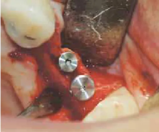
Insertion and removal of chisels and osteotomes (Fig. 4) was performed in a straight path with light malleting (10). The procedure was completed when the conical osteotomes had prepared adequate implants sites. Two types of implants had been used: trasmucosal ITI® (Straumann AG, Waldenburg, Switzerland) standard implants (2.8 mm neck height) and 3i Biomet® (Palm Beach Gardens, Florida, USA) submerged implants. When a high density bone quality was present, it had been necessary to adjust the shape of implant bed with the corresponding implant bur especially in the case of straight non self-tapping transmucosal implants. Transmucosal implants had been positioned with the neck’s shoulder aligned at the level of the crestal bone. Submerged implants had been placed into the osteotomy until they had reached 1 mm below the level of the crestal bone (10) to compensate the amount of physiological bone resorption after the surgical phase (Fig. 5). All implants had been inserted using handpiece implant driver. Autogenous bone chips harvested during the bone drilling phases plus bone from bovine sources (Tutodent® Chips 0.25 mm–1.0 mm - Tutogen Medical GmbH RTI Biologics Company, Neunkirchen a. Br., Germany) were used to fill the space created by expansion around the implants. A resorbable membrane (Pericardium Membrane - Tutogen® Medical GmbH RTI Biologics Company, Neunkirchen a.B., Germany) had been always placed to prevent mucosal penetration into the surgical site and to allow bone regeneration. All implants had been allowed to integrate with a submerged approach, avoiding incongruous loading due to removable provisional prosthesis, if present. Primary wound closure had been achieved with interrupted polyglycolic suture (Vicryl ® 4-0, Ethicon Inc., Johnson & Johnson, Somerville, NJ, USA). Second stage surgery had been performed after 4 months (21). A provisional fixed prosthesis was always used for 2 months with the aim of exerting a progressively increasing load on the implants (22). Subsequently definitive restorations had been delivered to the patients. All the definitive prostheses were fixed
fig. 4 sequence with larger instruments for a progressive and controlled bone expansion. fig. 5 split crest with submerged implants.
partial dentures, using an intraoral luting technique to achieve passive fit (23).
Photographs documentation had been performed during surgery to show the implants shoulders positioned following the protocol previously described. Radiographic examination was obtained using periapical radiographs taken with Rinn X-ray holders (Rinn®, Elgin, IL, USA) and the paralleling long-cone technique. In particular the radiographs had been performed at the 10 days recall for suture removal. The last photographic and radiographic evaluation was done during patients recall at the beginning of this retrospective study.
For each implant, the radiograph obtained at sutures removal (baseline) and the most recent radiograph obtained during patient’s visit recall examination (after a mean period of 6.2 years, min 4 years-max 8 years) were digitized and stored in a commercially available computer software (Sidexis, Sirona Dental GmbH, Salzburg, Austria), allowing gray value adaptation and calculation of the magnification factor of radiograph (calibration). In particular, for each radiograph the known implant length of ITI system (10 mm, 12 mm or 14 mm) and the known implant length of 3i Biomet system (10 mm, 11.5 mm or 13 mm) were used to calibrate images prior to bone level measurements. When the implant length was not available, the measure of implants diameter was used as a reference. Only radiographs showing the implants both mesially and distally, and the ones with an adequate quality with respect to contrast, were used.
absence of implants mobility, no radiolucency around implants in the radiographic control, no infection, no pain and other symptoms or complaints referred by patients) (19) and survival rate were calculated.
RESULTS
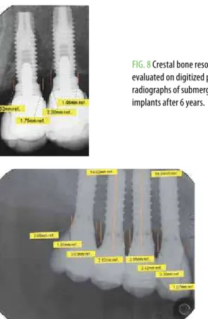
The total number of patients enrolled in the present retrospective study was 30, aged between 30 years and 70 years (mean 50,8 years) (13 male, 17 female). They were treated with split crest technique in the latero-posterior area of the upper jaw (from the canine to the first molar region) and 88 implants were placed. In particular, 64 ITI transmucosal standard implants (2.8 mm neck height) were inserted in 20 patients with a mean observation period of 6.4 years. After 2 years from the placement of the first implants, 24 3iBiomet submerged implants were also placed in 10 patients in order to achieve an easier management of soft tissues and aesthetics: the mean observation period was 5.6 years (Fig. 6 and 7). Globally the mean observation period for all patients treated with split crest technique was for both implants systems 6.2 years. The implants survival rate was 96.6%: 3 implants were lost during the unloaded healing period at second stage surgery (Table 1). Lost implants where replaced with larger diameter implants. At the moment of recall all the prostheses were in situ, so the prostheses survival rate was 100% and the patients did not refer any major complaint. Radiographic examination of implants showed that in transmucosal implants the mean bone resorption was mesially 2.5 mm (± 0.6 mm SD) and distally 2.7 mm (± 0.5 mm SD). In submerged implants the mean bone resorption was mesially 2.4 mm (± 0.5 mm SD) and distally 2.3 mm (± 0.6 mm SD) (Table 1) (Fig. 8 and 9).
DISCUSSION
As described in the international literature (11, 12, 15, 17), the split crest is a reproducible technique, but it depends upon the surgeon’s technical skills and it requires an adequate learning curve.
As a matter of fact, the main risk of split crest technique is the fracture of the labial cortical plate: this unlucky event may occur during the surgical phase especially if there is not cancellous bone between the reabsorbed cortical plates.
Indications for bone splitting are restricted to those sites that do not require vertical ridge augmentation (Cawood and Howell class IV atrophy). The rationale of split crest choice is to expand the residual ridge
obtaining quickly palatal and buccal bone walls ≥ 1
mm in order to position implants completely embedded into bone for long lasting osseointegration (1, 10). An important aspect of the split crest technique regards fig. 6 detail of prosthetic restoration onto submerged implants after 6 years.
fig. 7 detail of prosthetic restoration onto transmucosal implants after 6 years.
fig. 8 Crestal bone resorption evaluated on digitized periapical radiographs of submerged implants after 6 years.
the difficulty to plan a prosthetically driven implant placement. In fact, implants are placed between the labial and the palatal cortical plates following the angulation of bone ridge fracture. As a consequence in prosthetics steps often it is necessary to rectify abutment angulation to achieve an ideal aesthetic and functional result. Moreover, the rationale for full thickness flap is to improve visualization of bone ridge to be expanded, better controlling the labial cortical wall during splitting to reduce the risk of fracture. In this way, it is possible to perform and, if necessary, to position biomaterials and membranes before sutures. But, there was the possibility of blood supply reduction that should is taken into consideration.
In comparison with split crest technique, horizontal onlay block grafting allows to obtain a real 3D bone reconstruction with prosthetically favorable implant positioning. As reported by Aghaloo et al. in 2007, the implant survival rate positioned after onlay horizontal bone grafting is 90.4% and there are not long term follow up studies with exception of GBR (24). Also for ridge expansion techniques very few long-term, clinical studies have been published to date.
Simion et al. (11) reported on 5 patients in which a split-crest technique combined with guided tissue regeneration was performed.
A 5 years clinical study was carried out by Scipioni et al. (12) showing a survival rate ranging from 85% to 99%. Engelke et al. presented a clinical study placed with ridge splitting technique with micro-fixation reporting a survival rate of 86.2% after 5 years (15).
Sethi et al. showed a survival rate of 97% after an observation period of 5 years (17).
Bravi F. et al. reported a survival rate of 95.7% after 10 years (22).
Blus et al. reported a high survival rate of 96.5% with split crest technique (25).
In this retrospective study, clinical and radiographic
parameters showed that this surgical technique was safe and predictable when all inclusion criteria are carefully evaluated. With 96.6% of implants survival and 100% of prosthesis survival rate, the outcomes of the present investigation were consistent with the recent papers of Garcez-Filho et al. (26) and Santagata M et al. (27), with previously cited articles and with traditional implant placement procedure. Specifically, the 3 lost implants were placed in an area where a major bone expansion was performed, and this probably reduced the possibility of achieving an optimal primary stability. Radiographic bone levels measured mesially and distally to the implants inserted with the split crest technique were in the ranges of the bone levels reported in the literature around the implants positioned with the conventional standard technique (18). Both types of implants showed similar bone resorption (range: min 2.3 mm max 2.7 mm). This resorption was probably due to bone remodeling after the surgical expansion trauma and the bone blood supply interruption between the vestibular and palatal cortical plates. As a consequence, aesthetic outcomes were different in relation to the different implant-abutment connections and therefore, due to implant neck morphology. The aesthetic behaviors were observed during patient’s recall examination when this retrospective study started. In particular, in periodontal thin biotype the transmucosal smooth neck of implants was frequently exposed representing an aesthetic limit for the patient in comparison with submerged implants. Further research is needed to confirm these preliminary results.
CONCLUSION
Within the limitation of the present study, implants inserted in conjunction with split crest technique, regardless of the implant placement protocol (i.e.: transmucosal or submerged) showed good functional results. The one-stage approach should be considered predictable as long as selection of patients and surgical protocols described are carefully followed.
REFERENCES
1. Misch Ce. available bone and implant dentistry. in: Misch Ce. Contemporary implant dentistry. st. louis, MO: elsevier Mosby, 2008:105-129.
2. de wijs flJa, Cune Ms.immediate labial contour restoration for improved esthetics: a radiographic study on bone splitting in anterior single-tooth replacement. int J Oral Maxillofac implants 1997;12:686-696.
3. guirado Jl, yuguero Mr, Carrión del Valle MJ, Zamora gP.a maxillary ridge-splitting technique followed by immediate placement of implants: a case report. implant dent 2005;14:14-20.
4. Cawood Ji1, howell ra. a classiication of the edentulous jaws. int J Oral Maxillofac surg1988;17(4):232-6.
5. simion M, Trisi P, Piattelli a. Vertical ridge augmentation using a membrane technique associated with osseointegrated implants. int J Periodontics restorative dent 1994;14:496-511.
TaBle 1implants survival and implant success. Marginal bone resorption was measured at recall examination.
*Tsi = Transmucosal standard implants
ITI (TSI*) 3i BIOMET
Total number of implants 64 24
Total number of lost implants 2 1
survival rate 96.9 % 95.8%
global survival rate 96.6%
Mesial bone level change: mean 2.5 2.4
Mesial bone level change: sd 0.6 0.5
distal bone level change: mean 2.7 2.3
6. simion M, Jovanovic sa, Trisi P, scarano a, Piattelli a. Vertical ridge augmentation around dental implants using a membrane technique and autogenous bone or allografts in humans. int J Periodontics restorative dent 1998;18:8-23.
7. von arx T, hardt N, wallkamm B. The TiMe technique: a new method for localized alveolar ridge augmentation prior to placement of dental implants. int J Oral Maxillofac implants 1996;11:387-394.
8. Maiorana C, santoro f, rabagliati M, salina s. evaluation of the use of iliac cancellous bone and anorganic bovine bone in the reconstruction of the atrophic maxilla with titanium mesh: a clinical and histologic investigation. int J Oral Maxillofac implants 2001;16:427-432.
9. longoni s, sartori M, apruzzese d, Baldoni M. Preliminary clinical and histologic evaluation of a bilateral 3-dimensional reconstruction in an atrophic mandible: a case report. int J Oral Maxillofac implants 2007;22:478-483.
10. ferrigno N, laureti M. surgical advantages with iTi Te implants placement in conjunction with split crest technique. 18-month results of an ongoing prospective study. Clin Oral implants res 2005;16:147-155.
11. simion M, Baldoni M, Zafe d. Jawbone enlargement using immediate implant placement associated with a split-crest technique and guided tissue regeneration. int J Periodonticsrestorativedent 1992;12:462-473.
12. scipioni a, Bruschi gB, Calesini g. The edentulous ridge expansion technique: a ive-year study. int J Periodontics restorative dent 1994;14:451-459. 13. scipioni a, Bruschi gB, Calesini g, Bruschi e, de Martino C. Bone regeneration
in the edentulous ridge expansion technique: histologic and ultrastructural study of 20 clinical cases. int J Periodontics restorative dent 1999;19:269-277. 14. duncan JM, westwood rM. ridge widening for the thin maxilla: a clinical
report. int J Oral Maxillofac implants 1997;12:224-227.
15. engelke wg, diederichs Cg, Jacobs hg, deckwer i. alveolar reconstruction with splitting osteotomy and microixation of implants. int J Oral Maxillofac implants 1997;12:310-318.
16. Malchiodi l, scarano a, Quaranta M, Piattelli a. rigid ixation by means of titanium mesh in edentulous ridge expansion for horizontal ridge augmentation in the maxilla. int J Oral Maxillofac implants 1998;13:701-5.
17. sethi a, Kaus T. Maxillary ridge expansion with simultaneous implant placement: 5-year results of an ongoing clinical study. int J Oral Maxillofac implants 2000;15:491-499.
18. Misch Ce. an implant is not a tooth: a comparison of periodontal indexes. in: Misch Ce. Contemporary implant dentistry. st. louis, MO: elsevier Mosby, 2008:18-31.
19. albrektsson T, Zarb g, worthington P, eriksson ar. The long-term eicacy of currently used dental implants: a review and proposed criteria of success. int J Oral Maxillofac implants1986;1:11-25.
20. lekholm, u, Zarb ga. Patient selection and preparation. in: Brånemark Pi, Zarb ga, albrektsson T, eds. Tissue integrated Prostheses: Osseointegration in Clinical dentistry. Chicago: Quintessence Publ Co., 1985:199–209. 21. Basa s, Varol a, Turker N. alternative bone expansion technique for immediate
placement of implants in the edentulous posterior mandibular ridge: a clinical report. int J Oral Maxillofac implants 2004;19:554-558.
22. Bravi f, Bruschi gB, ferrini f. a 10-year multicenter retrospective clinical study of 1715 implants placed with the edentulous ridge expansion technique. int J Periodontics restorative dent 2007;27:557-565.
23. longoni s, sartori M, Maroni i, Baldoni M. intraoral luting: Modiied Prosthetic design to achieve Passivity, Precision of fit, and esthetics for a Cement-retained, implant-supported Metal-resin-fixed Complete denture. J Prosthodont 2010;19:166-170.
24. aghaloo Tl, Moy PK. which hard tissue augmentation techniques are the most successful in furnishing bony support for implant placement? int J Oral Maxillofac implants 2007;22 suppl:49-70.
25. Blus C, szmukler-Moncler s. split-crest and immediate implant placement with ultra-sonic bone surgery: a 3-year life-table analysis with 230 treated sites. Clin Oral implants res 2006;17:700-707.
26. garcez-filho J, Tolentino l, sukekava f, seabra M, Cesar-Neto JB, araújo Mg. long-term outcomes from implants installed by using split-crest technique in posterior maxillae: 10 years of follow-up. Clin Oral implants res 2015;26:326-31.