Effect of vertical placement angle on the insertion
torque of mini-implants in human alveolar bone
Rafael Ribeiro Maya1, Célia Regina Maio Pinzan-Vercelino2, Julio de Araujo Gurgel2
Objective: The aim of the present ex-vivo study was to evaluate the effect of the vertical placement angle of mini-implants on primary stability by analyzing maximum insertion torque (MIT). Methods: Mini-implants were placed in 30 human cadavers, inserted at either a 90° or 60° angle to the buccal surface of the maxillary first molar. Out of 60 self-drilling mini-implants used, half were of the cylindrical type and half were of the conical type. Primary stability was assessed by means of measuring the MIT. Data were subjected to analysis of variance (ANOVA) and Newman-Keuls tests. A significance level of 5% was adopted. Results: The MIT was higher for both mini-implant types when they were placed at a 90° angle (17.27 and 14.40 Ncm) compared with those placed at a 60° angle (14.13 and 11.40 Ncm). Conclu-sions: MIT values were differed according to the vertical mini-implant placement angle in the maxillary posterior area. Regardless of the type of mini-implant used, placement at a 90° angle resulted in a higher MIT.
Keywords:Anchorage. Torque. Orthodontics.
1 MSc in Orthodontics, Universidade Ceuma (UNICEUMA), S̃o Lús,
Maranh̃o, Brazil.
2 Professor, Universidade Ceuma (UNICEUMA), Masters Program in
Dentistry, S̃o Luis, Maranh̃o, Brazil.
DOI: http://dx.doi.org/10.1590/2177-6709.21.5.047-052.oar
How to cite this article: Maya RR, Pinzan-Vercelino CRM, Gurgel JA. Effect of vertical placement angle on the insertion torque of mini-implants in human alveolar bone. Dental Press J Orthod. 2016 Sept-Oct;21(5):47-52.
DOI: http://dx.doi.org/10.1590/2177-6709.21.5.047-052.oar
Submitted: November 05, 2015 - Revised and accepted: May 20, 2016
» The authors report no commercial, proprietary or financial interest in the products or companies described in this article.
Contact address: Julio de Araujo Gurgel
Rua Cel José Braz, 480 , Marilia, S̃o Paulo 17501570, Brazil E-mail: [email protected]
Objetivo: o propósito deste estudo foi avaliar a influência do ângulo vertical de inserç̃o dos mini-implantes sobre a estabi-lidade primária, a partir da análise do torque máximo de inserç̃o (TMI). Material e Métodos: os mini-implantes foram instalados em 30 cadáveres humanos, em angulaç̃o de 90° ou 60° em relaç̃o à face vestibular de primeiros molares superiores. Os mini-implantes autoperfurantes (n = 60) utilizados foram 30 do tipo ciĺndrico e 30 do tipo cônico. A estabilidade primária foi obtida por meio da quantificaç̃o do TMI. Os dados foram submetidos à análise de variância (ANOVA), seguida do teste de Newman-Keuls, considerando-se um ńvel de significância de 5%. Resultados: o TMI mostrou-se maior para ambos os tipos de mini-implante na inserç̃o em 90o (17,27 e 14,40 Ncm) em comparaç̃o à inclinaç̃o de 60o (14,13 e 11,40 Ncm). Conclusões: o valor do TMI mostrou-se diferente de acordo com o ângulo de inserç̃o vertical na regĩo posterior da maxila. Independentemente do tipo de mini-implante utilizado, a inserç̃o em 90° resultou em valores mais altos de TMI.
INTRODUCTION
Insertion and removal torques of mini-implants are numerical representations of the quality of primary stability achieved; therefore, such measures are im-portant factors in the success of orthodontic anchor-age by means of mini-implants.1 Primary stability of
mini-implants mainly relies on the device dimension and type, thickness of patient’s cortical bone and the insertion technique used.2,3,4 Among several factors
re-lated to mini-implant success, cortical bone thickness has been reported as a factor that afects the placement angle.5 The current trend is to use mini-implants
mea-suring between 1.4 and 2.0 mm because of the im-proved primary stability that results from placement into the inter-radicular space, as well as the improved mechanical characteristics of the interface between the mini-implant and the maxillary and mandibular corti-cal bone.6,7 Although reducing the placement angle has
been proven to increase the contact area between the screw and the cortical bone, angle reductions are not believed to provide greater mini-implant retention.8
The recommended insertion technique for mini-implants is placement at an angle relative to the long axis of teeth to help the screw reach the uppermost portion of the alveolar crest, which avoids proximity to the den-tal roots while placing the mini-implant in an area with more bone contact available because of the conical shape of dental roots. This angle provides adequate mechanical stability without damaging tooth roots.1, 8-11
The amount of maximum insertion torque (MIT) represents the quality of primary stability achieved. It is not the only factor related to the success of mini-implants, but it is a measure that can be compared; how-ever, it is important to study the variables that inluence MIT. Cortical thickness and age seem to be the patient-related factors that most inluence the amount of MIT. For self-tapering mini-implants, MIT has been reported to be between 5 and 10 Ncm. Because partial osseointe-gration should occur for mini-implants, the MIT value represents not only primary stability, but also the numer-ical quantiication of mini-implant stability.3
Technical diiculties in measuring the amount of accumulated stress and cortical bone tissue regeneration in humans has led to diferent types of in vitro studies. Studies of artiicial bone, inite elements, animals and cadavers have shown that increased mini-implant place-ment angle improves stability.12-16
It is at present unclear how the placement angle in-luences the MIT.17 However, more numerical evidence
will help understanding the insertion torque variability for diferent insertion angles in human cortical bone.
The aim of the present ex vivo study was to evalu-ate the primary stability of two types of mini-implants (cylindrical and conical) by means of measuring the MIT for their placement at 90° and 60° angulation rela-tive to the buccal surface of the maxillary irst molar.
MATERIAL AND METHODS
A total of 60 self-drilling mini-implants were used with diferent diameters, but with the same length (Table 1). Thirty of them had a cylindrical body of 1.6 × 9 mm, while the other 30 had a conical body with dimensions of 1.8 × 9 mm. Mini-implants were placed by a single operator in 30 human cadavers (23 males and 7 females) aged between 21 and 39 years old (mean age: 29.4 years), with the posterior maxillary bone and dentition preserved. This study received approval from the institutional review board of UNICEUMA (protocol #2011/0544). The sample was divided into groups according to mini-implant type (1.8-mm conical or 1.6-mm cylindrical) and placement angle, as follows: Group 1, cylindrical mini-implants placed at a 90° angle; Group 2, cylindrical mini-implants placed at a 60° angle; Group 3, conical mini-implants placed at a 90° angle; and Group 4, conical mini-implants placed at a 60° angle (Table 1).
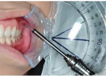
Because of the split-mouth design of this study, all mini-implants were placed manually on both sides of the maxilla of the same cadaver without pilot drilling. The insertion point was standardised by a millime-ter probe used to measure the height at 7 mm from the gingiva margin or the tip of the papilla between the second premolar and maxillary irst molar.18 MIT
the 60° or 90° lines that run from the protractor base line (Fig 1). Mini-implants of the same type were then placed into the same cadaver at a 90° angle on one side and a 60° angle on the other side.
To evaluate the hypothesis that the vertical place-ment angle would inluence MIT, the values obtained for screws with the same diameter were compared. Two-way ixed-efects analysis of variance (ANOVA) and Newman-Keuls tests were used to compare MIT values. All calculations were performed by means of Statistica sotware Version 10.0 (StatSot Inc., Tulsa, OK, USA). A signiicance level of p < 0.05 was adopted.
RESULTS
The mean MIT values difered among groups and varied between 11.40 and 17.27 Ncm. The mean MIT values for cylindrical mini-implants were 14.13 Ncm for the 60° angle and 17.27 Ncm for the 90° angle. The mean MIT values for the conical mini-implants were 11.40 Ncm for the 60° angle and 14.40 Ncm for the 90° angle (Table 2).
Evaluating the relationship of MIT values with the placement angles in the axial plane, we observed sig-niicant diferences between Groups 1 and 2 (p = 0.013) and Groups 3 and 4 (p = 0.018). Mini-implants placed at 90° in the cortical bone exhibited greater insertion torque than those placed at 60° relative to the cortical bone (Tables 2 and 3).
DISCUSSION
In order to add information to evaluate the vari-ables that afect mini-implant stability, this research focused on insertion torque to study angle efect as a surgery-related factor for stability. The present study re-veals that the vertical placement angle of mini-implants might interfere in the amount of MIT. Ex vivo placing of orthodontic mini-implants at a 90° angle resulted in Table 1 - Mini-implant specifications and codes.
Table 2 - Means and standard deviation (SD) of MITs (Ncm) for two types of mini-implants and placement angles (n = 15).
Figure 1 - The manual driver tip positioned beside the flat surface of the pro-tractor over the 60° angle.
Table 3 - Newman-Keuls test for placement angle comparisons for each type of mini-implant.
Different superscript letters indicate significant differences between groups.
* significant difference (p < 0.05).
Code Group Type Diameter Length Angle Manufacturer
CL 90 1 Cylindrical 1.6 mm 9 mm 90° Dewimed (Germany)
CL 60 2 Cylindrical 1.6 mm 9 mm 60° Dewimed (Germany)
CN 90 3 Conical 1.8 mm 9 mm 90° Conexão (Brazil)
CN 60 4 Conical 1.8 mm 9 mm 60° Conexão (Brazil)
Diameter Angle Torque (Ncm)
Mean SD
1.6 mm 60° 14.13
b 3.93
90° 17.27a 3.22
1.8 mm 60° 11.40
c 1.99
90° 14.40b 2.06
Comparison p value
(CL 60 vs. CL 90) 0.013 *
increased MIT. It needs to be emphasized that our re-sults are related to human maxillary alveolar bone, and not to all maxillomandibular areas. For example, for the posterior mandibular area, the vertical placement angle seems necessary to increase the contact area between the screw and the cortical bone.8
The literature reports that axial angles from 45° to 70° are the most appropriate for preventing the screw from contacting the dental roots and increasing the amount of bone surrounding the screw.1,9 However,
such angles seem to compromise screw insertion depth and cortical bone integrity, despite providing better contact with the bone of the inter-radicular space compared with greater angles. In other words, it was not related to the 90° insertion angle. Therefore, in our study, the comparison between 90° and 60° angles was proposed because a 60° angle represents a mean point of the rate for vertical angle placement recommended in the literature, which is between 45° to 70°.1,9
An-other study in human alveolar bone did not ind any inluence regarding placement angle; nevertheless, this clinical study was performed in multiple maxilloman-dibular areas with three diferent types of screw.2
The placement area between the second premolar and maxillary irst molar was chosen because it pro-vides the widest maxillary inter-radicular space and it is, therefore, a safe space for mini-implant placement.8,18-22
The standardization of insertion height at 7 mm away from the interdental papilla made it possible to place mini-implants in the attached gingiva, and also to have a suicient inter-radicular space.18,19,21,23,24
Although routinely used in dental studies, human cadavers present some restrictions in clinical applica-tions. There was some concern regarding variability in the post-mortem interval among cadavers; therefore, the experiment used newly deceased cadavers (up to 24 hours post-mortem), so that the cortical bone den-sity of the sample components could be compared.24, 25
For greater reliability of the obtained results and to re-duce standard deviation, we used more cadavers than the average commonly reported in the literature for this type of study.14,26-31
Similarly to our indings, studies using several ex-perimental models have shown that a mini-implant angle of 90° relative to the cortical bone is advantageous compared with other angles indicated for technical advantages.13 Placing orthodontic mini-implants to the
alveolar process bone surface at angles less than 90° did not ofer force anchorage resistance advantages.14
The 1.6-mm screws placed at a 90° angle displayed the greatest insertion torque, suggesting that the in-creased mini-implant diameter had a signiicant efect on insertion torque. Greater structural preservation of the cortical bone may have resulted from the lower pressure of the smaller-diameter mini-implant because larger-diameter mini-implants and greater cortical bone thickness require more insertion force.1,9,12
Therefore, variations in screw design and diameter lead to changes in the MIT value.12 However, in this
study, it was found that 1.6-mm mini-implants exhib-ited a MIT value higher than 1.8 mm for the same in-sertion angle. In addition, the commercial brands used in our study had diferent diameters and types, which did not allow statistical analysis between the types of mini-implant. Nevertheless, we were able to compare the inluence of placement angle for mini-implants of the same type. Variation in placement angle may lead to reduced strain on the cortical bone, thus overcoming the increased tendency towards damage associated with increased mini-implant diameter.13
The insertion torque values found were similar to those observed in human cadavers 28 and in
anoth-er clinical study.3 Thus, the variations found when
placement angles were compared represent changes that may also occur in patients. Furthermore, when two brands of mini-implants with different designs and diameters were used, the values differed accord-ing to whether they were placed at 60° or 90°. Re-gardless of the type of mini-implant used, the MIT value was higher when the implants were placed at 90°. This finding means that, for self-drilling mini-implants, placement at a 90° angle should be priori-tized to reduce stress on the cortical bone.
The cylindrical mini-implant (1.6 mm) exhibited a higher MIT value, probably in regard to the cylin-drical design of the screws and not as a consequence of the diameter. This reinforces a previous report in the literature, in which conical mini-implants induced more microdamage than the cylindrical ones.12 In
an-other study, the range for MIT in human bone was from 5 to 10 Ncm.3 The self-drilling mini-implant
Conlicting indings concerning factors that inlu-ence MIT values have yielded no evidinlu-ence to suggest that speciic MIT levels result in higher success rates for mini-implants.17 Therefore, it is not possible to
under-stand the high torque values obtained here as overload of the cortical bone. Furthermore, our results obtained in human cortical bone will help to provide better as-sociation and quantitative records to identify a speciic relationship with mini-implant primary stability. In previous human studies, the mini-implant system used increased MIT values with self-drilling insertion when compared with self-tapping.32,33 Also, variations in the
MIT value for human cortical bone have been present-ed, possibly as a consequence of the diferent devices (mechanical and digital) used to record torque during mini-implant placement.34 In our research, we used a
digital instead of mechanical torquimeter to provide more accurate values.17,35
Histological studies have shown that mini-implant design afects the amount of damage caused to the cortical bone and may be useful in clarifying the types of changes in the area of contact between the mini-implant and the cortical bone associated with MIT.29,36
The quantity and quality of cortical bone on the failure force of mini-implants have been shown when comparing maxillae and mandibles. In our study, we analyzed the mini-implant/cortical bone interface related to the posterior maxillary alveolar region.
The same results should not be extrapolated to other areas, such as the posterior mandibular cortical bone. Cortical bone thickness and bone hardness of man-dibles are different when compared with maxillae, mainly in the posterior region.1,7,11
An in vitro study, which did not take into con-sideration different cortical bone thickness, reported that angled insertion provides greater MIT as a con-sequence of increased contact in the mini-implant– cortical bone interface.37 The results of this present
study suggest that the characteristics of the alveo-lar cortical bone should be taken into consideration when determining a suitable placement angle for mini-implant insertion.
Future studies should analyze whether damage to the cortical bone surface and/or reduced screw inser-tion depth are associated with the vertical placement angle of the mini-implant. Additionally, further re-search should be conducted to investigate mini-implant placement in other sites, especially those with different buccal cortical bone thicknesses.
CONCLUSION
REFERENCES
1. Watanabe H, Deguchi T, Hasegawa M, Ito M, Kim S, Takano-Yamamoto T.
Orthodontic miniscrew failure rate and root proximity, insertion angle, bone contact length, and bone density. Orthod Craniofac Res. 2013 Feb;16(1):44-55.
2. Park HS, Jeong SH, Kwon OW. Factors afecting the clinical success of screw implants used as orthodontic anchorage. Am J Orthod Dentofacial Orthop. 2006 July;130(1):18-25.
3. Motoyoshi M, Yoshida T, Ono A, Shimizu N. Efect of cortical bone thickness and implant placement torque on stability of orthodontic mini-implants. Int J Oral Maxillofac Implants. 2007 Sept-Oct;22(5):779-84.
4. Chen YJ, Chang HH, Huang CY, Hung HC, Lai EH, Yao CC. A retrospective
analysis of the failure rate of three diferent orthodontic skeletal anchorage systems. Clin Oral Implants Res. 2007 Dec;18(6):768-75.
5. Santos RF, Ruellas ACO, Fernandes DJ, Elias CN. Insertion torque versus mechanical resistance of mini-implants inserted in diferent cortical thickness. Dental Press J Orthod. 2014 May-June;19(3):90-4.
6. Motoyoshi M, Uemura M, Ono A, Okazaki K, Shigeeda T, Shimizu N. Factors
afecting the long-term stability of orthodontic mini-implants. Am J Orthod Dentofacial Orthop. 2010 May;137(5):588.e1-5; discussion 588-9.
7. Monnerat-Aylmer C, Restle L, Mucha JN. Tomographic mapping of
mandibular interradicular spaces for placement of orthodontic mini-implants. Am J Orthod Dentofacial Orthop. 2009 Apr;135(4):428-37. 8. Lim JE, Lee SJ, Kim YJ, Lim WH, Chun YS. Comparison of cortical bone
thickness and root proximity at maxillary and mandibular interradicular sites for orthodontic mini-implant placement. Orthod Craniofac Res. 2009 Nov;12(4):299-304.
9. Wilmes B, Su YY, Drescher D. Insertion angle impact on primary stability of orthodontic mini-implants. Angle Orthod. 2008 Nov;78(6):1065-70. 10. Poggio PM, Incorvati C, Velo S, Carano A. “Safe zones”: a guide for
miniscrew positioning in the maxillary and mandibular arch. Angle Orthod. 2006 Mar;76(2):191-7.
11. Baumgaertel S, Hans MG. Buccal cortical bone thickness for mini-implant placement. Am J Orthod Dentofacial Orthop. 2009 Aug;136(2):230-5. 12. Lim SA, Cha JY, Hwang CJ. Insertion torque of orthodontic miniscrews
according to changes in shape, diameter and length. Angle Orthod. 2008 Mar;78(2):234-40.
13. Petrey JS, Saunders MM, Kluemper GT, Cunningham LL, Beeman CS. Temporary anchorage device insertion variables: efects on retention. Angle Orthod. 2010 July;80(4):446-53.
14. Woodall N, Tadepalli SC, Qian F, Grosland NM, Marshall SD, Southard TE. Efect of miniscrew angulation on anchorage resistance. Am J Orthod Dentofacial Orthop. 2011 Feb;139(2):e147-52.
15. Jasmine MI, Yezdani AA, Tajir F, Venu RM. Analysis of stress in bone and microimplants during en-masse retraction of maxillary and mandibular anterior teeth with diferent insertion angulations: a 3-dimensional inite element analysis study. Am J Orthod Dentofacial Orthop. 2012 Jan;141(1):71-80.
16. Lee SJ, Jang SY, Chun YS, Lim WH. Three-dimensional analysis of tooth movement after intrusion of a supraerupted molar using a mini-implant with partial-ixed orthodontic appliances. Angle Orthod. 2013 Mar;83(2):274-9. 17. Meursinge Reynders RA, Ronchi L, Ladu L, van Etten-Jamaludin F, Bipat S. Insertion torque and success of orthodontic mini-implants: a systematic review. Am J Orthod Dentofacial Orthop. 2012 Nov;142(5):596-614.e5. 18. Park J, Cho HJ. Three-dimensional evaluation of interradicular spaces
and cortical bone thickness for the placement and initial stability of microimplants in adults. Am J Orthod Dentofacial Orthop. 2009 Sept;136(3):314.e1-12; discussion 314-5.
19. Silvestrini Biavati A, Tecco S, Migliorati M, Festa F, Marzo G, Gherlone E, et al. Three-dimensional tomographic mapping related to primary stability and structural miniscrew characteristics. Orthod Craniofac Res. 2011 May;14(2):88-99.
20. Kim HJ, Yun HS, Park HD, Kim DH, Park YC. Soft-tissue and cortical-bone thickness at orthodontic implant sites. Am J Orthod Dentofacial Orthop. 2006 Aug;130(2):177-82.
21. Santiago RC, de Paula FO, Fraga MR, Picorelli Assis NM, Vitral RW. Correlation between miniscrew stability and bone mineral density in orthodontic patients. Am J Orthod Dentofacial Orthop. 2009 Aug;136(2):243-50.
22. Martinelli FL, Luiz RR, Faria M, Nojima LI. Anatomic variability in alveolar sites for skeletal anchorage. Am J Orthod Dentofacial Orthop. 2010 Sept;138(3):252.e1-9.
23. Kuroda S, Sugawara Y, Deguchi T, Kyung HM, Takano-Yamamoto T. Clinical use of miniscrew implants as orthodontic anchorage: success rates and postoperative discomfort. Am J Orthod Dentofacial Orthop. 2007 Jan;131(1):9-15.
24. Choi JH, Park CH, Yi SW, Lim HJ, Hwang HS. Bone density measurement in interdental areas with simulated placement of orthodontic miniscrew implants. Am J Orthod Dentofacial Orthop. 2009 Dec;136(6):766.e1-12; discussion 766-7.
25. Kribbs PJ. Comparison of mandibular bone in normal and osteoporotic women. J Prosthet Dent. 1990 Feb;63(2):218-22.
26. Kingsmill VJ, Boyde A. Variation in the apparent density of human mandibular bone with age and dental status. J Anat. 1998 Feb; 192 (Pt 2):233-44.
27. Friberg B, Sennerby L, Roos J, Lekholm U. Identiication of bone quality in conjunction with insertion of titanium implants. A pilot study in jaw autopsy specimens. Clin Oral Implants Res. 1995 Dec;6(4):213-9.
28. Brettin BT, Grosland NM, Qian F, Southard KA, Stuntz TD, Morgan TA, et al. Bicortical vs monocortical orthodontic skeletal anchorage. Am J Orthod Dentofacial Orthop. 2008 Nov;134(5):625-35.
29. Pickard MB, Dechow P, Rossouw PE, Buschang PH. Efects of miniscrew orientation on implant stability and resistance to failure. Am J Orthod Dentofacial Orthop. 2010 Jan;137(1):91-9.
30. Suzuki EY, Suzuki B, Aramrattana A, Harnsiriwattanakit K, Kowanich N. Assessment of miniscrew implant stability by resonance frequency analysis: a study in human cadavers. J Oral Maxillofac Surg. 2010 Nov;68(11):2682-9. 31. Lemieux G, Hart A, Cheretakis C, Goodmurphy C, Trexler S, McGary C,
et al. Computed tomographic characterization of mini-implant placement pattern and maximum anchorage force in human cadavers. Am J Orthod Dentofacial Orthop. 2011 Sept;140(3):356-65.
32. Suzuki EY, Suzuki B. Placement and removal torque values of
orthodontic miniscrew implants. Am J Orthod Dentofacial Orthop. 2011 May;139(5):669-78.
33. Chaddad K, Ferreira AF, Geurs N, Reddy MS. Inluence of surface characteristics on survival rates of mini-implants. Angle Orthod. 2008 Jan;78(1):107-13.
34. Schätzle M, Golland D, Roos M, Stawarczyk B. Accuracy of mechanical torque-limiting gauges for mini-screw placement. Clin Oral Implants Res. 2010 Aug;21(8):781-8.
35. Crismani AG, Bertl MH, Celar AG, Bantleon HP, Burstone CJ. Miniscrews in orthodontic treatment: review and analysis of published clinical trials. Am J Orthod Dentofacial Orthop. 2010 Jan;137(1):108-13.
36. Lee NK, Baek SH. Efects of the diameter and shape of orthodontic mini-implants on microdamage to the cortical bone. Am J Orthod Dentofacial Orthop. 2010 July;138(1):8.e1-8; discussion 8-9.