Pathway-Focused PCR Array Profiling of
Enriched Populations of Laser Capture
Microdissected Hippocampal Cells after
Traumatic Brain Injury
Deborah R. Boone, Maria-Adelaide Micci, Isabella G. Taglialatela, Judy L. Hellmich, Harris A. Weisz, Min Bi, Donald S. Prough, Douglas S. DeWitt, Helen L. Hellmich*
Department of Anesthesiology, The University of Texas Medical Branch, 301 University Boulevard, Galveston, Texas 77555–0830, United States of America
Abstract
Cognitive deficits in survivors of traumatic brain injury (TBI) are associated with irreversible neurodegeneration in brain regions such as the hippocampus. Comparative gene expres-sion analysis of dying and surviving neurons could provide insight into potential therapeutic targets. We used two pathway-specific PCR arrays (RT2 Profiler Apoptosis and Neurotro-phins & Receptors PCR arrays) to identify and validate TBI-induced gene expression in dying (Fluoro-Jade-positive) or surviving (Fluoro-Jade- negative) pyramidal neurons ob-tained by laser capture microdissection (LCM). In the Apoptosis PCR array, dying neurons showed significant increases in expression of genes associated with cell death, inflamma-tion, and endoplasmic reticulum (ER) stress compared with adjacent, surviving neurons. Pro-survival genes with pleiotropic functions were also significantly increased in dying neu-rons compared to surviving neuneu-rons, suggesting that even irreversibly injured neuneu-rons are able to mount a protective response. In the Neurotrophins & Receptors PCR array, which consists of genes that are normally expected to be expressed in both groups of hippocam-pal neurons, only a few genes were expressed at significantly different levels between dying and surviving neurons. Immunohistochemical analysis of selected, differentially ex-pressed proteins supported the gene expression data. This is the first demonstration of pathway-focused PCR array profiling of identified populations of dying and surviving neu-rons in the brain after TBI. Combining precise laser microdissection of identifiable cells with pathway-focused PCR array analysis is a practical, low-cost alternative to microarrays that provided insight into neuroprotective signals that could be therapeutically targeted to ame-liorate TBI-induced neurodegeneration.
OPEN ACCESS
Citation:Boone DR, Micci M-A, Taglialatela IG, Hellmich JL, Weisz HA, Bi M, et al. (2015) Pathway-Focused PCR Array Profiling of Enriched Populations of Laser Capture Microdissected Hippocampal Cells after Traumatic Brain Injury. PLoS ONE 10(5): e0127287. doi:10.1371/journal.pone.0127287
Academic Editor:Alexandre Hiroaki Kihara, Universidade Federal do ABC, BRAZIL
Received:November 25, 2014
Accepted:April 13, 2015
Published:May 27, 2015
Copyright:© 2015 Boone et al. This is an open access article distributed under the terms of the
Creative Commons Attribution License, which permits unrestricted use, distribution, and reproduction in any medium, provided the original author and source are credited.
Data Availability Statement:All relevant data are within the paper and its Supporting Information files.
Funding:This work was supported by RO1 NS052532 (HLH), and in part by the department of Anesthesiology (UTMB) and a Moody Foundation grant to DSD. The funders had no role in study design, data collection and analysis, decision to publish, or preparation of the manuscript.
Introduction
Presently, there are no approved treatments that can be administered after traumatic brain in-jury (TBI) to mitigate the progression of brain damage and improve functional outcome. Dur-ing the past several decades, thousands of gene expression studies have provided much insight into the pathogenesis of TBI, and led to therapeutic strategies to inhibit genes and cellular path-ways associated with cell death but these have not translated into effective treatments [1–4]; to date, hundreds of clinical trials of brain injury have failed to advance past Phase 2 trials [5,6]. This failure is reflected in the clinical management of TBI patients; once they are stabilized, there is little other than supportive care that can be done to limit the cascades of neuronal in-flammation, oxidative stress and cell death [7–9]. Therefore, it is imperative to continue explor-ing the molecular underpinnexplor-ings of TBI, particularly, the injury-induced changes in the molecular signals that are critically important for survival, regeneration and recovery of the injured brain.
In a recent, comparative, genome-wide gene expression analysis of dying and surviving neu-rons obtained by laser capture microdissection (LCM), we gained several insights into mecha-nisms of cell survival in the hippocampus of rats subjected to TBI [10]. Specifically, we showed evidence of pre- and post-injury stochasticity in gene expression in both dying and surviving neurons, thus suggesting that random fluctuations in pro-survival gene expression likely influence the effects of TBI on vulnerable brain cells, i.e., determine whether a neuron dies or survives following TBI. Since these microarray studies were costly, labor-intensive, and a bioin-formatics challenge, in separate studies we investigated whether gene expression in limited numbers of laser captured neurons could also be delineated using pathway-focused PCR arrays. Because the majority of molecular biology laboratories have access to thermal cyclers capable of generating quantitative, real-time PCR data, we tested a qPCR-based method (RT2Profiler PCR arrays) with the features of a miniarray. Since these PCR arrays are comprised of func-tionally related genes in disease-associated and/or known cell signaling pathways, they are de-signed to interrogate expression of groups of genes that are functionally and coordinately regulated, and they have the added benefit of providing immediate information about activa-tion or inhibiactiva-tion of key canonical pathways without resorting to extensive bioinformatics analysis. The broad diversity of known cellular pathways covered by PCR arrays allows us to test our hypotheses about the biological roles of specific genes in identified populations of neu-rons. Although previous studies have used, for example, cDNA arrays [11], genome-wide ar-rays [12], Taqman pathway PCR arar-rays [13] or single-nucleotide polymorphism arar-rays [14] to examine gene expression in laser captured cells from salivary gland, breast, prostate, and glio-blastoma tissues, respectively, to our knowledge this is the first study to use pathway-focused arrays for analysis of laser capture microdissected hippocampal neurons from brain injured rats.
Experimental procedures
Animal preparations for fluid percussion brain injury
The Institutional Animal Care and Use Committee of The University of Texas Medical Branch approved all experimental protocols. Adult, male, Charles River Sprague-Dawley rats (300–400 g) were anesthetized with 4% isoflurane, intubated, then mechanically ventilated. A craniotomy was performed laterally to the sagittal suture, midway between the lambda and bregma struc-tures. The fluid percussion device was attached, and the animal was subjected to lateral fluid percussion traumatic brain injury (TBI) as previously described [15]. Each animal was injured at a moderate-to-severe level. The rats were sacrificed 24 hours after injury. Animals were eu-thanized using methods consistent with the AVMA Guidelines for the Euthanasia of Animals, 2013 Edition, Section S2.2.2.1, Inhaled Agents. Animals were deeply anesthetized in a small chamber containing 5% isoflurane delivered by a precision vaporizer at a flow rate sufficient to saturate the chamber in approximately one minute. Animals remained in the chamber for more than 8 minutes to ensure deep anesthesia. Death was confirmed by decapitation, brains dissected out, placed on dry ice for 10 minutes, and stored at -80°C until LCM was performed.
Tissue preparation
Brains were removed from -80°C and allowed to warm up to -25°C in a cryostat. The tissue was embedded in Tissue-Tek O.C.T. (Sakura, Hayward CA) mounting medium and prepared for cryosectioning. 10μM serial coronal sections of the hippocampus were collected on
pre-cleaned slides (VWR, West Chester PA). Sections were fixed in 75% ethanol for 1 min, rinsed in RNase free water for 1 min, counterstained with 1% cresyl violet for 15 to 20 sec, rinsed in RNase free water 2X30 sec, stained with 0.001% Fluoro-jade (Histo-Chem, Inc., Jefferson AR) for 4 min, rinsed in RNase free water 3X1min, dehydrated in 95% ethanol for 30 sec, 100% eth-anol for 30 sec, then cleared in xylene 2X3 min each. Sections were then air dried in a fume hood for approximately 10 min before proceeding to LCM as previously described [10].
Laser capture microdissection (LCM)
LCM was performed using a PixCell IIe laser capture microscope with an infrared diode laser (Arcturus Engineering, Mountain View CA) as previously described [10]. Based on an estimate of neuronal cell size of 20–30μm, we determined that three captured neurons from three 10μm
thick sections contained the total RNA equivalent of a single cell. Thus, 1500 captured cells, the equivalent of 500 dying/injured (Fluoro-Jade positive) neurons were captured onto the ther-moplastic film of a CapSure Macro LCM Cap (Arcturus Engineering, Mountain View CA) from the CA1/CA3 subfields of the hippocampus; each biological replicate of cells were pooled from 3 TBI brains (9 brains total). The smallest laser spot size (7.5um) was used, and the power set to 65–80mW with pulse duration of 0.40–0.50 (these last 2 settings were adjusted for opti-mal capture of single neurons). The cells were lysed in 100μl of lysis buffer (Ambion, Austin
TX) and vortexed before storing at -80°C. 500 adjacent uninjured (Fluoro-Jade negative) neu-rons were also captured, lysed, and stored in this same manner. We should point out that slight contamination of dying cells with some surviving cells and/or processes of supporting glial cells is virtually unavoidable despite the small size of the laser.
RNA isolation
level of biological variation is high compared to technical variation on the array [24]. There were three such biological pools of injured and uninjured samples (from a total of 9 TBI rats) that were assayed with individual PCR arrays. Total RNA from each injured/uninjured pool was isolated using the RNAqueous-Micro kit (Ambion, Austin TX), following the manufactur-er’s RNAqueous-Micro Protocol for LCM. Genomic DNA was removed from each sample by treatment with rDNase at 37°C for 20 minutes (Ambion, Austin TX). The injured/uninjured samples were run through the Agilent Bioanalyzer (Agilent Technologies, Santa Clara CA) to check both RNA quality and quantity. Our RIN values for our samples ranged from as low as 4 to as high as 9 but averaged between 5 and 7, which is in line with values reported in the litera-ture [25,26].Afterwards, approximately 1.2 ng of each sample were precipitated with 3M Sodi-um Acetate and 100% ethanol, and resuspended in 10μl of nuclease-free water in preparation
for theRT2Nano PreAMP cDNA synthesiskit (SA Biosciences, Frederick MD).
cDNA synthesis and amplification
1.2 ng of injured and uninjured total RNA were reverse transcribed using the first strand cDNA synthesis kit (SA Biosciences, Frederick MD), omitting the gDNA elimination buffer step. Following the manufacturer’s protocol, 5μl of cDNA was then preamplified using either
the Neurotrophin and Receptors or Apoptosis array-specific primers and theRT2Nano Pre-AMP cDNA synthesiskit (SA Biosciences, Frederick MD). Cycling conditions for the preampli-fication of each cDNA sample was as follows: 1 cycle (10min) at 95°C, 12 cycles of (15sec at 95°C, then 2 min at 60°C). Samples were then placed on ice where 2ul of a side reaction reducer was added, incubated at 37°C for 15 minutes, then heat inactivated at 95°C for 5 minutes.
PCR Arrays
The amplified cDNA was then diluted with nuclease-free water and added to theRT2qPCR SYBR green Master Mix(SA Biosciences, Frederick MD). 25μl of the experimental cocktail
was added to each well of the ratNeurotrophin and Receptor PCR array(SA Biosciences, Fred-erick MD) or the ratApoptosis PCR array(SA Biosciences, Frederick MD). Real-Time PCR was performed on the Mx3000P QPCR System (Stratagene, La Jolla CA) and used SYBR green detection with the following thermal profile: segment 1–1 cycle: 95°C for 10 minutes, segment 2–40 cycles: 95°C for 15 seconds followed by 60°C for 1 minute, segment 3 (dissociation curve)–95°C for 1 minute, 55°C 30 seconds, and 95°C for 30 seconds. All data from the PCR was collected by the MXPro software (provided by Stratagene with purchase of Mx3000 Multi-plex Quantitative PCR System) and analyzed by SA Bioscience’s PCR Array Data Analysis Web Portal.
Analysis of Real-Time PCR array data
Each array contained 5 separate housekeeping genes (RPLP1,HPRT,RPL13A,LDHA, and
ACTB) that were used for normalization of the sample data. Normalization to the house keep-ing genes (HKG) was performed by calculatkeep-ing theΔCt for each gene of interest (GOI) in the plate (Ct value of GOI-Ct value of HKG). Any Ctvalue>35 was considered to be a negative
call. If the Ctvalue of the genomic DNA control was>30, then no genomic DNA was
software allows for the ability to define the best reference genes for normalization with guid-ance and recommendations from the recommendations made in the following publication by Vandesompele et al., [28]. Arikawa et al, compared the results of the original MicroArray Qual-ity Control (MAQC) study with the RT Profiler platform and reported the PCR arrays deliver gene expression data that is highly comparable with TaqMan PCR and high-density microar-rays [29].
Statistical analysis of PCR array data
The RT2Profiler PCR Array Data Analysis software does not perform any statistical analysis beyond the calculation of p-values using a Student’s t-test (two tail distribution and equal vari-ances between the two samples) based on the triplicate 2^(-ΔC
T) values for each gene in the
in-jured group compared to the surviving group. The Microarray Quality Control (MAQC) published results indicating that a ranked list of genes based on fold-change and such a p-value calculation was sufficient to demonstrate reproducible results across multiple microarray and PCR Arrays including the RT2Profiler PCR Arrays [30,31].
Immunofluorescence analysis
Rats were anesthetized with sodium pentobarbital (70 mg/kg, i.p.) 24 hours after TBI, transcar-dially perfused and fixed with freshly prepared ice-cold 4% paraformaldehyde (PFA) in 0.1 M phosphate-buffered saline (PBS, pH 7.4). The brain was removed, post-fixed in 4% PFA and cryoprotected by infiltration in 20% sucrose solution in PBS overnight at 4°C. The tissue was rapidly frozen in O.C.T. embedding medium (Tissue Tek, Sakura, Tokyo). Frozen serial sec-tions (10μm thick) were cut on a cryostat (Leica CM1850, Wetzlar, Germany), placed on plus
slides (VWR, West Chester, Pennsylvania), and stored at–20°C until needed. Frozen sections were blocked and permeabilized for 30 minutes at room temperature with 0.3% Triton X-100 in PBS containing 5% normal goat serum. After washing in PBS, sections were incubated with primary antibodies diluted in PBS overnight at 4°C. After washing in PBS, sections were incu-bated for 1 hour at room temperature with Alexa-conjugated secondary antibodies (Alexa-594: 1:400 dilution; Molecular Probes, Eugene, Oregon). The sections were washed 2 times for 10 minutes each in PBS and once in tap water, and incubated in 0.06% potassium permanganate in tap water for 1 minute. The sections were then washed in tap water for 5 minutes and incu-bated with Fluor-Jade C (0.0001% in tap water with 0.1% acetic acid) for 10 minutes. After three more washes in tap water (1 minute each), sections were cover- slipped with acidic mounting media (0.1% acetic acid/80% glycerol in tap water).
For the detection of CD40, the rats were anesthetized and euthanized by decapitation. The brains were rapidly removed, quickly frozen on dry ice, and embedded in O.C.T. embedding medium. Frozen serial sections (10μm thick) were cut as described above and stored at–80°C
until needed. The sections were fixed in methanol pre-chilled at -20°C for 20 minutes before immunofluorescence analysis.
The slides were viewed with an Olympus BX51 fluorescence microscope equipped with a cooled CCD camera (Microfire by Optronics, Goleta, CA) and image acquisition software (Pic-tureFrame, Optronics, Goleta, CA).
Results
Laser capture microdissection of identified neurons
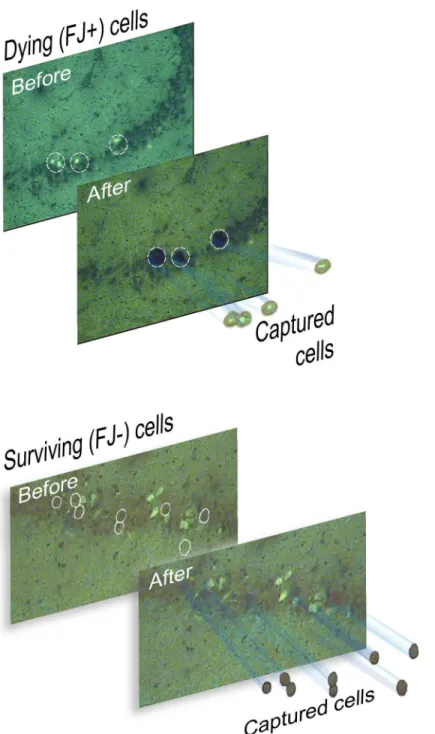
of surviving, FJ-negative pyramidal neurons immediately adjacent to FJ-positive neurons were also captured onto separate LCM caps (Fig 1B). The quality and quantity of isolated total RNA samples were assessed using the ultrasensitive pico assay on the Agilent Bioanalyzer (S1 Fig). Overall, the RNA quality from LCM procedures was excellent, in many cases matching the quality of total RNA isolated from hippocampal tissue samples using established TRIzol-based methods. This is in spite of the fact that RNA isolated from laser capture samples tends to give
Fig 1. Laser capture microdissection of rat hippocampal neurons after fluid percussion brain injury. (A). Dying, Fluoro-Jade-positive neurons in the CA3 subfield of the rat hippocampus 24 hr after TBI are shown, before and after LCM on the capture caps. (B). Surviving, Fluoro-Jade negative neurons were captured immediately adjacent to FJ positive, dying neurons.
lower RNA integrity (RIN) values; this typically reflects the long arduous laser microdissection techniques [25]. Moreover, in all our previous LCM studies, our analysis showed a similar uni-formity in RNA quality [10,17,18]. This gave us confidence that gene expression data from these experimental LCM RNA samples obtained using any established gene expression profil-ing platform would be reliable. Each biological sample from approximately 500 cells yielded several nanograms of total RNA, which was sufficient starting material for preamplification of the RNA samples [32] and subsequent real time PCR array analysis.
Expression of apoptosis-associated genes in dying neurons
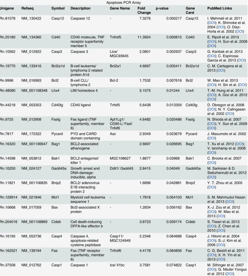
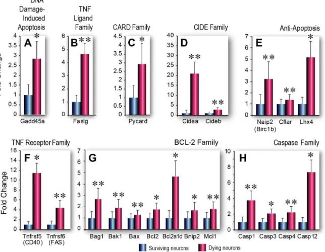
In the apoptosis PCR array, we found that, compared to adjacent, surviving FJ-negative neu-rons, dying, FJ-positive pyramidal neurons had significantly higher expression of many genes known to be associated with apoptotic cell death (Table 1,S1 Table, fold changes are shown as ratios of gene expression in dying vs surviving neurons). Among these were multiple genes as-sociated with the caspase family of cysteine proteases [33,34], the pro- and anti-apoptotic genes in theBcl-2family [35] and genes in theTNFreceptor family [36–38] that possess pro-survival and/or pro-death functions (Fig 2A–2H).In silicoanalysis of gene functions in the PCR array confirmed that the majority of these genes are associated with apoptotic cell death. Moreover, we found that expression of most of the differentially expressed genes (particularly those that were significantly different or of borderline significance) has been linked to TBI pa-thology (Table 1). Complete gene expression data and supporting references are provided inS1 andS2Tables and inS1 References, raw data shown inS3 Table. For instance, although
Gadd45awas expressed with borderline significance in our study, previous studies have shown that this gene is involved in oxidative stress-induced apoptotic cell death in hippocampal neu-rons [39]. Thus, increased expression of this gene in dying neuneu-rons is consistent with its known functions. Our observation that several significantly upregulated genes in dying neu-rons, such asPycard(also known asAsc) are known to be involved in inflammatory cell death cascades [40] is consistent with a large number of TBI studies showing that inflammatory pro-cesses play a prominent role in neuronal injury and death [41]. On the other hand, we noted that several genes, such asBnip2, that were increased significantly or with borderline signifi-cance have previously been shown to have multifunctional or pleiotropic roles in cell signaling processes [42]. The apoptosis array was initially chosen because we anticipated that if the array data was to be deemed biologically relevant, then apoptosis-related genes should be differen-tially expressed at significant levels in dying neurons compared to surviving neurons. Thus, our study serves as a training set for future studies of identified cell populations with postulated functions.
We noted that several prominent pro-survival genes, such asBcl-2andMcl1[35,43], were expressed at significantly higher levels in the dying neurons than in adjacent surviving neurons. The neuropeptide galanin and its receptors (GalR1andGalR2) have previously been shown to be involved in hippocampal neuron survival [44]; therefore, it was not surprising to find that
GalRexpression-the gene for galanin is not on the PCR array list-was significantly increased after TBI. We should like to emphasize that in our study, the use of LCM allowed us to demon-strate for the first time thatGalRexpression is significantly higher in dying neurons than sur-viving neurons.
Table 1. Differential expression of Apoptosis and Neurotrophin genes in dying and surviving neurons.
Apoptosis PCR Array Unigene Refseq Symbol Description Gene Name Fold
Change
p-value Gene Card
PubMed Links
Rn.81078 NM_130422 Casp12 Caspase 12 - 7.3276 0.000217 Casp12 I. Mehmeti et al. 2011
(DOI); K. Shimoke et al. 2004 (DOI); O. Diaz-Horta et al. 2002 (DOI) Rn.25180 NM_134360 Cd40 CD40 molecule, TNF
receptor superfamily member 5
Tnfrsf5 11.3924 0.000815 Cd40 E. Ripoll et al. 2013 (DOI); H. Sun et al. 2008 (DOI)
Rn.10562 NM_012922 Casp3 Caspase 3 Lice/
MGC93645 2.0801 0.002937 Casp3 G. Kanbak et al. 2013(DOI); C. Espinosa-Garcia et al. 2013 (DOI) Rn.19770 NM_133416 Bcl2a1d B-cell leukemia/
lymphoma 2 related protein A1d
Bcl2a1 4.6697 0.005411 Bcl2a1d C. M. Cartagena et al. 2013 (DOI)
Rn.9996 NM_016993 Bcl2 B-cell CLL/
lymphoma 2 Bcl-2 1.7532 0.007618 Bcl2 W. Mao et al. 2013(DOI); H. Sin et al. (DOI)
Rn.48080 NM_001108348 Lhx4 LIM homeobox 4 - 5.1575 0.01244 Lhx4 T.-M. Hung et al. 2011
(DOI); A. Goc et al. 2012 (DOI)
Rn.44218 NM_053353 Cd40lg CD40 ligand Tnfsf5 5.6438 0.013359 Cd40lg D. Obregon et al. 2008 (DOI); N. Y. Calingasan et al. 2002 (DOI) Rn.9725 NM_012908 Faslg Fas ligand (TNF
superfamily, member 6)
Apt1Lg1/ CD95-L/ Fasl/ Tnfsf6
4.6482 0.020486 Faslg N. Shioda et al. 2007 (DOI); Y. Sun et al. 2009 (DOI)
Rn.7817 NM_172322 Pycard PYD and CARD domain containing
Asc 2.9349 0.023679 Pycard J. Masumoto et al. 2002 (DOI)
Rn.16320 NM_001106647 Bag1 BCL2-associated athanogene
- 2.6697 0.026695 Bag1 T. Xu et al. 2012 (DOI);
V. lanchamp et al. 2008 (DOI)
Rn.14598 NM_053812 Bak1 BCL2-antagonist/
killer 1 MGC108627 1.8877 0.03968 Bak1 C. Brooks et al. 2007(DOI) Rn.10250 NM_024127 Gadd45a Growth arrest and
DNA-damage-inducible, alpha
Ddit1/ Gadd45 2.8415 0.04049 Gadd45a M. Sarkisian & D. Siebzhenrubl et al. 2012 (DOI)
Rn.11821 NM_001106835 Bnip2 BCL2/ adenovirus E1B interacting protein 2
- 1.6896 0.042881 Bnip2 Y. T. Zhou et al. 2005
(DOI)
Rn.129914 NM_021846 Mcl1 Myeloid cell leukemia
sequence 1 - 1.7818 0.054103 Mcl1 S. M. Mahmudul Hasanet al. 2013 (DOI) Rn.10668 NM_017059 Bax Bcl2-associated X
protein - 1.2834 0.056162 Bax X.-J. Zou et al. 2012(DOI); W. Mao et al. 2013 (DOI) Rn.204016 NM_001108869 Cideb Cell death-inducing
DFFA-like effector b
- 3.6723 0.059174 Cideb S. Tiwari et al. 2013
(DOI); Z. Chen et al. 2010 (DOI) Rn.16195 NM_053736 Casp4 Caspase 4,
apoptosis-related cysteine peptidase
Casp11/
MGC124949 2.2346 0.064688 Casp4 J. Hitomi et al. 2004(DOI); S.-J. Kim et al. 2006 (DOI)
Rn.162521 NM_139194 Fas Fas (TNF receptor superfamily, member 6)
Tnfrsf6 4.4178 0.064856 Fas C. G. Besirli et al. 2011 (DOI); X. H. Yin et al. 2013 (DOI)
Rn.37508 NM_012762 Casp1 Caspase 1 Ice/ Il1bc 3.7581 0.074822 Casp1 M. Sifringer et al. 2007 (DOI); G. Nilufer Yonguc et al. 2012 (DOI)
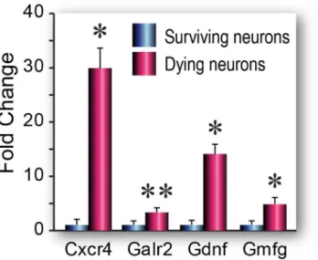
fewer numbers of differentially expressed genes (Fig 3,Table 1,S2 Table). On the other hand, we found that the few genes from this array that were upregulated in dying neurons were either associated with neurodegenerative disorders when expressed at high levels, e.g., glia maturation factor gamma (Gmfγ) is a proinflammatory gene previously linked to Alzheimer’s pathology
[45], or were known to promote pro-survival functions after brain injury (e.g.,Gdnf)[46]. As we found in the apoptosis array, several of the genes upregulated in dying neurons are known to have pleiotropic functions—e.g., increased expression of chemokine receptors such as CXCR4 have been linked to hippocampal damage and neuronal loss in HIV patients [47], but this gene has also been shown to modulate hippocampal plasticity and neurogenesis [48].
Table 1. (Continued)
Rn.204752 NM_057138 Cflar CASP8 and FADD-like apoptosis regulator
Flip/
MGC108616 1.3566 0.081339 Cflar K. Järvinen et al. 2011(DOI); Y. Matsumori et al. 2006 (DOI) Rn.8171 NM_001170467 Cidea Cell death-inducing
DFFA-like effector a - 26.1125 0.091347 Cidea N. Omae et al. 2012(DOI); M. Ito et al. 2011 (DOI)
Rn.92423 XM_226742 Naip2 NLR family, apoptosis inhibitory protein 2
Birc1/ Birc1a/ Birc1b/ Naip
3.2266 0.095072 Naip2 M. Ito et al. 2011 (DOI)
Rn.83633 NM_130426 Tnfrsf1b Tumor necrosis factor receptor superfamily, member 1b
Tnfr2 3.3096 0.098788 Tnfrsf1b M. S. Weinberg et al. 2013 (DOI)
Neurotrophins & Receptors PCR Array Unigene RefSeq Symbol Description Gene Name Fold
Change
p-value GeneCard PubMed Links
Rn.55036 NM_019139 Gdnf Glial cell derived
neurotrophic factor - 14.0367 0.011829 Gdnf Y.-.M Yoo et al.2012(DOI); I. Kanter-Schlifke et al. 2009 (DOI); J. E. Minnich et al. 2012 (DOI); L.-F. Wong et al. 2006 (DOI)
Rn.973 NM_181091 Gmfg Glia maturation
factor, gamma - 4.8159 0.017405 Gmfg H. Tsuiki et al. 2000(DOI) Rn.24822 NM_019172 Galr2 Galanin receptor 2 - 3.3122 0.025542 Galr2 R. Toifghi et al. 2008
(DOI); Y. Yang et al. 2006 (DOI) Rn.137580 NM_022205 Cxcr4 Chemokine (C-X-C
motif) receptor 4 MGC108696 29.8805 0.036531 Cxcr4 A. J. Shepherd et al.2012 (DOI); V. Ödemis et al. 2002 (DOI); X. Liu et al. 2013 (DOI) Rn.34398 NM_030997 Vgf VGF nerve growth
factor inducible
- 2.0202 0.060712 Vgf J. Adler et al. 2003
(DOI); G.-L. Ferri et al. 2011 (DOI)
Rn.204252 NM_022196 Lif Leukemia inhibitory
factor - 11.6678 0.078427 Lif B. E. Deverman & P. H.Patterson et al. 2012 (DOI)
Differentially expressed genes that were deemed significant (p<0.05) or of borderline significance (p<0.1) are shown. The complete datasets are shown in S1andS2Tables. Hyperlinks to the GeneCard entry for each gene and Digital Object Identifier (DOI) for at least one supporting published reference are included. GeneCards is a searchable, integrated, database of human genes that provides concise genomic related information, on all known and predicted human genes.
Fold changes are ratios of gene expression levels (dying/surviving neurons).
Immunohistochemical validation of gene expression data
Double immunofluorescence analysis using specific antibodies against a selected pool of pro-teins, whose mRNA was significantly increased in FJ-positive neurons as compared to adjacent FJ-negative neurons, was performed in order to confirm the PCR array data (Fig 4). Our data show that, 24 hours following TBI, the TNF receptor CD40 is highly expressed in FJ-positive pyramidal neurons of the hippocampus, although it is also detected, albeit at low levels, in FJ-negative neurons (Fig 4A). We found that both CASP12 and CXCR4were highly expressed in some but not all FJ-positive cells, thus indicating that a certain degree of stochasticity exists at the protein level (Fig 4B and 4C). In order to confirm that FJ-positive cells are indeed irrevers-ibly injured, we performed immunofluorescence analysis using an antibody that specifically recognizes the active form of CASP3, an enzyme that plays a critical role in the activation of the apoptotic cascade. Our results show that active caspase 3 is expressed in FJ-positive neu-rons but not in FJ-negative neuneu-rons (Fig 4D), confirming the PCR array results, i.e., a signifi-cant increase in the expression ofCasp3mRNA in degenerating neurons as compared to adjacent uninjured neurons.
Fig 2. Apoptosis-related gene expression in dying and surviving neurons.A-H. Functional groups of genes involved in programmed cell death that are upregulated with significant (p<0.05) or borderline significance (0.05<p<0.1) in dying vs surviving neurons. (A) DNA Damage-induced Apoptosis. (B) TNF Ligand Family. (C) CARD Family. (D) CIDE Family. (E) Anti-apoptosis. (F) TNF Receptor Family. (G) Bcl-2 Family. (H) Caspase Family. Data are shown as fold changes in mRNA expression in dying compared to surviving cells, mean±SEM (n = 3 biological pools each of Fluoro-Jade positive or Fluoro-Jade negative cells). Statistical analysis was performed using Student’s t-test,*p<0.05,**p<0.1
We also sought to confirm, using immunofluorescence analysis, the differential expression of the protein products of some of the genes for which differential expression was of borderline significance or not statistically significant, e.g., LIF, DAD1, STAT3 and TGFα. Our reasoning was that mRNA expression often correlates poorly with protein expression [49] and some of the statistically insignificant changes in gene expression (due to high variability in the RNA samples) may actually correspond to significant differences at the level of protein expression. However, our results were equivocal; specific staining for each of these proteins was detected in the injured rat hippocampus, but it could not be unambiguously determined whether expres-sion of these proteins was higher in dying compared to surviving neurons (S2 Fig). Therefore, the immunohistochemical data neither confirmed nor refuted the gene expression data and further studies may be needed to validate individual gene expression data.
Discussion
This is the first report of a study utilizing pathway-focused PCR-based arrays to examine TBI-induced gene expression in dying and surviving hippocampal neurons obtained by LCM. Ge-nome-wide microarray analysis is a hybridization-based method which works well for moder-ate to relatively abundant transcripts but is not ideal for detecting genes expressed at low levels. On the other hand, PCR is an amplification-based method and because of this, PCR arrays could technically be far more sensitive with a greater dynamic range than microarrays. Previ-ous studies have reported use of pathway specific PCR arrays to examine gene expression in breast tissue after pregnancy to ascertain mechanisms of cancer risk [50] and to identify genetic markers of angiogenesis in laser dissected blood vessels [51]. PCR arrays have also been used to determine molecular mechanisms of brain damage and neuroprotection in manually microdis-sected samples of the ipsilateral cortex containing the ischemic penumbra after experimental transient focal ischemia [52] and to study the effects of progesterone treatment after TBI [53]. However, none of these brain injury studies involved LCM. As these arrays can generate gene expression data from as little as 1 nanogram of total RNA, our study shows that they are ideal tools for gene expression studies of small numbers of laser captured cells.
Fig 3. Neurotrophins & Receptors PCR array gene expression in dying and surviving neurons.Data are shown as fold changes (mean±SEM) (n = 3 biological pools each of Fluoro-Jade positive or Fluoro-Jade negative cells) in dying cells compared to surviving cells. Statistical analysis was performed using Student’s t test,*p<0.05,**p<0.1
In addition to providing support for the use of PCR arrays for limited numbers of laser cap-tured cells, the second purpose of this study was to gain insight into the prosurvival mecha-nisms inducedin vivoin injured neurons after TBI. We reasoned that knowing the identity of genes involved in endogenous protection will help in design of pharmacotherapeutic strategies targeted towards increasing the expression or activity of these genes and possibly mitigating the effects of TBI on the injured brain cells. Previous studies have usedin vitroapproaches to identify neuroprotective genes, such as the ones involved in the endogenous protective re-sponse induced by preconditioning paradigms [54]. We suggest that our approach is more bio-logically relevant, as we are precisely determining TBI-induced gene expressionin situin defined populations of neurons.
Comparing our PCR results with microarray data [10], we were surprised that only 9 genes on the PCR arrays were also found on the filtered microarray dataset (S4 Table). However, be-cause the microarray data has the potential for significant numbers of false positives, in our pre-vious study we had used a stringent 5-fold cutoff to study gene expression differences between dying and surviving neurons and we suggest that this, along with the greater dynamic range of the PCR-based method as well as the pre-selected (thus, biased) gene lists, could explain the lack of extensive concordance between the microarrays and PCR arrays. On the other hand, the trends (increase or decrease) in gene expression in all 9 common genes were concordant in both the microarray and PCR array datasets. We also surmised another positive outcome of this study, that despite the pathway-focused and limited gene sets, the overall sensitivity of the PCR arrays allowed us to detect gene expression differences that were not apparent in the transcrip-tome analysis.
We chose to profile dying and surviving hippocampal neurons as proof-of-concept because we could easily identify these two populations of neurons with a well characterized fluorescent marker of neurodegeneration, Fluoro-Jade [20]. Additionally, by using LCM to capture en-riched pools of dying or surviving neurons, we have greatly increased the resolution of the gene expression analysis over earlier studies of laser captured neurons that sampled broad swaths of neurons from injured brains and contained a mix of dying and surviving cell types [55]. We show that profiling distinct populations of identified neurons can provide valuable information about mechanisms of cell death by contrasting our cell-specific results with that of a recent arti-cle about the neurodegenerative effects of blast-induced neurotrauma in the rat hippocampus [56]. In that study, the authors showed that expression of apoptotic genesBaxandCasp3were increased in the injured hippocampus, evidenced by overall increased Fluoro-Jade B staining throughout the hippocampus. However, unlike our study, the authors did not assess gene ex-pression in selected, identified populations of dying and surviving neurons. Thus, their results merely imply that dying cells express apoptotic genes, whereas our study shows clearly that ap-optotic gene expression is higher in the identified dying cells compared with surviving cells.
The value of using precise LCM techniques for identified cells cannot be overstated. A cDNA array study of apoptosis-related genes showed thatMcl-1was down-regulated in the cerebella ofIgf-1transgenic mice. However, subsequent Northern analysis and LCM of cerebel-lar granule neurons followed by qPCR analysis showed that expression of this gene was actually increased in cerebellar granule neurons compared to non-transgenic littermates [57]. Thus, when gene expression analysis of a distinct cell type was performed, the results were completely
Fig 4. Immunohistochemical validation of gene expression data.Representative images of rat brain sections double-stained, using Fluoro-Jade C (FJ, in green) to identify injured neurons, and specific antibodies against CD40 (A), CASP 12 (B), CXCR4 (C) and active caspase 3 (D). Arrows point to cells co-expressing Fluoro-Jade C and the protein of interest. Arrowheads point to cells co-expressing low levels of CASP12 (B) and CXCR4 (C). (E) Gene expression in dying and surviving neurons, expressed as fold changes in dying to surviving neurons (normalized to a value of 1) are shown as a point of reference for the protein expression data. Calibration bars = 50 mm
the opposite of that gained from analysis of a heterogeneous mix of brain cell types in the cere-bellum. On the other hand, because we were able to examine gene expression in relatively pure populations of defined neurons, we have greater confidence in the definitive conclusions we made about the causative role of specific apoptosis related genes in neuronal cell death. Studies similar to ours could be performed using identified groups of immunolabeled cells obtained by LCM [58]. Because of the great heterogeneity of cell types in the mammalian brain, as many as 2500–5000 cell types [59], gene expression studies of identifiable and functionally distinct cell populations that can be obtained by LCM are essential in efforts to elucidate disease-associated pathogenesis.
Our study results also bolstered our previous observation that a strong, protective response is induced in both dying and surviving neurons after TBI [10]. Interestingly, in the present study we found that several, well known genes associated with cell survival were actually ex-pressed significantly higher in dying neurons compared to surviving neurons. This seems to be counterintuitive, but the prominent expression of many other deleterious genes in the dying cells appears to counter the beneficial effects.In silicoanalysis of genes that were differentially expressed revealed that many of these also possessed pleiotropic functions, i.e., immune-modu-latory genes such asCxcr4had functional roles in critical cellular processes such as synaptic plasticity but also were implicated in hippocampal injury [47,48]. Bcl-2-associated athanogene-1 (Bag1), a co-chaperone forHsp70/Hsc70, is also a multifunctional protein that has been shown to suppress apoptosis and enhance neuronal differentiation. The TBI-induced increases inBag1andCasp12, (the former reduces endoplasmic reticulum [ER] stress and apoptosis [60] while the latter increases ER stress and apoptosis [61]), show that multiple pro-survival- and pro-death-associated genes are upregulated in both dying and surviving cells. Thus, the differ-ence in cell fate is likely due to a preponderance of prosurvival gene expression in surviving neurons and a preponderance of prodeath genes in dying neurons. This is the same conclusion that we drew in our microarray study, thus showing that use of different gene expression plat-forms can lead to similar insights.
Immunohistochemical analysis of differentially expressed gene products clearly showed that protein expression was a continuum rather than an all or none phenomenon. The expression of any one gene was also variable from cell to cell, indicating that the stochastic nature of TBI-induced gene expression is also reflected in protein expression. This was not unexpected since the poor correlation of gene and protein expression is well known [49]. Protein expression of the genes that were not statistically significant or were of borderline significance was particular-ly variable, and we were unable to correlate the higher expression in dying neurons at the mRNA level with protein expression. However, we noted that only the expression of LIF had borderline significance; protein expression of the other three non-significant genes was exam-ined because their mRNA expression was higher in dying neurons and each had been function-ally implicated in pro- or anti-apoptotic functions [62–64]. Interestingly, like several other genes with borderline significance (S1 References), LIF is known to possess pleiotropic func-tions in both prosurvival and pathogenic pathways [65]. Because of this functional duality, even at the resolution of single cells, it cannot be determined whether LIF is playing a beneficial or deleterious role in dying cells.
the accuracy and precision of the LCM experiments [66,67]. On the other hand, we should point out that even pro-death associated genes, such as CD40, have also been shown to have prominent roles in neuronal differentiation and proliferation [68]. This indicates that the inter-pretation of cell-specific gene expression of any apoptosis–related gene is entirely dependent on the context, and we should be careful about drawing definitive conclusions about the func-tional role of any gene without addifunc-tional supporting evidence. However, the validation of gene expression data at the protein level gave us confidence in the qPCR array results, and has spurred current studies of drug effects using other pathway-focused PCR arrays.
Pathway-specific PCR arrays have several advantages for different applications. One, they can quickly provide clinically relevant data without the need for the extensive bioinformatics analysis necessary to decipher the results of genome-wide gene arrays. For instance, Hansel et al. showed that PCR array analysis could distinguish an allergy-associated gene expression profile of CD4+ T cells in allergic patients compared to the more cumbersome data obtained from Affymetrix oligonucleotide arrays [69]. Second, as shown in our present study, PCR array analysis of LCM samples provides information about specific cellular mechanisms that cannot be gleaned from profiling heterogeneous tissues comprised of multiple cell types. Marciano et al. compared expression in caspase positive dying neurons to that of uninjured neurons from uninjured mice [70]. Using LCM to procure pure or enriched cell populations, we were able to refine the original experimental design in the Marciano study and focus on why neurons subjected to the same injury as adjacent dying neurons in the same injured animal are able to survive TBI. In future studies, we plan to design and use custom PCR arrays of identified neu-roprotective genes to test our hypothesis that TBI induces a significant protective response in the injured brain and that drugs that enhance this protective response will improve functional outcome after TBI.
Several therapeutic agents in clinical use for other indications might be able to boost the ex-pression of endogenous neuroprotective genes such as the ones identified in our study. For in-stance, antidepressants have been shown to increase expression of GDNF in cultured cells [71]. Because depression is a common comorbidity in TBI patients [72], it is possible that those who are treated with antidepressants may be benefiting from the neuroprotective effects of these treatments. Notably, edaravone, a free radical scavenger drug currently in clinical trials for vari-ous neurological disorders, has been shown to be neuroprotective by enhancing expression of BDNF, Bcl-2 and suppressing caspase-3 activity [73]. Drugs targeted to single mechanisms of brain injury have failed to consistently improve outcome in clinical trials, so one current line of thinking is that pharmacotherapeutic agents with multiple mechanisms of action (i.e., antioxi-dative and anti-inflammatory, etc.) are more likely to be successful [74]. Indeed, our study shows that genes involved in multiple pathological and prosurvival processes are significantly induced in dying neurons, and multifunctional drugs that target these genes may have more ef-ficacy than drugs with single targets in reducing cell death after TBI.
Supporting Information
S1 Fig. Analysis of total RNA isolated from laser capture microdissected hippocampal neu-rons from traumatically brain injured rats using the pico assay on the Agilent Bioanalyzer. Total RNA from each pool (500 cells) of dying or surviving neurons is assayed in duplicate. (TIF)
appear to validate the lack of significance in PCR arrays. (TIF)
S1 Table. Apoptosis PCR Array. (DOCX)
S2 Table. Neurotrophins &Receptors PCR Array. (DOCX)
S3 Table. Apoptosis and Neurotrophins & Receptors data (DOCX)
S4 Table. Comparison of genes in Agilent microarrays and pathway-specific PCR arrays. (DOCX)
S1 References. Lists of published reports supporting the functional roles of each detectable gene on both arrays.
(DOC)
Acknowledgments
We thank Maggie Parsley and Jeremy Cowart for assistance with the fluid percussion brain in-jury, Andy Hall and Christine Courteau-Butler for editorial assistance, Dr. Stacy Sell for read-ing the manuscript, and Christy Perry for her excellent work on all illustrations and figures.
Author Contributions
Conceived and designed the experiments: HLH DRB MAM DSP. Performed the experiments: DRB MAM JLH IGT. Analyzed the data: HLH DRB HAW MB MAM JLH IGT. Contributed reagents/materials/analysis tools: HLH DSD DSP. Wrote the paper: HLH DRB HAW MAM DSD.
References
1. McIntosh TK, Saatman KE, Raghupathi R, Graham DI, Smith DH, Lee VM, et al. (1998) The Dorothy Russell Memorial Lecture. The molecular and cellular sequelae of experimental traumatic brain injury: pathogenetic mechanisms. Neuropathol Appl Neurobiol 24: 251–267. PMID:9775390
2. Stoica BA, Faden AI (2010) Cell Death Mechanisms and Modulation in Traumatic Brain Injury. Neu-rotherapeutics 7: 3–12. doi:10.1016/j.nurt.2009.10.023PMID:20129492
3. Cernak I (2006) Recent advances in neuroprotection for treating traumatic brain injury. Expert Opin Investig Drugs 15: 1371–1381. PMID:17040197
4. Loane DJ, Faden AI (2010) Neuroprotection for traumatic brain injury: translational challenges and emerging therapeutic strategies. Trends Pharmacol Sci 31: 596–604. doi:10.1016/j.tips.2010.09.005 PMID:21035878
5. Schouten JW (2007) Neuroprotection in traumatic brain injury: a complex struggle against the biology of nature. Curr Opin Crit Care 13: 134–142. PMID:17327733
6. Xiong Y, Mahmood A, Chopp M (2009) Emerging treatments for traumatic brain injury. Expert Opin Emerg Drugs 14: 67–84. doi:10.1517/14728210902769601PMID:19249984
7. Adamides AA, Winter CD, Lewis PM, Cooper DJ, Kossmann T, Rosenfeld JV (2006) Current controver-sies in the management of patients with severe traumatic brain injury. ANZ J Surg 76: 163–174. PMID: 16626360
8. Adelson PD, Bratton SL, Carney NA, Chesnut RM, du Coudray HE, Goldstein B, et al. (2003) Guide-lines for the acute medical management of severe traumatic brain injury in infants, children, and adoles-cents. Chapter 1: Introduction. Pediatr Crit Care Med 4: S2–S4. PMID:12847337
10. Rojo DR, Prough DS, Boone DR, Micci MA, Kahrig KM, Crookshanks JM, et al. (2011) Influence of sto-chastic gene expression on the cell survival rheostat after traumatic brain injury. PLoS One 6: e23111. doi:10.1371/journal.pone.0023111PMID:21853077
11. Chen W, Zhang HL, Shao XJ, Jiang YG, Zhao XG, Gao X, et al.(2007) Gene expression profile of sali-vary adenoid cystic carcinoma associated with perineural invasion. Tohoku J Exp Med 212: 319–334.
PMID:17592219
12. Aaltonen KE, Ebbesson A, Wigerup C, Hedenfalk I (2011) Laser capture microdissection (LCM) and whole genome amplification (WGA) of DNA from normal breast tissue—optimization for genome wide
array analyses. BMC Res Notes 4: 69. doi:10.1186/1756-0500-4-69PMID:21418556
13. Nonn L, Vaishnav A, Gallagher L, Gann PH (2010) mRNA and micro-RNA expression analysis in laser-capture microdissected prostate biopsies: valuable tool for risk assessment and prevention trials. Exp Mol Pathol 88: 45–51. doi:10.1016/j.yexmp.2009.10.005PMID:19874819
14. Kuga D, Mizoguchi M, Guan Y, Hata N, Yoshimoto K, Shono T, et al.(2008) Prevalence of copy-number neutral LOH in glioblastomas revealed by genomewide analysis of laser-microdissected tissues. Neuro Oncol 10: 995–1003. doi:10.1215/15228517-2008-064PMID:18697953
15. Shimamura M, Garcia JM, Prough DS, Hellmich HL (2004) Laser capture microdissection and analysis of amplified antisense RNA from distinct cell populations of the young and aged rat brain: effect of trau-matic brain injury on hippocampal gene expression. Mol Brain Res 17: 47–61.
16. Hellmich HL, Garcia JM, Shimamura M, Shah SA, Avila MA, Uchida T, et al.(2005) Traumatic brain inju-ry and hemorrhagic hypotension suppress neuroprotective gene expression in injured hippocampal neurons. Anesthesiology 102: 806–814. PMID:15791111
17. Boone DR, Sell SL, Micci MA, Crookshanks JM, Parsley MA, Uchida T, et al.(2012) Traumatic Brain In-jury-induced Dysregulation of the Circadian Clock. PLoS One 7: e46204. doi:10.1371/journal.pone. 0046204PMID:23056261
18. Hellmich HL, Rojo DR, Micci MA, Sell SL, Boone DR, Crookshanks JM, et al.(2013) Pathway analysis reveals common pro-survival mechanisms of metyrapone and carbenoxolone after traumatic brain inju-ry. PLoS One 8: e53230. doi:10.1371/journal.pone.0053230PMID:23326402
19. Boone DR, Sell SL, Hellmich HL (2012) Laser capture microdissection of enriched populations of neu-rons or single neuneu-rons for gene expression analysis after traumatic brain injury. Journal of Visualized Experiments 74: 1–7.
20. Schmued LC, Albertson C, Slikker W Jr. (1997) Fluoro-Jade: a novel fluorochrome for the sensitive and reliable histochemical localization of neuronal degeneration. Brain Res 751: 37–46. PMID:9098566
21. Schmued LC, Stowers CC, Scallet AC, Xu L (2005) Fluoro-Jade C results in ultra high resolution and contrast labeling of degenerating neurons. Brain Res 1035: 24–31. PMID:15713273
22. Clark RE, Squire LR (2013) Similarity in form and function of the hippocampus in rodents, monkeys, and humans. Proc Natl Acad Sci U S A 110 Suppl 2: 10365–10370. doi:10.1073/pnas.1301225110 PMID:23754372
23. Squire LR, Stark CE, Clark RE (2004) The medial temporal lobe. Annu Rev Neurosci 27: 279–306. PMID:15217334
24. Kendziorski CM, Zhang Y, Lan H, Attie AD (2003) The efficiency of pooling mRNA in microarray experi-ments. Biostatistics 4: 465–477. PMID:12925512
25. Wang S, Wang L, Zhu T, Gao X, Li J, Wu Y, et al.(2010) Improvement of tissue preparation for laser capture microdissection: application for cell type-specific miRNA expression profiling in colorectal tu-mors. BMC Genomics 11: 163. doi:10.1186/1471-2164-11-163PMID:20219115
26. Torres Mena JE, Sanchez RR, Quintanar J, V, Mojica ER, Del Pozo YL, Melendez ZJ, et al. (2014) Laser capture microdissection after gamma-glutamyl transferase histochemistry: An optimization for gene expression analysis. Anal Biochem 447: 126–132. doi:10.1016/j.ab.2013.11.015PMID: 24269708
27. Livak KJ, Schmittgen TD (2001) Analysis of relative gene expression data using real-time quantitative PCR and the 2(-Delta Delta C(T)) Method. Methods 25: 402–408. doi:10.1006/meth.2001.1262; S1046-2023(01)91262-9 [pii]. PMID:11846609
28. Vandesompele J, De PK, Pattyn F, Poppe B, Van RN, De PA, Speleman F (2002) Accurate normaliza-tion of real-time quantitative RT-PCR data by geometric averaging of multiple internal control genes. Genome Biol 3: RESEARCH0034.
30. Shi L, Reid LH, Jones WD, Shippy R, Warrington JA, Baker SC, et al.(2006) The MicroArray Quality Control (MAQC) project shows inter- and intraplatform reproducibility of gene expression measure-ments. Nat Biotechnol 24: 1151–1161. nbt1239 [pii];doi:10.1038/nbt1239PMID:16964229 31. Shi L, Campbell G, Jones WD, Campagne F, Wen Z, Walker SJ, et al.(2010) The MicroArray Quality
Control (MAQC)-II study of common practices for the development and validation of microarray-based predictive models. Nat Biotechnol 28: 827–838. nbt.1665 [pii];doi:10.1038/nbt.1665PMID:20676074 32. Ciotti P, Garuti A, Ballestrero A, Cirmena G, Chiaramondia M, Baccini P, et al.(2009) Reliability and re-producibility of a RNA preamplification method for low-density array analysis from formalin-fixed paraf-fin-embedded breast cancer samples. Diagn Mol Pathol 18: 112–118. doi:10.1097/PDM.
0b013e3181831320PMID:19430292
33. Knoblach SM, Nikolaeva M, Huang X, Fan L, Krajewski S, Reed JC, et al.(2002) Multiple caspases are activated after traumatic brain injury: evidence for involvement in functional outcome. J Neurotrauma 19: 1155–1170. PMID:12427325
34. Cao G, Luo Y, Nagayama T, Pei W, Stetler RA, Graham SH, et al.(2002) Cloning and characterization of rat caspase-9: implications for a role in mediating caspase-3 activation and hippocampal cell death after transient cerebral ischemia. J Cereb Blood Flow Metab 22: 534–546. PMID:11973426 35. Adams JM, Cory S (1998) The Bcl-2 protein family: arbiters of cell survival. Science 281: 1322–1326.
PMID:9735050
36. Lenzlinger PM, Morganti-Kossmann MC, Laurer HL, McIntosh TK (2001) The duality of the inflammato-ry response to traumatic brain injuinflammato-ry. Mol Neurobiol 24: 169–181. PMID:11831551
37. Tamatani M, Che YH, Matsuzaki H, Ogawa S, Okado H, Miyake S, et al.(1999) Tumor necrosis factor induces Bcl-2 and Bcl-x expression through NFkappaB activation in primary hippocampal neurons. J Biol Chem 274: 8531–8538. PMID:10085086
38. So T, Croft M (2013) Regulation of PI-3-Kinase and Akt Signaling in T Lymphocytes and Other Cells by TNFR Family Molecules. Front Immunol 4: 139. doi:10.3389/fimmu.2013.00139PMID:23760533 39. Choi HJ, Kang KS, Fukui M, Zhu BT (2011) Critical role of the JNK-p53-GADD45alpha apoptotic
cas-cade in mediating oxidative cytotoxicity in hippocampal neurons. Br J Pharmacol 162: 175–192. doi: 10.1111/j.1476-5381.2010.01041.xPMID:20955365
40. Satoh T, Kambe N, Matsue H (2013) NLRP3 activation induces ASC-dependent programmed necrotic cell death, which leads to neutrophilic inflammation. Cell Death Dis 4: e644. doi:10.1038/cddis.2013. 169PMID:23703389
41. Nortje J, Menon DK (2004) Traumatic brain injury: physiology, mechanisms, and outcome. Curr Opin Neurol 17: 711–718. PMID:15542980
42. Pan CQ, Low BC (2012) Functional plasticity of the BNIP-2 and Cdc42GAP Homology (BCH) domain in cell signaling and cell dynamics. FEBS Lett 586: 2674–2691. doi:10.1016/j.febslet.2012.04.023 PMID:22710163
43. Mori M, Burgess DL, Gefrides LA, Foreman PJ, Opferman JT, Korsmeyer SJ, et al.(2004) Expression of apoptosis inhibitor protein Mcl1 linked to neuroprotection in CNS neurons. Cell Death Differ 11: 1223–1233. PMID:15286683
44. Elliott-Hunt CR, Pope RJ, Vanderplank P, Wynick D (2007) Activation of the galanin receptor 2 (GalR2) protects the hippocampus from neuronal damage. J Neurochem 100: 780–789. PMID:17263796 45. Zaheer S, Thangavel R, Wu Y, Khan MM, Kempuraj D, Zaheer A (2013) Enhanced expression of glia
maturation factor correlates with glial activation in the brain of triple transgenic Alzheimer's disease mice. Neurochem Res 38: 218–225. doi:10.1007/s11064-012-0913-zPMID:23086473
46. Ikeda T, Xia XY, Xia YX, Ikenoue T, Han B, Choi BH (2000) Glial cell line-derived neurotrophic factor protects against ischemia/hypoxia-induced brain injury in neonatal rat. Acta Neuropathol 100: 161–
167. PMID:10963363
47. Petito CK, Roberts B, Cantando JD, Rabinstein A, Duncan R (2001) Hippocampal injury and alterations in neuronal chemokine co-receptor expression in patients with AIDS. J Neuropathol Exp Neurol 60: 377–385. PMID:11305873
48. Williamson LL, Bilbo SD (2013) Chemokines and the hippocampus: A new perspective on hippocampal plasticity and vulnerability. Brain Behav Immun 30: 186–194. doi:10.1016/j.bbi.2013.01.077PMID:
23376170
49. Gygi SP, Rochon Y, Franza BR, Aebersold R (1999) Correlation between protein and mRNA abun-dance in yeast. Mol Cell Biol 19: 1720–1730. PMID:10022859
51. Slevin M, Turu MM, Rovira N, Luque A, Baldellou M, Krupinski J, et al.(2010) Identification of a 'snap-shot' of co-expressed angiogenic markers in laser-dissected vessels from unstable carotid plaques with targeted arrays. J Vasc Res 47: 323–335. doi:10.1159/000265566PMID:20016206
52. Haines B, Li Pa (2012) Overexpression of mitochondrial uncoupling protein 2 inhibits inflammatory cy-tokines and activates cell survival factors after cerebral ischemia. PLoS One 7: e31739. doi:10.1371/ journal.pone.0031739PMID:22348126
53. Hua F, Wang J, Ishrat T, Wei W, Atif F, Sayeed I, et al.(2011) Genomic profile of Toll-like receptor path-ways in traumatically brain-injured mice: effect of exogenous progesterone. J Neuroinflammation 8: 42. doi:10.1186/1742-2094-8-42PMID:21549006
54. Dai C, Liang D, Li H, Sasaki M, Dawson TM, Dawson VL (2010) Functional identification of neuropro-tective molecules. PLoS One 5: e15008. doi:10.1371/journal.pone.0015008PMID:21124846 55. Lefebvre dC Harry GJ (2005) Molecular profiles of mRNA levels in laser capture microdissected murine
hippocampal regions differentially responsive to TMT-induced cell death. J Neurochem 93: 206–220. PMID:15773920
56. Sajja VS, Galloway MP, Ghoddoussi F, Thiruthalinathan D, Kepsel A, Hay K, et al.(2012) Blast-induced neurotrauma leads to neurochemical changes and neuronal degeneration in the rat hippocampus. NMR Biomed 25: 1331–1339. doi:10.1002/nbm.2805PMID:22549883
57. Zhang J, D'Ercole AJ (2004) Expression of Mcl-1 in cerebellar granule neurons is regulated by IGF-1 in a developmentally specific fashion. Developmental Brain Research 152: 255–263. PMID:15351513 58. Murakami H, Liotta L, Star RA (2000) IF-LCM: laser capture microdissection of immunofluorescently
defined cells for mRNA analysis rapid communication. Kidney Int 58: 1346–1353. PMID:10972700 59. Bota M, Dong HW, Swanson LW (2003) From gene networks to brain networks. Nat Neurosci 6: 795–
799. PMID:12886225
60. Wu H, Liu Y, Zhou Y, Long L, Cheng X, Ji L, et al.(2013) Changes in the BAG1 expression of Schwann cells after sciatic nerve crush. J Mol Neurosci 49: 512–522. doi:10.1007/s12031-012-9910-6PMID:
23108487
61. Larner SF, Hayes RL, McKinsey DM, Pike BR, Wang KK (2004) Increased expression and processing of caspase-12 after traumatic brain injury in rats. J Neurochem 88: 78–90. PMID:14675152
62. Sugimoto A, Hozak RR, Nakashima T, Nishimoto T, Rothman JH (1995) dad-1, an endogenous pro-grammed cell death suppressor in Caenorhabditis elegans and vertebrates. EMBO J 14: 4434–4441. PMID:7556086
63. Dziennis S, Alkayed NJ (2008) Role of signal transducer and activator of transcription 3 in neuronal sur-vival and regeneration. Rev Neurosci 19: 341–361. PMID:19145989
64. Ebadi M, Bashir RM, Heidrick ML, Hamada FM, Refaey HE, Hamed A, et al.(1997) Neurotrophins and their receptors in nerve injury and repair. Neurochem Int 30: 347–374. PMID:9106250
65. Nogueira-Silva C, Piairo P, Carvalho-Dias E, Peixoto FO, Moura RS, Correia-Pinto J (2012) Leukemia inhibitory factor in rat fetal lung development: expression and functional studies. PLoS One 7: e30517. doi:10.1371/journal.pone.0030517PMID:22291973
66. Ruan Y, Rabizadeh S, Camerini D, Bredesen DE (1997) Expression of CD40 induces neural apoptosis. J Neurosci Res 50: 383–390. PMID:9364323
67. Hernandez-Baltazar D, Mendoza-Garrido ME, Martinez-Fong D (2013) Activation of GSK-3beta and Caspase-3 Occurs in Nigral Dopamine Neurons during the Development of Apoptosis Activated by a Striatal Injection of 6-Hydroxydopamine. PLoS One 8: e70951. doi:10.1371/journal.pone.0070951 PMID:23940672
68. Hou H, Obregon D, Lou D, Ehrhart J, Fernandez F, Silver A, et al.(2008) Modulation of neuronal differ-entiation by CD40 isoforms. Biochem Biophys Res Commun 369: 641–647. doi:10.1016/j.bbrc.2008. 02.094PMID:18312851
69. Hansel NN, Cheadle C, Diette GB, Wright J, Thompson KM, Barnes KC, et al.(2008) Analysis of CD4+ T-cell gene expression in allergic subjects using two different microarray platforms. Allergy 63: 366–
369. doi:10.1111/j.1398-9995.2007.01540.xPMID:18269679
70. Salminen A, Tapiola T, Korhonen P, Suuronen T (1998) Neuronal apoptosis induced by histone deace-tylase inhibitors. Brain Res Mol Brain Res 61: 203–206. PMID:9795219
71. Hisaoka K, Takebayashi M, Tsuchioka M, Maeda N, Nakata Y, Yamawaki S (2007) Antidepressants in-crease glial cell line-derived neurotrophic factor production through monoamine-independent activation of protein tyrosine kinase and extracellular signal-regulated kinase in glial cells. J Pharmacol Exp Ther 321: 148–157. PMID:17210798
73. Wang G, Su J, Li L, Feng J, Shi L, He W, et al.(2013) Edaravone alleviates hypoxia-acidosis/reoxygen-ation-induced neuronal injury by activating ERK1/2. Neurosci Lett 543: 72–77. doi:10.1016/j.neulet.
2013.02.067PMID:23562504