Hippocampal CA1 Pyramidal Neurons by Physiological
Levels of Choline in the Presence of PNU-120596
Bopanna I. Kalappa, Alexander G. Gusev, Victor V. Uteshev*
Department of Pharmacology, Southern Illinois University School of Medicine, Springfield, Illinois, United States of America
Abstract
Background:The level of expression of functionala7-containing nicotinic acetylcholine receptors (nAChRs) in hippocampal CA1 pyramidal neurons is believed to be very low compared to hippocampal CA1 interneurons, and for many years this expression was largely overlooked. However, high densities of expression of functional a7-containing nAChRs in CA1 pyramidal neurons may not be necessary for triggering important cellular and network functions, especially if activation of
a7-containing nAChRs occurs in the presence of positive allosteric modulators such as PNU-120596.
Methodology/Principal Findings: An approach previously developed for a7-containing nAChRs expressed in tuberomammillary neurons was applied to investigate functional CA1 pyramidala7-containing nAChRs using rat coronal hippocampal slices and patch-clamp electrophysiology. The majority (,71%) of tested CA1 pyramidal neurons expressed
low densities of functional a7-containing nAChRs as evidenced by small whole-cell responses to choline, a selective endogenous agonist ofa7 nAChRs. These responses were potentiated by PNU-120596, a novel positive allosteric modulator of a7 nAChRs. The density of functional a7-containing nAChRs expressed in CA1 pyramidal neurons (and thus, the normalized net effect of activation, i.e., response net charge per unit of membrane capacitance per unit of time) was estimated to be,5% of the density observed in CA1 interneurons. The results of this study demonstrate that despite low
levels of expression of functional pyramidala7-containing nAChRs, physiological levels of choline (,10mM) are sufficient to activate these receptors and transiently depolarize and even excite CA1 pyramidal neurons in the presence of PNU-120596. The observed effects are possible because in the presence of 10mM choline and 1–5mM PNU-120596, a single opening of an individual pyramidala7-containing nAChR ion channel appears to transiently depolarize (,4 mV) the entire pyramidal
neuron and occasionally trigger action potentials.
Conclusions: 1) The majority of hippocampal CA1 pyramidal neurons express functional a7-containing nAChRs. In the absence of PNU-120596, a positive allosteric modulator ofa7 nAChRs, a lack of responsiveness of some hippocampal CA1 pyramidal neurons to focal application of 0.5–1 mM choline does not imply a lack of expression of functionala7-containing nAChRs in these neurons. Rather, it may indicate a lack of detection ofa7-containing nAChR-mediated currents by patch-clamp electrophysiology. 2) PNU-120596 can serve as a powerful tool for detection and enhancement of responsiveness of low densities of functionala7-containing nAChRs such as those present in hippocampal CA1 pyramidal neurons. 3) In the presence of PNU-120596, physiological concentrations of choline activate functional CA1 pyramidala7-containing nAChRs and produce step-like currents that cause repetitive step-like depolarizations, occasionally triggering bursts of action potentials in CA1 pyramidal neurons. Therefore, the results of this study suggest that in the presence of PNU-120596 and possibly other positive allosteric modulators, endogenous choline may persistently activate CA1 pyramidala7-containing nAChRs, enhance the excitability of CA1 pyramidal neurons and thus act as a potent therapeutic agent with potential neuroprotective and cognition-enhancing properties.
Citation:Kalappa BI, Gusev AG, Uteshev VV (2010) Activation of Functionala7-Containing nAChRs in Hippocampal CA1 Pyramidal Neurons by Physiological Levels of Choline in the Presence of PNU-120596. PLoS ONE 5(11): e13964. doi:10.1371/journal.pone.0013964
Editor:Kenji Hashimoto, Chiba University Center for Forensic Mental Health, Japan
ReceivedJuly 23, 2010;AcceptedOctober 22, 2010;PublishedNovember 12, 2010
Copyright:ß2010 Kalappa et al. This is an open-access article distributed under the terms of the Creative Commons Attribution License, which permits unrestricted use, distribution, and reproduction in any medium, provided the original author and source are credited.
Funding:This study was supported by the NIH grants DA021216 (http://www.nida.nih.gov/) and DK082625 (http://www2.niddk.nih.gov/) to VU. The funders had no role in study design, data collection and analysis, decision to publish, or preparation of the manuscript.
Competing Interests:The authors have declared that no competing interests exist. * E-mail: [email protected]
Introduction
The hippocampus is a brain region that supports essential cognitive functions and memory formation [1]. Hippocampal CA1 pyramidal neurons are important for learning, short-term memory and memory consolidation [2,3,4] and are particularly vulnerable to neurodegenerative processes associated with Alzheimer’s
CA1 pyramidal neurons may benefit patients with various forms of dementia and brain trauma.
The hippocampus receives cholinergic inputs from the basal forebrain and expresses multiple pre- and postsynaptic nAChR subunits [12,13]. Nicotine and other nicotinic agents including selective a7 nAChR agonists have been shown to provide neuroprotection and reduce cognitive decline associated with Alzheimer’s disease, schizophrenia, brain trauma and aging [9,14,15,16,17,18,19,20,21,22,23,24,25,26,27,28,29,30,31]. Although the majority of hippocampal neurons express a 7-containing (i.e.,a7*) nAChR subunits [32], functional hippocam-pala7* nAChRs are less common. In fact, for many years the expression of functional a7* nAChRs in hippocampal CA1 pyramidal neurons was largely overlooked. Although functional nAChRs have been detected on both excitatory (i.e., glutamater-gic) and inhibitory (i.e., GABAerglutamater-gic) pre-synaptic terminals, postsynaptic functionala7* nAChRs were initially believed to be expressed largely on the inhibitory GABAergic interneurons and not on the principal pyramidal neurons [33,34,35,36]. This position has recently been challenged by at least two studies where whole-cell responses of CA1–3 pyramidal neurons to ACh or nicotine were detected in electrophysiological and fluorescent Ca2+ imaging experiments in hippocampal slices [37,38]; and activation of post-synaptic, likely dendritic,a7* nAChRs in CA1 pyramidal neurons supported long term potentiation [38]. A failure of early studies to consistently detecta7* nAChR-mediated responses in CA1 pyramidal neurons may be explained by the relatively low density of expression and a potential distal/dendritic location of functional pyramidala7* nAChRs.
PNU-120596, a novel positive allosteric modulator of a7* nAChRs, increases the potency of nicotinic agonists [39,40,41, 42,43] enhancing the responsiveness of functional a7* nAChRs and producing therapeutic benefits in animal models [40]. In rats, intravenous injections of 1 mg/kg PNU-120596 elevated the concentration of PNU-120596 in the cerebrospinal fluid to
,1.5mM [40]. This value falls near the EC50 for potentiating
effects of PNU-120596 (EC50,1.5mM) [44,45]. Concentrations
slightly lower than the EC50(i.e., 1mM PNU-120596) have been
shown to enhance the effects of sub-threshold concentrations of choline, a selective endogenous agonist ofa7* nAChRs, allowing physiological levels of choline (i.e., 5–10mM) to become effective
in activation of nativea7* nAChRs in the absence of exogenous nicotinic agents [43]. Although choline is found in the cerebro-spinal fluid inin vivopreparations at concentrations near 5–10mM [46,47,48,49,50,51,52,53], isolated brain slices are likely choline-depleted and may contain only sub-mM levels of choline
(V. Uteshev, R. Papke and L. Prokai, unpublished). Therefore, to model the effects of endogenous choline on CA1 pyramidal a7* nAChRs in hippocampal slices, physiological concentrations of choline (i.e., ,10mM) need to be added to artificial
cerebrospinal fluid (ACSF).
Additional challenges for detection of whole-cella7* nAChR-mediated responses arise from the extremely short mean open time (,100ms) of individuala7* nAChR ion channels [54]. Therefore, to produce a clearly detectable whole-cell current, activation of individuala7* nAChRs has to be well-synchronized which may be difficult to achieve in whole-cell experiments in brain slices. PNU-120596 has been shown to increase the mean open time of a7 nAChR ion channels without producing significant changes in ion channel selectivity, Ca2+permeability and single channel conduc-tance [40]. PNU-120596 does not activate native functionala7* nAChRs in the absence of nicotinic agonists [43], instead, it lowers the energy barrier and allows lower concentrations of nicotinic agonists to activatea7* nAChRs [55]. When administered alone
intravenously, low concentrations of PNU-120596 (i.e., 0.1– 1.5mM) were effective in reversing the amphetamine-induced auditory gating deficit in rats [40] likely via potentiating the effects of endogenous choline on nativea7* nAChRs.
The concentration of endogenous choline (i.e., 5–10mM) in the cerebrospinal fluid (CSF) is too low to activate nativea7* nAChRs [43,56], especially in hippocampal CA1 pyramidal neurons where expression ofa7* nAChRs appears limited [33,34,38]. Therefore, choline is not usually considered an effective therapeutic agent. However, recently this laboratory has proposed that in the presence of PNU-120596, 5–10mM choline may become effective in
producing a weak but persistent activation of a7* nAChRs in hypothalamic tuberomammillary (TM) neurons known to express high densities of functional a7* nAChRs [43]. In addition to producing a weak persistent activation, synergistic action of 1mM
PNU-120596 plus 5–10mM choline generated whole-cell responses that were reminiscent of and postulated to be singlea7* nAChR ion channel openings detectable in whole-cell patch-clamp configura-tion [43]. Since in the presence of PNU-120596, a single opening of a7* nAChR ion channel was sufficient for significant depolarization and even excitation of the entire TM neuron, high densities of expression of functionala7* nAChRs may not be necessary for a considerable enhancement of excitability ofa7* nAChR-expressing neurons under similar conditions. The present study tests the hypothesis that the majority of hippocampal CA1 pyramidal neurons express functionala7* nAChRs although at relatively low densities compared to TM neurons or hippocampal CA1 interneu-rons. Secondly, this study demonstrates that in the presence of PNU-120596, physiological levels of choline produce a weak persistent activation of CA1 pyramidala7* nAChRs and enhance excitability of hippocampal CA1 pyramidal neurons. This weak persistent activation ofa7* nAChRs may be neuroprotective [9,14,16,22,57] and cognitively beneficial [15,23,25,26,27,28,29,58].
Materials and Methods
Animals
Young adult male and female Sprague-Dawley rats (P18–30) were used in experiments. Hippocampal slices from male and female rats were found to be consistently similar and expressed functionala7* nAChRs with similar properties. However, this study did not specifically investigate gender dependence of functionala7* nAChRs in the hippocampus. Animal care was in accordance with the Guide for the Care and Use of Laboratory Animals (NIH 865-23, Bethesda, MD). Full details of the study were approved by the Animal Care and Use Committee of Southern Illinois University (Protocols 197-06-011 and 197-07-018).
Preparation of brain slices
To exclude potential drug interference, general or local anesthetics were not used and animals were euthanized via a rapid decapitation. Brains were rapidly removed and transferred to an ice-cold sucrose-rich solution of the following composition (in mM): sucrose 250, KCl 3, NaH2PO41.23, MgCl25, CaCl20.5,
NaHCO326, glucose 10 (pH 7.4), when bubbled with carbogen
(95% O2and 5% CO2). Three to four coronal whole-brain slices
(260mm thick) containing the hippocampus were cut in a
sucrose-rich solution at 3uC using Vibratom-1000+(Vibratom, St. Louis, MO). Slices were transferred to a temporary storage chamber where they were maintained for ,30 min at 30uC in an
oxygenated artificial cerebral-spinal fluid (ACSF) of the following composition (in mM): NaCl 125, KCl 3, NaH2PO41.23, MgCl21,
CaCl2 2, NaHCO326, glucose 10 (pH 7.4), when bubbled with
carbogen. Slices were then transferred back into the storage
chamber and maintained at room temperature bubbled with carbogen. The majority of experiments were conducted within
,6 hours after the tissue preparation. However, experiments
occasionally lasted longer than expected and slices were incubated in carbogenated ACSF for up to 10 hours. Regardless of the duration of incubation, if slices and/or neurons showed signs of damage (e.g., slices lost their original shape, texture or color; or neurons became granulated, shapeless or borderless with clearly seen nuclei), then slices were discarded and a new tissue preparation was conducted or the work was postponed until the next day.
Drugs
PNU-120596 (N-(5-Chloro-2,4-dimethoxyphenyl)-N ’-(5-methyl-3-isoxazolyl)-urea) was purchased from Tocris Bioscience (Ellis-ville, MO). Gabazine (6-Imino-3-(4-methoxyphenyl)-1(6H )-pyrida-zinebutanoic acid hydrobromide), DNQX (6,7-dinitroquinoxa-line-2,3-dione), AP-5-Na+ (DL-2-Amino-5-phosphonopentanoic
acid sodium salt) and tetrodotoxin (TTX) were purchased from Ascent Scientific (Bristol, UK). Other chemicals were purchased from Sigma-Aldrich (St. Louis, MO). Choline-containing solutions were freshly made immediately prior to each experiment from a 1 M choline stock solution which was kept frozen at 220uC. Previous studies reported that, in heterologous systems, the EC50
for potentiating effects of PNU-120596 was near 1.5mM [44,45].
Therefore, in the present study, 2mM and 5mM PNU-120596 were tested. These concentrations of PNU-120596 may be therapeutically relevant because intravenous administration of 1 mg/kg PNU-120596 has been shown to elevate the concentra-tion of PNU-120596 in the brains of rats to similar values (,1.5mM) producing therapeutically beneficial effects [40].
In the majority of experiments, ACSF contained the following antagonists to inhibit GABAA, AMPA, NMDA, muscarinic ACh
and GABAA/glycine receptors, respectively: 20mM gabazine,
15mM DNQX, 50mM AP-5, 10mM atropine and 40mM picrotoxin. In voltage-clamp experiments, 0.3mM TTX was
typically added to ACSF to inhibit voltage-gated sodium ion channels.
Patch-clamp recordings
For patch-clamp experiments, slices were transferred to the recording chamber perfused with ACSF at a rate of 1 ml/min using a perfusion pump 2232 Microperpex S (LK.B, Upsalla, Sweden). Hippocampal CA1 pyramidal neurons and CA1 interneurons were selected under visual control using an infrared Olympus BX-51WI microscope (Olympus America Inc, Center Valley, PA). Electrophysiological patch-clamp recordings were made using a MultiClamp-700B amplifier equipped with Digi-data-1440A A/D converter (Molecular Devices, Sunnyvale, CA). Data were filtered at 4–8 kHz, sampled at 20–50 kHz and stored on a personal computer for offline analysis. For recordings, patch pipettes were pulled using a Sutter P-97 horizontal puller (Sutter Instruments, Novato, CA). The pipette resistance was,4–6 MV
when filled with the internal solution (see below). After formation of a stable gigaseal (.2 GV), the whole-cell configuration was established. Choline (0.5–1 mM) was applied via picospritzer pipettes (application pressure 5–8 psi, Parker Hannifin Instru-mentation, Cleveland, OH, USA) identical to those used for patch-clamp recordings. The tip of application pipette was positioned
,15mm from the recorded neuron. PNU-120596 (1–5mM) was
applied to ACSF. Whole-cell recordings were conducted at room temperature. The membrane voltage in voltage-clamp experi-ments was -60 mV, unless otherwise specified. The extracellular solution was identical to ACSF which was used for the brain tissue preparation. The intracellular electrode solution contained
(in mM): K-gluconate 140, NaCl 1, MgCl2 2, Mg-ATP 2,
Na-GTP 0.3, HEPES 10, KOH 0.42 (pH 7.38). To reduce the recording noise, in experiments where responses ofa7* nAChRs in CA1 pyramidal neurons and CA1 interneurons were compared, a Cs-methanesulfonate-based internal solution was used with the following composition (in mM): CsMeSO3140, NaCl 6, MgCl22,
Mg-ATP 2, Na-GTP 0.3, HEPES 10, CsOH 0.3 (pH 7.38). Membrane voltages were not corrected for the liquid junction potentials: VLJ(K-gluconate) = 16.2 mV and VLJ(CsMeSO3)
= 9.8 mV. When necessary, a GENIE Plus syringe pump (Kent Scientific, Torrington, CT) was used to add 0.3mM tetrodotoxin
(TTX, a Na+
ion channel antagonist) or 20 nM methyllycaconi-tine (MLA, a selectivea7 nAChR antagonist) to the ACSF just before it entered the recording chamber. The final drug concentrations in the chamber were then calculated based on the known concentrations of drug stock solutions and adjustable rates of all pumps. The application solutions containing various concentrations of choline were prepared fresh daily.
Analysis
The analysis of current-deviations and action potentials was conducted using Clampfit-10.1 software program. A threshold search event detection protocol (Clampfit-10.1) was used to evaluate the frequencies of step-like current deviations and action potentials. To measure current net charge, choline and PNU-120596 were added to ACSF for at least 20 min and step-like responses of hippocampal CA1 pyramidal neurons in rat brain slices were continuously recorded in voltage-clamp in 10 min intervals and analyzed offline. The experimental results are presented as the mean6S.D., unless otherwise indicated. Curve fitting was done using ProStat analysis program (Poly Software International, Pearl River, NY).
Results
Patch-clamp electrophysiological recordings were conducted using coronal hippocampal slices of young adult rats to test and confirm the hypothesis that in the presence of PNU-120596, physiological concentrations of choline (i.e.,,10mM) are effective
in eliciting a weak but persistent activation of functional hippocampal CA1 pyramidala7* nAChRs and that high densities of expression of these receptors are not required to cause significant effects on excitability of hippocampal CA1 pyramidal neurons. The majority of experiments were conducted in the absence of glutamatergic, gabaergic/glycinergic and muscarinic AChR-mediated inputs. A total of 321 hippocampal CA1 pyramidal neurons and 72 hippocampal interneurons were studied. The recorded neurons were identified by their morphol-ogy and location within the slice. Specifically, hippocampal CA1 pyramidal neurons had a distinct pyramidal shape (Figure 1A) and were located in the CA1 Stratum Pyramidale region of the hippocampus (Figure 1A). Typically, these neurons did not exhibit robust responsiveness to pressure-applied 0.5–1 mM choline in voltage-clamp whole-cell experiments (Figure 1C–D). By contrast, hippocampal CA1 interneurons had a more rounded shape (Figure 1B) and were located in the CA1Stratum Radiatumregion of the hippocampus (Figure 1B). Typically, these neurons responded to pressure-applied 0.5–1 mM choline with robust currents in voltage-clamp whole-cell experiments (see below and [33,35]).
Expression of native functionala7* nAChRs in hippocampal CA1 pyramidal neurons
administered to the recorded hippocampal CA1 pyramidal neurons via a picospritzer pipette identical to that used for patch-clamp recordings. In these experiments, the tip of application pipette was positioned within ,15mm from the recorded cell soma (white arrows, Figure 1A) and brief (1–2 s) puffs of choline were delivered every 2–3 min. Pressure-applied 0.5–1 mM choline (,5 psi pressure) elicited either no responses
(Figure 1C) or small whole-cell currents (,30 pA in amplitude, Figure 1D). In this study, pressure-applied 0.5–1 mM choline produced detectable responses (i.e., current peak/noise .0.5) in 52 out of 73 tested CA1 pyramidal neurons (,71%) (e.g.,
Figure 1D). The remaining 21 neurons (,29%) did not respond to
0.5–1 mM choline by clearly detectable current deviations (e.g., Figure 1C).
As expected, addition of 1–5mM PNU-120596 to ACSF
potentiated responses of CA1 pyramidal neurons to pressure-applied 0.5–1 mM choline (Figure 1E). These responses were completely and reversibly blocked by 20 nM methyllycaconitine (MLA; n = 11), a selective antagonist ofa7 nAChRs (Figure 1F– G), supporting the involvement of functional CA1 pyramidala7* nAChRs. In the presence of PNU-120596, some whole-cell current deviations were step-like (arrows, Figure 1E) and reminiscent of openings of individual ion channels as reported previously for hypothalamic tuberomammillarya7* nAChRs [43]. To quantify these effects an identical experimental protocol was used in four CA1 pyramidal neurons (a-d; Figure 1H): (a) 0.5 mM choline was pressure-applied 3–4 times in the absence of PNU-120596; (b) PNU-120596 (1mM) was then added to ACSF for at
least 50 min and responses to pressure-applied 0.5 mM choline were recorded again; (c) MLA (20 nM) was then applied to ACSF for ,15 min resulting in a complete block of choline-evoked
responses and (d) MLA was removed from ACSF resulting in a partial reversal of responses after,50 min of washout. Net charge
of step-like currents over the first 10 s after the beginning of choline puff was measured for each of the four cells tested, the results were averaged and plotted (Figure 1H). Addition of 1mM PNU-120596 to ACSF significantly enhanced responses of hippocampal CA1 pyramidal neurons to pressure-applied 0.5 mM choline (p,0.017; n = 4; Figure 1H, columns (a,b)). All net charge values (i.e., b–d) in each given experiment were normalized to net charge of the control response (i.e.,a).
In some cases, a lack of responses of CA1 pyramidal neurons to focal administration of 0.5–1 mM choline (Figures 2A and 2D) may have reflected an inability of whole-cell patch-clamp recordings to detect a7* nAChR-mediated currents in hippocampal CA1 pyramidal neurons in slices rather than a lack of expression of functionala7* nAChRs in these neurons. To test this hypothesis, 1– 5mM PNU-120596 was added to ACSF in experiments where CA1 pyramidal neurons did not respond to 0.5–1 mM choline (Figures 2A and 2D). Indeed, 17 out of 18 tested CA1 pyramidal
neurons that did not initially respond to pressure-applied choline (Figures 2A and 2D) began responding to choline after addition of PNU-120596 to ACSF (Figures 2B and 2E).
To quantify these effects, an identical experimental procedure was used in six experiments in which in the absence of PNU-120596 hippocampal CA1 pyramidal neurons did not respond to pressure-applied 0.5 mM choline (e.g., Figures 2A and 2D). Addition of 1mM PNU-120596 to ACSF enhanced the respon-siveness ofa7* nAChRs expressed in these neurons resulting in step-like responses (e.g., Figures 2B and 2E). Current net charge over the first 10 s after the beginning of choline puff was measured and plotted for three experimental phases: (a) in the absence of PNU-120596; (b) ,24 min after addition of PNU-120596 to
ACSF; and (c),45 min after addition of 1mM PNU-120596 to ACSF (Figure 2C). Net charge values measured at,24 min and ,45 min after PNU-120596 administration were significantly
different (p,0.02, n = 6; Figure 2C). Potentiated responses were also completely and reversibly blocked by 20 nM MLA (n = 3; Figure 2E–G). These results demonstrated that a lack of responsiveness of CA1 pyramidal neurons to 0.5 mM choline does not necessarily reflect a lack of expression of functionala7* nAChRs.
The responses of hippocampal CA1 pyramidala7* nAChRs to 0.5–1 mM choline were resistant to 0.3mM TTX, 20mM gabazine, 15mM DNQX, 50mM AP-5, 10mM atropine and 40mM picrotoxin which were routinely added to ACSF to inhibit Na+ voltage-gated ion channels, GABA
A, AMPA, NMDA,
muscarinic ACh and GABAA/glycine receptors, respectively.
These initial voltage-clamp experiments used focal pressure application of relatively high concentrations of choline (0.5–1 mM) to demonstrate that: 1) Hippocampal CA1 pyramidal neurons express functional a7* nAChRs as previously reported [37,38]; and 2) A lack of responsiveness of some hippocampal CA1 pyramidal neurons to focal application of 0.5–1 mM choline in the absence of PNU-120596 does not imply a lack of expression of functionala7* nAChRs in these neurons. Rather, it may indicate a lack of detection ofa7* nAChR-mediated currents by patch-clamp electrophysiology.
Comparison of the net effects of activation of native functionala7* nAChRs in CA1 pyramidal neurons and CA1 interneurons
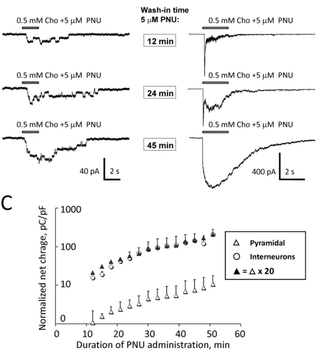
zer pipettes positioned in the vicinity of recorded CA1 pyramidal neurons or CA1 interneurons (Figure 1A–B). Choline was applied every 3 min during the wash-in phase of 5mM PNU-120596 added to ACSF. Net charge of responses of pyramidal neurons (Figure 3A) and interneurons (Figure 3B) was measured over a 20 s interval from the moment of choline application between the 12th and the 51st min of the PNU-120596 wash-in. If the quality of recordings declined during this 39 min time window (i.e., 14 individual applications), then, the data were discarded. Net charge from successful recordings was then normalized to (i.e., divided by) the neuronal capacitance to account for differences in neuronal size among tested neurons. The normalized net charge was then plotted as a function of time from the start of PNU-120596 wash-in (Figure 3C). The results of these experiments demonstrate that the density of expression of functional a7* nAChRs in CA1 pyramidal neurons comprises of,5% (i.e.,,1/20) of the density
of functionala7* nAChRs expressed in CA1 interneurons (n = 7; Figure 3C). This finding implies that the normalized net effects of activation (i.e., response net charge per unit of membrane capacitance per unit of time) ofa7* nAChRs expressed in CA1 pyramidal neurons is ,20-fold weaker than the net response of
activation of functionala7* nAChRs expressed in CA1 interneu-rons. In these experiments, ACSF contained 20mM gabazine,
15mM DNQX, 50mM AP-5, 40mM picrotoxin and 0.3mM TTX. The internal solution contained CsMeSO3(see Methods).
One limitation of this approach is that it does not fully account for potential differences in the distribution of functional a7* nAChRs in CA1 pyramidal neurons and CA1 interneurons. For instance, upon focal pressure application of 0.5 mM choline, dendritic a7* nAChRs (i.e., distal location relative to the application tip) will be exposed to a somewhat lower concentration of choline than somatic a7* nAChRs (i.e., proximal location relative to the application tip) because of the mostly somatic position of the application pipette tip (white arrows, Figure 1A–B). Therefore, in this approach, the contribution of dendritic a7* nAChRs to the net response may have been somewhat underestimated.
The effects of physiological levels of choline in the presence of PNU-120596
Administration of physiological concentrations of choline (i.e.,
,10mM) to ACSF is a more adequate model of the effects of endogenous choline and systemic drug administration than focal pressure application of 0.5–1 mM choline. Administration of 10mM choline alone to ACSF (n = 6; Figure 4A) or 2mM PNU-120596 alone to ACSF (n = 6; Figure 4C) did not elicit detectable responses in CA1 pyramidal neurons when the drugs were administered for over 1 hr. It is worth mentioning that in one experiment, two step-like current deviations (850 ms and 900 ms in duration) were detected within 1 min from one another after
,94 min of administration of 2mM PNU-120596 in the absence
of exogenous choline (not shown). These unusual openings were attributed to a potential presence in hippocampal slices of
extremely small amounts of endogenous a7* nAChR agonists (e.g., ACh and/or choline). This rare ion channel activity in the absence of exogenous choline was not investigated in this study.
By contrast, the addition of 2mM PNU-120596 to ACSF containing 10mM choline (Figure 4B), or the addition of 10mM choline to ACSF containing 2mM PNU-120596 (Figure 4D)
resulted in repetitive step-like current deviations in 13 out of 16 voltage-clamp whole-cell experiments (,81%; Table 1). Step-like
responses were reminiscent of openings of individual ion channels and were completely and reversibly blocked by 20 nM MLA supporting the involvement ofa7* nAChRs (n = 5; Figure 4E–F). The mean duration, the frequency and the mean net charge of step-like current deviations are presented in Table 1. To evaluate net charge, the area under step-like current deviations was measured over a 10 min interval at least 20 min after adminis-tration of 2mM PNU-120596 to ACSF. At the time of application
of PNU-120596, 10mM choline was already present in ACSF for
at least 20 min.
A similar protocol was used in 12 experiments where 5mM
PNU-120596 (instead of 2mM) was added to ACSF containing 10mM choline and step-like responses were observed in 10 out of
12 voltage-clamp whole-cell experiments (Table 1). In these experiments, the frequency of step-like current deviations and the mean net charge (but not the mean open time of current deviations) were significantly greater than the corresponding values measured in experiments with 2mM PNU-120596 (Table 1). These results demonstrate that, in the presence of PNU-120596, the enhanced responsiveness of pyramidal a7* nAChRs to choline results from a synergistic action of choline and PNU-120596 and that the potentiation of 10mM choline-mediated responses by PNU-120596 is concentration dependent. In these experiments, ACSF contained 20mM gabazine, 15mM
DNQX, 50mM AP-5, 10mM atropine, 40mM picrotoxin and 0.3mM TTX.
The current-voltage relationship. To build the
current-voltage relationship of step-like current deviations, 0.5 mM choline plus 5mM PNU-120596 were added to ACSF and step-like responses were recorded in voltage-clamp at various membrane potentials between290 mV and+30 mV with a step of 30 mV (n = 6; Figure 4G). The current-voltage relationship was inwardly rectified and lacked the outward current component (n = 6; Figure 4H), a typical feature of a7* nAChRs in the presence of external and internal Mg2+[59]. These results further supported the enhanced sustained activation of CA1 pyramidal a7* nAChRs by 10mM choline in the presence of PNU-120596.
The effects of 10mM choline plus 2–5mM PNU-120596 on the excitability of CA1 pyramidal neurons
In the absence of choline and PNU-120596, and in the presence of 20mM gabazine, 15mM DNQX, 50mM AP-5, 40mM picrotoxin and 10mM atropine, the mean resting potential of hippocampal
CA1 pyramidal neurons was estimated to be 260.062.2 mV (n = 64). Under these conditions, CA1 pyramidal neurons were not Figure 2. Improved detection ofa7* nAChR-mediated currents in hippocampal CA1 pyramidal neurons in the presence of
PNU-120596.In some patch-clamp experiments conducted in hippocampal slices, focal pressure administration of 0.5 mM choline to hippocampal CA1 pyramidal neurons did not elicit detectable whole-cell currents (A and D). Traces A and D were obtained from different CA1 pyramidal neurons. Trace D is the same as trace C in Figure 1. The same neurons began to respond to choline after addition of 1mM PNU-120596 to ACSF (B and E). These responses were completely and reversibly blocked by 20 nM MLA (F–G) supporting the expression of functionala7* nAChRs. C) To quantify these effects, in six experiments in which in the absence of PNU-120596 hippocampal CA1 pyramidal neurons did not respond to pressure-applied 0.5 mM choline, addition of 1mM PNU-120596 to ACSF resulted in robust responses. Current net charge over the first 10 s after the beginning of choline puff was measured for three experimental phases: (C,a) in the absence of PNU-120596; (C,b),24 min after addition of PNU-120596 to ACSF; and (C,c)
spontaneously active and did not exhibit step-like voltage deviations during current-clamp recordings (n = 4, Figure 5A). By contrast, upon administration of 10mM choline plus 2–5mM PNU-120596
for at least 20 min, step-like voltage (Figure 5B) or current (Figure 5C)
deviations were observed in current- or voltage-clamp experiments, respectively. Traces shown in Figures 5B and 5C were obtained from the same CA1 pyramidal neuron in current- and voltage-clamp configurations, respectively,,1 min apart.
Figure 3. Comparison of the levels of expression of native functionala7* nAChRs in CA1 pyramidal neurons and CA1 interneurons. The densities of expression of functionala7* nAChRs in CA1 pyramidal neurons and CA1 interneurons were compared in voltage-clamp whole-cell experiments in hippocampal slices. PNU-120596 (5mM) was added to ACSF to enhance the detectability ofa7* nAChR-mediated responses. In the presence of 5mM PNU-120596 in ACSF, 0.5 mM choline was pressure-applied to CA1 pyramidal neurons or CA1 interneurons via a picospritzer pipette every 3 min. Representative traces (A–B) illustrate responses that were obtained from CA1 pyramidal neurons (A) and CA1 interneurons (B) 12, 24 and 45 min (framed time marks) after addition of 5mM PNU-120596 to ACSF. Responses of CA1 pyramidal neurons (A) and interneurons (B) were analyzed within a time window between the 12thand the 51stmin after the start of wash-in phase of PNU-120596. Net charge of current responses from both types of neurons was measured over a 20 s interval from the moment of choline application. Net charge was then normalized to the neuronal capacitance to account for differences in the neuronal size and plotted as a function of time (C). The results are presented as the mean6
S.E.M. Open triangles illustrate the data points obtained from CA1 pyramidal neurons (n = 7); while open circles represent the data points obtained from CA1 interneurons (n = 7). Closed triangles illustrate values obtained by multiplying the pyramidal response net charge (i.e., open triangles) by a factor of 20.
doi:10.1371/journal.pone.0013964.g003
In current-clamp experiments, in the presence of 10mM choline plus 2mM PNU-120596 added to ACSF for at least 20 min, step-like voltage deviations triggered action potentials ina7 out of 24
(,29%) CA1 pyramidal neurons tested (Figure 5D). In the
remaining 17 cells, step-like voltage-deviations did not trigger action potentials (Figure 5B). Traces shown in Figures 5D and 5E Figure 4. Responses of hippocampal CA1 pyramidal neurons to physiological concentrations of choline in ACSF in the presence of 2mM PNU-120596.Physiological concentrations of choline alone (i.e., 10mM; A) or 2mM PNU-120596 alone (C) added to ACSF, did not elicit responses in CA1 pyramidal neurons in voltage-clamp whole cell experiments. By contrast, addition of 2mM PNU-120596 to ACSF (containing 10mM choline) for at least 20 min (B), or addition of 10mM choline to ACSF (containing 2mM PNU-120596) for at least 20 min (D) elicited repetitive step-like current deviations completely and reversibly inhibited by 20 nM MLA (E–F). The current-voltage relationship (n = 6) built for step-like current deviations was inwardly rectified and lacked the outward component – a typical property ofa7* nAChRs (G–H).
were obtained from the same CA1 pyramidal neuron in current-and voltage-clamp configurations, respectively,,1 min apart.
When 10mM choline plus 5mM PNU-120596 were added to
ACSF, action potentials were triggered by step-like voltage deviations in over twice as many CA1 pyramidal neurons: 12 out of 17 (,70.6%) cells tested (Figure 5F). The average number of
action potentials elicited by step-like voltage-deviations was estimated to be 6.866.0 APs/min (n = 7) in the presence of 10mM choline plus 2mM PNU-120596 and 24.5618.0 APs/min (n = 10) in the presence of 10mM choline plus 5mM PNU-120596 (Table 1) – a statistically significant difference (p,0.025). Traces shown in Figures 5F and 5G were obtained from the same CA1 pyramidal neuron in current- and voltage-clamp configurations, respectively,,1 min apart.
These results demonstrate that in the presence of PNU-120596 in ACSF, individual openings ofa7* nAChR ion channels appear to be detectable in whole-cell voltage- and current-clamp recordings as step-like current and voltage deviations, respectively. In current-clamp experiments, these putative single channel openings resulted in small repetitive step-like depolarizations that can occasionally excite hippocampal CA1 pyramidal neurons. Therefore, in the presence of PNU-120596, physiological concentrations of choline appear to enhance the excitability of CA1 pyramidal neurons and these effects are concentration-dependent.
Evaluation of the first latency time and wash-out rates of the effects of choline plus PNU-120596 applied to ACSF
The first latency time (i.e., time between the start of drug application to ACSF and the first step-like current deviation) was 6.861.6 min (n = 6) upon addition of 5mM PNU-120596 to
ACSF that contained 0.5 mM choline for at least 30 min. The first latency time was 3.160.2 min (n = 4) upon addition of 0.5 mM choline to ACSF that contained 5mM PNU-120596 for at least 30 min. Therefore, the onset of effects of 0.5 mM choline (in the continued presence of 5mM PNU-120596) was approximately 2.2-fold faster than the onset of effects of 5mM PNU-120596 (in the continued presence of 0.5 mM choline). The first latency times likely reflected the method of drug administration employed in this study (drugs were added to ACSF), the speed of ACSF perfusion (1 ml/min), the volume of the recording chamber (,2 ml), the
location of recorded neurons within the slice and the location of slices within the recording chamber. However, the 2.2-fold difference in the first latency times for wash-in of choline and PNU-120596 likely reflected differences in the rates of equilibra-tion of these drugs in hippocampal tissues and was determined by the drug lipophilicity, molecular size, diffusion coefficient, receptor binding properties and other biophysical properties.
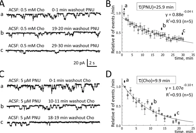
To determine the washout rates of choline and PNU-120596, 0.5 mM choline plus 5mM PNU-120596 were added to ACSF for
,1 hour to elicit repetitive step-like responses in voltage-clamp
experiments (Figures 6Aa and 6Ca). Choline or PNU-120596 was then removed from ACSF one at a time, while the other compound remained in ACSF. As the effects of choline plus PNU-120596 were synergistic (Figure 4A–D), a removal of either of the two components led to a reduction in the frequency of step-like current deviations (Figures 6Ab-c and 6Cb-c). The frequency of step-like responses was measured in events per minute and plotted as a function of time (Figures 6B and 6D). The data were fitted with single exponential functions and the decay time constants were determined. The washout time constant of 5mM PNU-120596 (in the continued presence of 0.5 mM choline in ACSF) was 25.9 min (n = 5; Figure 6B; an exponential fit (R2= 0.93): y = 0.88exp(20.04 t); whereas the washout time constant of 0.5 mM choline (in the continued presence of 5mM PNU-120596 in ACSF) was 9.9 min (n = 5; Figure 6D; an exponential fit (R2= 0.93): y = 1.07exp(20.10 t), where t is the washout time in minutes. Therefore, the washout of 0.5 mM choline from hippocampal tissue (in the continued presence of 5mM PNU-120596 in ACSF) was ,2.6-fold faster than the
washout of 5mM PNU-120596 (in the continuing presence of
0.5 mM choline in ACSF). Thus, the rates of both wash-in and washout of PNU-120596 appear to be considerably slower than the rates of wash-in and washout of choline, respectively. Again, these differences likely reflect differences in the rates of equilibration of these drugs within hippocampal tissue. In these experiments, ACSF always contained 20mM gabazine, 15mM DNQX, 50mM AP-5, 40mM picrotoxin and 0.3mM TTX.
Discussion
Deficits in hippocampal a7* nAChR activation are a key accompanying factor in certain cognitive disorders and enhancing this activation by nicotinic agonists has been shown to produce cognitive benefits. Choline is an endogenous selectivea7 nAChR agonist but present in the cerebrospinal fluid (CSF) at much lower concentrations (,10mM) relative to its EC50(i.e.,,0.5–1.5 mM;
[60,61]). Therefore, endogenous choline may not be effective in activation of native hippocampal CA1 pyramidala7* nAChRs in the absence of positive allosteric modulators, such as PNU-120596. In fact, this is true even for hypothalamic tuberomam-millary neurons that express much higher densities ofa7* nAChRs [43] than CA1 pyramidal neurons. This conclusion also complements previous reports demonstrating that choline exhibits a greater potency for desensitization (IC50 ,40mM, [56]) than
activation ofa7* nAChRs (EC50,0.5–1.5 mM; [60,61]).
PNU-120596 reduces desensitization ofa7 nAChRs and thus, increases
Table 1.Properties of currents elicited by 10mM choline plus 2mM or 5mM PNU-120596.
ACSF: #cells
Mean open time (s)
Frequency of step-like current deviations (events/min)
Net charge (pC/min)
Frequency of action potentials (APs/min)
10mM choline+2mM PNU-120596 13 1.160.7 1.8±1.7 9.3±9.6 6.8±6.0&(n = 7)
10mM choline+5mM PNU-120596 10 1.060.3 112.2±41.6* 53.4±23.6* 24.5±18.0*
The results of experiments where 10mM choline plus either 2mM (top row) or 5mM PNU-1205096 (bottom row) were applied to ACSF for at least 20 min and step-like current deviations in voltage-clamp or action potentials in current-clamp were recorded and analyzed. Significant differences were observed among the frequencies (p = 0.0001) and net charge (p = 0.0001) of step-like current deviations, but not the mean open time (p.0.67). Significant differences were also observed between the frequencies of action potentials (p,0.025).
&) Note that in the presence of 10
mM choline plus 2mM PNU-120596, action potentials were observed in only 7 out of 13 CA1 pyramidal neurons. doi:10.1371/journal.pone.0013964.t001
the potency ofa7 nAChR agonists including choline. Accordingly, this study tested and confirmed the hypothesis that in the presence of PNU-120596, physiological levels of choline (,10mM) become effective in activation of native functionala7* nAChRs expressed in hippocampal CA1 pyramidal neurons in relatively low densities. In the presence of PNU-120596, physiological concentrations of choline were found to elicit a weak persistent activation of CA1 pyramidal a7* nAChRs and enhance the excitability of CA1 pyramidal neurons – effects that inin vivo settings may produce neuroprotection [14,22,62,63] and cognitive benefits [11,64].
In the initial series of experiments, focal pressure application of 0.5–1 mM choline was used to demonstrate that: 1) The majority of hippocampal CA1 pyramidal neurons express functional a7* nAChRs; 2) A lack of responsiveness of some hippocampal CA1 pyramidal neurons to focal application of 0.5–1 mM choline in the absence of PNU-120596 does not imply a lack of expression of functionala7* nAChRs in these neurons, but may indicate a lack of detection of a7* nAChR-mediated currents by patch-clamp electrophysiology; and 3) PNU-120596 can serve as a powerful
tool for detection of low level expression and activation of functionala7* nAChRs such as those expressed in hippocampal CA1 pyramidal neurons.
To model the effects of endogenous choline on CA1 pyramidal a7* nAChRs and neurons, physiological concentrations of choline (i.e.,,10mM) were added to ACSF and whole-cell voltage- and
current-clamp recordings were conducted in the presence and absence of 2–5mM PNU-120596 (Figures 4–5). These
experi-ments revealed that in the presence of PNU-120596 and 10mM
choline, low densities of expression of CA1 pyramidal a7* nAChRs generate persistent step-like currents which cause transient depolarizations and occasionally, may trigger bursts of action potentials (Figure 5). It is this capability of as few as only one individual functionala7* nAChR to depolarize and excite the entire neuron that makes it possible for a low density expression of functional a7* nAChRs to be effective in enhancing the excitability of hippocampal CA1 pyramidal neurons in the presence of PNU-120596. Therefore, high levels of expression of CA1 pyramidala7* nAChRs and synchronization of their activity
Figure 6. Washout rates of choline and PNU-120596 in hippocampal slices.To evaluate the rates of washout of choline and PNU-120596 in hippocampal slices, 0.5 mM choline and 5mM PNU-120596 were added to ACSF for,1 hr and step-like current deviations were recorded as controls (Aa and Ca). One of the compounds (i.e., choline or PNU-120596) was then removed from ACSF while the other remained. When PNU-120596 was removed from the mix (Ab–Ac), the frequency of step-like current deviations (events/min) dropped exponentially with the time constant, 25.9 min (n = 5, A–B). When choline was removed from the mix (Cb–Cc), the frequency of step-like current deviations also dropped exponentially, but with the time constant, 9.9 min (n = 5, C–D), i.e.,,2.6 times faster.
doi:10.1371/journal.pone.0013964.g006
In some current-clamp experiments, step-like voltage deviations elicited by 10mM choline plus 2mM PNU-120596 triggered full action potentials (D). Action potentials shown in F) and in the framed insert to D) were truncated. Traces shown in D and E were obtained from the same cell,1 min apart. Similar results were obtained when 10mM choline plus 5mM (instead of 2mM) PNU-120596 were applied to ACSF (F–G and Table 1). Traces shown in F and G were recorded from the same cell,1 min apart. Short horizontal bars in front of voltage traces correspond to the membrane voltage of260 mV.
doi:10.1371/journal.pone.0013964.g005
are not required for significant depolarizing and excitatory effects of physiological concentrations of choline plus PNU-120596 on CA1 pyramidal neurons. This may be important because deficits in activation of hippocampala7* nAChRs have been linked to certain cognitive disorders and enhancing a7* nAChR activation by systemic administration of nicotinic agonists has been shown to produce neuroprotection and cognitive benefits [15,16,17,18,19, 21,28,29,30,57,65]. For example, a weak but persistent activation of a7* nAChRs can be achieved with low concentrations of nicotinic agonists (e.g., ,1–6mM 4OH-GTS-21 (i.e., 3-(4-hydroxy,
2-methoxy-benzylidene)anabaseine), a selective a7 nAChR agonist [56,66]) and has been shown to be neuroprotective in the NGF/ serum-withdrawal toxicity model in pheochromocytoma-12 cells expressing functionala7* nAChRs [22]. Therefore, the results of this study suggest that in the presence of PNU-120596, endogenous choline may produce a weak but persistent activation of functional a7* nAChRs in hippocampal CA1 pyramidal neurons and thus, may act as a potent therapeutic agent with potential neuroprotective and cognition-enhancing properties.
In the absence of PNU-120596, the mean open time of hippocampal a7* nAChRs is very short (,100ms, [54]) and
therefore, only a highly synchronized activity ofa7* nAChRs can be confidently detected in voltage-clamp whole-cell experiments. Indeed, in this study, fairly synchronous, although small, responses were occasionally observed when high concentrations of choline (0.5–1 mM) were focally applied via a picospritzer pipette to recorded CA1 pyramidal neurons (Figure 1D). A lack of substantial delay between pressure applications of choline and whole-cell responses of CA1 pyramidal neurons suggests that at least some pyramidala7* nAChRs were expressed somatically (Figures 1D–E and 2E). However, when considerably lower concentrations of choline (e.g., 10mM) were administered to ACSF in the absence of PNU-120596, responses of pyramidal a7* nAChRs were not detectable in whole-cell experiments (Figure 4A). By contrast, in the presence of PNU-120596, 10mM choline applied to ACSF elicited
clearly detectable responses (Figures 4B and 4D). These effects are directly attributable to the synergistic action of 10mM choline and 2mM PNU-120596 allowing sub-threshold concentrations of choline to activate a7* nAChRs [43]. The mean open time of current deviations elicited by 10mM choline in the presence of 2–5mM PNU-120596 was estimated to be ,1.0 s (Table 1).
Therefore, in the presence of PNU-120596, a weaka7* nAChR activity has a better chance of being detected in voltage- or current-clamp whole-cell experiments than in the absence of PNU-120596, and the synchronization ofa7* nAChR activity is not necessary for detection. Indeed, co-administration of 10mM choline and 2–5mM PNU-120596 elicited clearly detectable step-like whole-cell respons-es in voltage- and current-clamp experiments (Figurrespons-es 4 and 5). These responses were completely and reversibly blocked by 20 nM MLA supporting the activation ofa7* nAChRs (Figure 4E–F).
Intriguingly, current and voltage deviations recorded in voltage-and current-clamp, respectively, in the presence of 10mM choline plus 2–5mM PNU-120596 were step-like and thus, reminiscent of single ion channel openings similar to step-like responses observed previously in hypothalamic tuberomammillary neurons which in contrast to hippocampal CA1 pyramidal neurons express very high densities of functional a7* nAChRs [43]. Therefore, it is likely that step-like responses elicited by synergistic action of 10mM choline and 2–5mM PNU-120596 result from openings of
individual a7* nAChR ion channels detectable in whole-cell patch-clamp experiments.
Detecting activity of individual a7* nAChR ion channels in whole-cell patch-clamp experiments appears to be possible if the probability of ion channel openings is sufficiently low; and if the
channels remain open for a prolonged period of time during which the ionic gradient across the membrane and thus, the ionic current, remain relatively constant. These requirements appear to be fulfilled fora7* nAChRs activated by physiological concentra-tions of choline in the presence of 1–5mM PNU-120596 in
hippocampal CA1 pyramidal neurons (this study), hippocampal CA1 interneurons (Kalappa and Uteshev, unpublished observa-tions) and hypothalamic TM a7* nAChRs [43]. In fact, the probability of openings (Popen) ofa7* nAChRs expressed in TM
neurons has been estimated to be so low that in the presence of 10mM choline plus 1mM PNU-120596, only NTMPopen,0.27 a7* nAChR ion channels appeared to be open in the entire TM neuron at any given time [43], where NTMis the total number of
detectablea7* nAChRs in a TM neuron.
In this study, the mean amplitude of current deviations and the mean net charge per min generated by pyramidala7* nAChR ion channels in response to 10mM choline plus 2mM PNU-120596
were estimated to be ,5.5 pA (Figure 4H) and ,9.3 pC/
min = 0.16 pA (Table 1). Therefore, under these conditions, the mean number ofa7* nAChR ion channels opened in the entire hippocampal CA1 pyramidal neuron at any given time would be estimated to be NpyrPopen,0.029 ( = 0.16pA/5.5pA), where Npyris
the total number of detectable a7* nAChRs in a pyramidal neuron. Note that in experiments with TM neurons, 10mM choline plus 1mM PNU-120596 were used [43], whereas in this
study, the concentration of PNU-120596 was increased to 2mM
because of the substantially lower levels of expression of functional a7* nAChRs in hippocampal CA1 pyramidal neurons compared to TM neurons. However, the exact subunit compositions of functional a7* nAChRs expressed in CA1 pyramidal and TM neurons remain unknown and may be different.
Nativea7* nAChRs are highly permeable to Ca2+ions with the permeability ratio PCa/PNa ,6.0 [67,68]. This value translates
into the fractional Ca2+current, Pf(a7*),10%, at260 mV [68].
Therefore, the persistent activation of CA1 pyramidal a7* nAChRs in the presence of 10mM choline plus 2mM
PNU-120596 would be expected to result in a persistent influx of Ca2+ ions at a rate of,0.016 pA (i.e., 10% of 0.16 pA). Although small,
this persistent Ca2+current may be physiologically relevant and support neuroprotection [14,16,21]. Moreover, if injected in a small volume of a pre-synaptic terminal expressinga7* nAChRs, it may prime the terminal for plastic changes and thus, may assist in modulation of neurotransmitter release. However, a possibility that this relatively low, but persistent, rate of Ca2+ influx is cytotoxic cannot be presently ruled out.
One limitation of this study is that only relatively short-term effects (,3 h) were investigated. Prolonged exposure of CA1 pyramidala7* nAChRs to choline and PNU-120596 may cause neurotoxicity due to a decrease in receptor desensitization and the high Ca2+permeability ofa7* nAChRs. Another limitation is that all experiments in this study were conducted at room temperature and the results may be somewhat altered by physiological temperatures. One additional limitation comes from the fact that in this study pyramidal neurons were pharmacologically isolated and the majority of inhibitory (glycine/GABAA) and excitatory
(AMPA/NMDA) inputs were blocked by an inhibitory cocktail (see Methods). Finally, the number of step-like current deviations may have been underestimated because not all openings of individuala7* nAChR ion channels may have been detectable in whole-cell experiments. For example, responses of a7* nAChRs located in distal dendritic regions may not have been detected even in the presence of PNU-120596, due to electrotonic filtering. As a result, the estimated value of NpyrPopen may have been
Hippocampal CA1 pyramidal neurons directly contribute to generation of the hippocampal output and express two predom-inant types of pre- and post-synaptic functional nAChRs:a7- and a4b2-containing. The location and timely activation of these receptors by pressure-applied ACh have been shown to modulate and determine the sign of synaptic plasticity and the hippocampal output [38,69]. CA1 pyramidal neurons receive inhibitory GABAergic inputs from CA1 interneurons that express high densities of a7* nAChRs (Figure 3B–C; [33,35,70]). CA1 interneurons may directly inhibit CA1 pyramidal neurons via GABAergic synaptic inputs, or may excite CA1 pyramidal neurons by inhibiting other CA1 interneurons (i.e., via disinhibition) [69]. Although PNU-120596 would be expected to enhance activation of a7* nAChRs in pyramidal neurons and interneurons propor-tionally (Figure 3), the net effect of this activation remains unclear and is likely to be concentration-dependent. Moreover, in the presence of PNU-120596, the activation ofa7* nAChRs by ACh would be expected to be substantially enhanced, while the activation of non-a7* nAChRs should remain unchanged. Therefore, in the presence of PNU-120596, the net effect on the hippocampal output of activation of CA1a7* nAChRs will likely depend on the strength, timing and location of cholinergic terminals and the relative densities of expression of pre- and postsynaptica7* and non-a7* subtypes of nAChRs, as discussed previously [38,69]. In the present study, the net effect of activation of CA1 a7* nAChRs was not investigated, as the majority of excitatory and inhibitory inputs to pyramidal neurons were blocked by an inhibitory cocktail (see Methods) and the effects of activation of muscarinic AChRs were eliminated by using choline and atropine. However, future studies may use the presented in this study protocols to answer an important question as to how a concurrent activation of CA1 pyramidal neurons and CA1 interneurons by physiological concentrations of choline and 1–5mM PNU-120596 affects the excitability of pyramidal neurons and thus, the hippocampal output.
Although hippocampal CA1 pyramidal neurons express func-tional a7* nAChRs, it remains to be determined whether cholinergic terminals from the basal forebrain directly innervate these neurons by establishing functional cholinergica7* nAChR-containing synapses, or activate pyramidala7* nAChRs indirectly, i.e., via volume transmission. It is also unknown whether endogenously released ACh can reach pyramidal a7* nAChRs
at concentrations sufficient for generating physiologically signifi-cant effects in the absence or presence of PNU-120596. Nevertheless, the presented results support the hypothesis that despite low densities of expression of functionala7* nAChRs in hippocampal CA1 pyramidal neurons, in the presence of PNU-120596 endogenous choline may persistently activate these receptors and enhance the excitability of hippocampal CA1 pyramidal neurons in the absence of exogenous nicotinic agents and endogenously released ACh. Therefore, in the presence of PNU-120596, endogenous choline may serve as an efficacious therapeutic agent in cholinergic therapies aimed at recovering the deficiency in hippocampal a7* nAChR activation. Treatments involving endogenous choline may also be safer than those involving exogenousa7* nAChR agonists.
The expression of functionala7* nAChRs in brain tissues can be estimated by [125I] a-bungarotoxin binding, a selective a7* nAChR antagonist [71]. The results of [125I] a-bungarotoxin binding in the hippocampal CA1 pyramidal region demonstrated that the level of expression ofa7* nAChRs is age-dependent [71], peaks on postnatal days 2–5 (i.e., P2–5) and then declines to the adult level by P20. In the present study, the majority of experiments were conducted using P22–26 rats, therefore the expression of functional hippocampala7* nAChRs was near its young adult levels [71]. Interestingly, only,10% of hippocampal a7 proteins are surface-expressed [32] and therefore, the CA1 hippocampal region may contain a large pool of unused a7 proteins. It is intriguing to speculate that under certain endogenous conditions this pool of dormanta7 proteins can be recruited to become functional and cell surface-expressed. It is also feasible that certain endogenous compounds are capable of enhancing a7* nAChR function in a manner similar to PNU-120596. Finding these endogenous conditions and mechanisms of regulation ofa7* nAChR surface expression and function in the hippocampus and other brain regions may have a tremendously positive impact on the future of cholinergic therapies aimed at restoring and boosting cognitive performance.
Author Contributions
Conceived and designed the experiments: VVU. Performed the experi-ments: BIK AGG VVU. Analyzed the data: BIK AGG VVU. Contributed reagents/materials/analysis tools: VVU. Wrote the paper: VVU.
References
1. Kandel ER (2009) The biology of memory: a forty-year perspective. J Neurosci 29: 12748–12756.
2. Jiang Z, Belforte JE, Lu Y, Yabe Y, Pickel J, et al. (2010) eIF2alpha Phosphorylation-dependent translation in CA1 pyramidal cells impairs hippo-campal memory consolidation without affecting general translation. J Neurosci 30: 2582–2594.
3. Chen C, Tonegawa S (1997) Molecular genetic analysis of synaptic plasticity, activity-dependent neural development, learning, and memory in the mamma-lian brain. Annu Rev Neurosci 20: 157–184.
4. Tsien JZ, Huerta PT, Tonegawa S (1996) The essential role of hippocampal CA1 NMDA receptor-dependent synaptic plasticity in spatial memory. Cell 87: 1327–1338.
5. Kim HJ, Chae SC, Lee DK, Chromy B, Lee SC, et al. (2003) Selective neuronal degeneration induced by soluble oligomeric amyloid beta protein. FASEB J 17: 118–120.
6. Bobinski M, de Leon MJ, Tarnawski M, Wegiel J, Reisberg B, et al. (1998) Neuronal and volume loss in CA1 of the hippocampal formation uniquely predicts duration and severity of Alzheimer disease. Brain Res 805: 267–269. 7. Kirino T (1982) Delayed neuronal death in the gerbil hippocampus following
ischemia. Brain Res 239: 57–69.
8. Pulsinelli WA, Brierley JB, Plum F (1982) Temporal profile of neuronal damage in a model of transient forebrain ischemia. Ann Neurol 11: 491–498. 9. Shimohama S, Kihara T (2001) Nicotinic receptor-mediated protection against
beta-amyloid neurotoxicity. Biol Psychiatry 49: 233–239.
10. Disterhoft JF, Oh MM (2007) Alterations in intrinsic neuronal excitability during normal aging. Aging Cell 6: 327–336.
11. Kaczorowski CC, Disterhoft JF (2009) Memory deficits are associated with impaired ability to modulate neuronal excitability in middle-aged mice. Learn Mem 16: 362–366.
12. Son JH, Winzer-Serhan UH (2008) Expression of neuronal nicotinic acetylcholine receptor subunit mRNAs in rat hippocampal GABAergic interneurons. J Comp Neurol 511: 286–299.
13. Albuquerque EX, Pereira EF, Alkondon M, Rogers SW (2009) Mammalian nicotinic acetylcholine receptors: from structure to function. Physiol Rev 89: 73–120.
14. Egea J, Rosa AO, Sobrado M, Gandia L, Lopez MG, et al. (2007) Neuroprotection afforded by nicotine against oxygen and glucose deprivation in hippocampal slices is lost in alpha7 nicotinic receptor knockout mice. Neuroscience 145: 866–872.
15. Guseva MV, Hopkins DM, Scheff SW, Pauly JR (2008) Dietary choline supplementation improves behavioral, histological, and neurochemical outcomes in a rat model of traumatic brain injury. J Neurotrauma 25: 975–983. 16. Shimohama S, Greenwald DL, Shafron DH, Akaike A, Maeda T, et al. (1998)
Nicotinic a`7 receptors protect against glutamate neurotoxicity and neuronal ischemic damage. Brain Res 779: 359–363.
17. Verbois SL, Scheff SW, Pauly JR (2003) Chronic nicotine treatment attenuates alpha 7 nicotinic receptor deficits following traumatic brain injury. Neurophar-macology 44: 224–233.
18. Buccafusco JJ (2004) Neuronal Nicotinic Receptor Subtypes: DEFINING THERAPEUTIC TARGETS. MolInterv 4: 285–295.
19. Buccafusco JJ, Letchworth SR, Bencherif M, Lippiello PM (2005) Long-lasting cognitive improvement with nicotinic receptor agonists: mechanisms of pharmacokinetic-pharmacodynamic discordance. Trends Pharmacol Sci 26: 352–360.
20. Akaike A, Takada-Takatori Y, Kume T, Izumi Y (2010) Mechanisms of neuroprotective effects of nicotine and acetylcholinesterase inhibitors: role of alpha4 and alpha7 receptors in neuroprotection. J Mol Neurosci 40: 211–216. 21. Shimohama S (2009) Nicotinic receptor-mediated neuroprotection in
neurode-generative disease models. Biol Pharm Bull 32: 332–336.
22. Li Y, Papke RL, He YJ, Millard B, Meyer EM (1999) Characterization of the neuroprotective and toxic effects of a`7 nicotinic receptor activation in PC12 cells. Brain Res 81: 218–225.
23. Kem WR (2000) The brain alpha7 nicotinic receptor may be an important therapeutic target for the treatment of Alzheimer’s disease: studies with DMXBA (GTS-21). Behav Brain Res 113: 169–181.
24. Meyer EM, Tay ET, Papke RL, Meyers C, Huang G, et al. (1997) Effects of 3-[2,4-dimethoxybenzylidene]anabaseine (DMXB) on rat nicotinic receptors and memory-related behaviors. Brain Res 768: 49–56.
25. Buccafusco JJ, Terry AV, Jr., Decker MW, Gopalakrishnan M (2007) Profile of nicotinic acetylcholine receptor agonists ABT-594 and A-582941, with differential subtype selectivity, on delayed matching accuracy by young monkeys. Biochem Pharmacol 74: 1202–1211.
26. Kitagawa H, Takenouchi T, Azuma R, Wesnes KA, Kramer WG, et al. (2003) Safety, pharmacokinetics, and effects on cognitive function of multiple doses of GTS-21 in healthy, male volunteers. Neuropsychopharmacology 28: 542–551. 27. Ren K, Thinschmidt J, Liu J, Ai L, Papke RL, et al. (2007) alpha7 Nicotinic receptor gene delivery into mouse hippocampal neurons leads to functional receptor expression, improved spatial memory-related performance, and tau hyperphosphorylation. Neuroscience 145: 314–322.
28. Thomsen MS, Hansen HH, Timmerman DB, Mikkelsen JD (2010) Cognitive improvement by activation of alpha7 nicotinic acetylcholine receptors: from animal models to human pathophysiology. Curr Pharm Des 16: 323–343. 29. Leiser SC, Bowlby MR, Comery TA, Dunlop J (2009) A cog in cognition: how
the alpha 7 nicotinic acetylcholine receptor is geared towards improving cognitive deficits. Pharmacol Ther 122: 302–311.
30. Olincy A, Stevens KE (2007) Treating schizophrenia symptoms with an alpha7 nicotinic agonist, from mice to men. Biochem Pharmacol 74: 1192–1201. 31. Ross RG, Stevens KE, Proctor WR, Leonard S, Kisley MA, et al. (2010)
Research review: Cholinergic mechanisms, early brain development, and risk for schizophrenia. J Child Psychol Psychiatry 51: 535–549.
32. Mielke JG, Mealing GA (2009) Cellular distribution of the nicotinic acetylcholine receptor alpha7 subunit in rat hippocampus. Neurosci Res 65: 296–306.
33. Jones S, Yakel JL (1997) Functional nicotinic ACh receptors on interneurones in the rat hippocampus. J Physiol 504(Pt 3): 603–610.
34. McQuiston AR, Madison DV (1999) Nicotinic receptor activation excites distinct subtypes of interneurons in the rat hippocampus. J Neurosci 19: 2887–2896.
35. Frazier CJ, Rollins YD, Breese CR, Leonard S, Freedman R, et al. (1998) Acetylcholine activates an alpha-bungarotoxin-sensitive nicotinic current in rat hippocampal interneurons, but not pyramidal cells. J Neurosci 18: 1187–1195. 36. Frazier CJ, Buhler AV, Weiner JL, Dunwiddie TV (1998) Synaptic potentials mediated via alpha-bungarotoxin-sensitive nicotinic acetylcholine receptors in rat hippocampal interneurons. J Neurosci 18: 8228–8235.
37. Grybko M, Sharma G, Vijayaraghavan S (2010) Functional distribution of nicotinic receptors in CA3 region of the hippocampus. J Mol Neurosci 40: 114–120.
38. Ji D, Lape R, Dani JA (2001) Timing and location of nicotinic activity enhances or depresses hippocampal synaptic plasticity. Neuron 31: 131–141.
39. Faghih R, Gfesser GA, Gopalakrishnan M (2007) Advances in the discovery of novel positive allosteric modulators of the alpha7 nicotinic acetylcholine receptor. Recent Patents CNS Drug Discov 2: 99–106.
40. Hurst RS, Hajos M, Raggenbass M, Wall TM, Higdon NR, et al. (2005) A novel positive allosteric modulator of the alpha7 neuronal nicotinic acetylcholine receptor: in vitro and in vivo characterization. J Neurosci 25: 4396–4405. 41. Lopez-Hernandez GY, Thinschmidt JS, Morain P, Trocme-Thibierge C,
Kem WR, et al. (2009) Positive modulation of alpha7 nAChR responses in rat hippocampal interneurons to full agonists and the alpha7-selective partial agonists, 4OH-GTS-21 and S 24795. Neuropharmacology.
42. Roncarati R, Seredenina T, Jow B, Jow F, Papini S, et al. (2008) Functional properties of alpha7 nicotinic acetylcholine receptors co-expressed with RIC-3 in a stable recombinant CHO-K1 cell line. Assay Drug Dev Technol 6: 181–193. 43. Gusev AG, Uteshev VV (2009) Physiological concentrations of choline activate native {alpha}7-containing nicotinic acetylcholine receptors in the presence of PNU-120596. J Pharmacol Exp Ther.
44. Young GT, Zwart R, Walker AS, Sher E, Millar NS (2008) Potentiation of alpha7 nicotinic acetylcholine receptors via an allosteric transmembrane site. Proc Natl Acad Sci USA 105: 14686–14691.
45. Gronlien JH, Hakerud M, Ween H, Thorin-Hagene K, Briggs CA, et al. (2007) Distinct profiles of alpha7 nAChR positive allosteric modulation revealed by structurally diverse chemotypes. Mol Pharmacol 72: 715–724.
46. Jope RS, Gu X (1991) Seizures increase acetylcholine and choline concentra-tions in rat brain regions. Neurochem Res 16: 1219–1226.
47. Scremin OU, Jenden DJ (1991) Time-dependent changes in cerebral choline and acetylcholine induced by transient global ischemia in rats. Stroke 22: 643–647.
48. Bertrand N, Ishii H, Spatz M (1996) Cerebral ischemia in young and adult gerbils: effects on cholinergic metabolism. Neurochem Int 28: 293–297. 49. Grimm D, Kern A, Rittner K, Kleinschmidt JA (1998) Novel tools for
production and purification of recombinant adenoassociated virus vectors. Hum Gene Ther 9: 2745–2760.
50. Zapata A, Capdevila JL, Trullas R (1998) Region-specific and calcium-dependent increase in dialysate choline levels by NMDA. J Neurosci 18: 3597–3605.
51. Rao AM, Hatcher JF, Dempsey RJ (2000) Lipid alterations in transient forebrain ischemia: possible new mechanisms of CDP-choline neuroprotection. J Neurochem 75: 2528–2535.
52. Sarter M, Parikh V (2005) Choline transporters, cholinergic transmission and cognition. Nat Rev Neurosci 6: 48–56.
53. Parikh V, Sarter M (2006) Cortical choline transporter function measured in vivo using choline-sensitive microelectrodes: clearance of endogenous and exogenous choline and effects of removal of cholinergic terminals. J Neurochem 97: 488–503.
54. Mike A, Castro NG, Albuquerque EX (2000) Choline and acetylcholine have similar kinetic properties of activation and desensitization on the alpha7 nicotinic receptors in rat hippocampal neurons. Brain Res 882: 155–168.
55. Barron SC, McLaughlin JT, See JA, Richards VL, Rosenberg RL (2009) The allosteric modulator of {alpha}7 nicotinic receptors, PNU-120596, causes conformational changes in the extracellular ligand binding domain similar to acetylcholine. Mol Pharmacol.
56. Uteshev VV, Meyer EM, Papke RL (2003) Regulation of neuronal function by choline and 4OH-GTS-21 through alpha 7 nicotinic receptors. Journal of Neurophysiology 89: 1797–1806.
57. Meyer E, Kuryatov A, Gerzanich V, Lindstrom J, Papke RL (1998) Analysis of 40H-GTS-21 Selectivity and Activity at Human and Rat a`7 Nicotinic Receptors. J Pharmacol Exp Ther 287: 918–925.
58. Arendash GW, Sanberg PR, Sengstock GJ (1995) Nicotine enhances the learning and memory of aged rats. Pharmacol Biochem Behav 52: 517–523. 59. Alkondon M, Reinhardt S, Lobron C, Hermsen B, Maelicke A, et al. (1994)
Diversity of nicotinic acetylcholine receptors in rat hippocampal neurons. II. The rundown and inward rectification of agonist-elicited whole cell currents and identification of receptor subunits by in situ hybridization. J Phamacol Exp Ther 271: 494–506.
60. Alkondon M, Pereira EF, Cortes WS, Maelicke A, Albuquerque EX (1997) Choline is a selective agonist of alpha7 nicotinic acetylcholine receptors in the rat brain neurons. Eur J Neurosci 9: 2734–2742.
61. Papke RL, Porter Papke JK (2002) Comparative pharmacology of rat and human alpha7 nAChR conducted with net charge analysis. Br J Pharmacol 137: 49–61.
62. Kihara T, Shimohama S, Sawada H, Kimura J, Kume T, et al. (1997) Nicotinic receptor stimulation protects neurons against beta-amyloid toxicity. Ann Neurol 42: 159–163.
63. Akaike A, Tamura Y, Yokota T, Shimohama S, Kimura J (1994) Nicotine-induced protection of cultured cortical neurons against N-methyl-D-aspartate receptor-mediated glutamate cytotoxicity. Brain Res 644: 181–187.
64. Disterhoft JF, Oh MM (2006) Learning, aging and intrinsic neuronal plasticity. Trends Neurosci 29: 587–599.
65. Buccafusco JJ, Beach JW, Terry AV, Jr., Doad GS, Sood A, et al. (2004) Novel analogs of choline as potential neuroprotective agents. J Alzheimers Dis 6: S85–S92.
66. Uteshev VV, Meyer EM, Papke RL (2002) Activation and inhibition of native neuronal alpha-bungarotoxin-sensitive nicotinic ACh receptors. Brain Research 948: 33–46.
67. Castro NG, Albuquerque EX (1995) a`-Bungarotoxin-sensitive hippocampal nicotinic receptor channel has a high calcium permeability. Biophys J 68: 516–524.
68. Uteshev VV (2010) Evaluation of Ca2+permeability of nicotinic acetylcholine receptors in hypothalamic histaminergic neurons. Acta Biochim Biophys Sin (Shanghai) 42: 8–20.
69. Ji D, Dani JA (2000) Inhibition and disinhibition of pyramidal neurons by activation of nicotinic receptors on hippocampal interneurons. J Neurphysiol 83: 2682–2690.
70. Albuquerque EX, Alkondon M, Pereira EF, Castro NG, Schrattenholz A, et al. (1997) Properties of neuronal nicotinic acetylcholine receptors: pharmacological characterization and modulation of synaptic function. J Pharmacol Exp Ther 280: 1117–1136.