Contents lists available atScienceDirect
Applied Surface Science
j o u r n a l h o m e p a g e :w w w . e l s e v i e r . c o m / l o c a t e / a p s u s c
Synthesis of chitosan/hydroxyapatite membranes coated with hydroxycarbonate
apatite for guided tissue regeneration purposes
Alexandre Félix Fraga
a, Edson de Almeida Filho
c,∗, Eliana Cristina da Silva Rigo
b, Anselmo Ortega Boschi
aaFederal University of São Carlos, Department of Materials Engineering, DEMa, UFSCar, São Carlos, SP, Brazil bUniversity of São Paulo, Department of Basic Science – FZEA-ZAB, Pirassununga, SP, Brazil
cUniversity Estadual Paulista, Department of Physical Chemistry – IQ, Araraquara, SP, Brazil
a r t i c l e
i n f o
Article history:
Received 8 July 2010 Received in revised form 16 November 2010 Accepted 16 November 2010 Available online 9 December 2010
Keywords:
Biomimetic method Chitosan
Membranes and hydroxyapatite
a b s t r a c t
Chitosan, which is a non-toxic, biodegradable and biocompatible biopolymer, has been widely researched for several applications in the field of biomaterials. Calcium phosphate ceramics stand out among the so-called bioceramics for their absence of local or systemic toxicity, their non-response to foreign bodies or inflammations, and their apparent ability to bond to the host tissue. Hydroxyapatite (HA) is one of the most important bioceramics because it is the main component of the mineral phase of bone. The aim of this work was to produce chitosan membranes coated with hydroxyapatite using the modified biomimetic method. Membranes were synthesized from a solution containing 2% of chitosan in acetic acid (weight/volume) via the solvent evaporation method. Specimens were immersed in a sodium silicate solution and then in a 1.5 SBF (simulated body fluid) solution. The crystallinity of the HA formed over the membranes was correlated to the use of the nucleation agent (the sodium silicate solution itself). Coated membranes were characterized by means of scanning electron microscopy – SEM, X-ray diffraction – XRD, and Fourier transform infrared spectroscopy – FTIR. The results indicate a homogeneous coating covering the entire surface of the membrane and the production of a semi-crystalline hydroxyapatite layer similar to the mineral phase of human bone.
© 2010 Elsevier B.V. All rights reserved.
1. Introduction
Interest in the use of chitosan for medical and pharmaceutical applications has been growing, especially because of its remarkable properties such as biocompatibility, which makes it suitable for several medical applications[1,2].
The wide variety of shapes that chitosan can be delivered in, e.g., films, membranes, fibers, gel, paste, tablets, microspheres, as well as flakes, powders and solutions, enables it to be employed in several commercial, industrial, environmental and biomedical applications[3]. Until a few years ago, chitosan was used mainly to remove sediments and metallic ions from water, as well as in the food industry. Currently, chitosan is used in the production of cosmetics, medicines, food additives, and semi-permeable mem-branes, and in the development of biomaterials for medical and dental applications.
The development and use of several kinds of biomaterials in for tissue reconstruction procedures are growing. Calcium phosphate ceramics are used especially for this purpose due to their
biocom-∗Corresponding author.
E-mail address:edsonafilho@yahoo.com.br(E.d.A. Filho).
patibility and osteocompatibility and their structural, physical and chemical similarity to the bone mineral matrix[4].
The use of these bioceramics does not induce any undesirable immunological or toxic reaction. There is no risk of transmission of infectious or contagious pathologies or of protein degradation because of their characteristics and their high purity, which is a result of the rigid control of the manufacturing process according to the strictest standards[5].
Hydroxyapatite is noteworthy because it is the main constituent of the mineral phase of calcified tissues. HA is a ceramic calcium phosphate, or bioceramic, whose structure and composition are similar to the mineral phase of bones and teeth[6]. Synthetic HA is also biocompatible and osteointegrable, which makes it one of the most important substitutes for the human bone in implants and prostheses. In technological applications, HA is used as a coating for metallic implants and periodontal membranes[7].
Relatively recent studies consider the use of chitosan as a bio-material because of its compatibility with live organisms and also due to economic reasons, since it derives from chitin, which is very abundant in nature. In view of the increasing use of chitosan as a biomaterial, the production of an organic mineral containing chi-tosan and hydroxyapatite should be studied, particularly due to the possible interactions between these constituents. The aim of
Table 1
Ionic concentrations of the solutions used to coat apatites (mM)[9].
Na+ K+ Ca2+ Mg2+ HCO32− Cl− HPO42− SO42− SiO32−
Blood plasma 142.0 5.0 2.5 1.5 27.0 103.0 1.0 0.5 –
SBF 142.0 5.0 2.5 1.5 4.2 148.0 1.0 0.5 –
1.5 SBF 213.0 7.5 3.8 2.3 6.3 223.0 1.5 0.75 –
Na2SiO3 2.0 – – – – – – – 3.6
the present work is to study and produce chitosan membranes coated with hydroxyapatite by the modified biomimetic method for possible application as a biomaterial.
2. Materials and methods
All the reagents used here were of analytical grade. Commercial chitosan (Sigma) was extracted from crab, and contained at least 85% of deacetylated chitin.
2.1. Production of chitosan membranes
Chitosan membranes were produced from a solution contain-ing 2% of chitosan in acetic acid (weight/volume). This solution was stored for one week at 4◦C to allow for its complete solubilization
according to the method proposed by Beppu et al.[8]. The solution was vacuum filtered to eliminate air bubbles and to prevent the formation of macro-defects on the surface of the membranes after synthesizing. The membranes were prepared by pouring the chi-tosan solution into 2 cm×3 cm TeflonTMmolds with a controlled thickness of 3 mm. The molds containing the solution were then dried for 72 h at 40◦C, after which the membranes were immersed
in 1 mol/L NaOH solution for 24 h at 25◦C.
2.2. Coating of chitosan membranes with hydroxyapatite
In this work, we chose to use a modified version of the biomimetic method introduced by Abe et al.[5]. Rigo et al.[9]
substituted the treatment with G glass from the original method proposed by Abe and coworkers by immersion in a sodium silicate (SS) solution, which acts as a nucleation agent.[9,10]
The membranes were coated with and without treatment in SS, using the following routes: S1 – membranes were immersed in 1.5 SBF solution for 3 days at 37◦C and pH = 7.0; S2 – membranes were
immersed in the SS solution for 3 days at 37◦C and then in 1.5
SBF for 3 days at 37◦C and pH = 7.0, according to the procedure
described by Rigo et al.[9]; S3 – membranes were immersed in the SS solution for 3 days at 37◦C and then in 1.5 SBF for 7 days at 37◦C
and pH = 7.0.Table 1lists the ionic concentrations of blood plasma and all the solutions used in this process[10].
2.3. Scanning electron microscopy
A morphological analysis of the membranes was performed by scanning electron microscopy (SEM), using a Philips TMP micro-scope. Organic specimens were coated with gold to improve their electrical conductivity.
2.4. X-ray diffraction
The presence of crystalline phases in the coated membranes was analyzed by X-ray diffraction (XRD), using a Siemens D5005 diffrac-tometer with Cu (k␣1) radiation and angular scanning between 4◦ and 70◦.
2.5. Fourier transform infrared spectroscopy
The ionic groups were characterized by vibrational infrared spectroscopy, using a Nicolet Magna 550 FTIR spectrophotometer with DRIFT CollectorTMdiffuse reflectance.
3. Results and discussion
The kinetic of formation of apatites via the biomimetic process using SBF solution follows the sequence shown in Eq.(1) [11]. (ACP) Ca10−xH2x(PO4)6(OH)2→(OCP) Ca8H2(PO4)6·5H2O
→(HA)Ca10(PO4)6(OH)2 (1)
According to several authors, in the initial steps of apatite precipitation, low calcium content hydroxyapatite can become hydroxyapatite without transforming into octacalcium phosphate (OCP). Processing parameters such as pH (neutral or alkaline), tem-perature and solution concentration are very important for the crystallization of calcium phosphates until the HA phase is formed. Phenomena such as precipitation, solubilization, and slow and effective dehydration at a molecular level lead to the formation of a molecular arrangement that allows for the phase transformation of apatites[4].
Fig. 1 depicts the coatings produced in all the conditions employed in this study. The coating produced without the nucle-ation agent SS (condition S1) shows a dense homogeneous layer on the surface with an undefined morphology of ACP, which is one of the precursors of HA. The morphology of the HA coating pretreated with sodium silicate (condition S2) is dense and uniform, consist-ing of spherical particles of 1–5m, similar to those reported in the literature for HA coatings on metals[12,13]. The coating produced in condition S3 was dense. However, after 7 days of exposure to the 1.5 SBF solution, the coating showed surface cracks and some of its outer spherical particles were not bonded to inner particles. This indicates that the concentration at the surface of the coating became saturated when compared to the concentration of the 1.5 SBF solution[4]. After 4 days of exposure to the 1.5 SBF solution, the reaction at equilibrium changed from precipitation to dissolution of the HA layer.
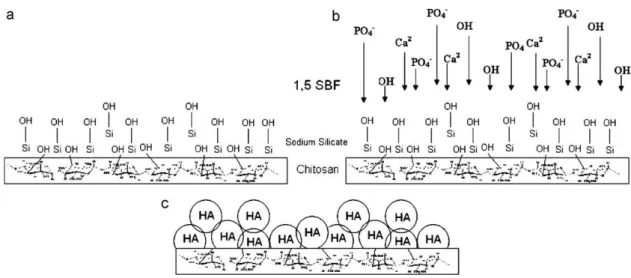
Fig. 2shows the proposed coating mechanism of CS membranes with HA by means of the biomimetic method. Carbon in position 6 – C6 in CS has a free hydroxyl group that can help activation of the membrane surface by silanol groups of the SS solution. The silicate ions in the SS solution are adsorbed on the CS substrate (Fig. 2a); HA nucleation of the adsorbed silicate ions begins (Fig. 2b); HA nucleons grow because of the saturated 1.5 SBF solution, leading to the coating of the substrate (Fig. 2c).
Hydroxyapatite Ca10(PO4)6(OH)2 has vibrational modes of phosphates and hydroxyls[14].Table 2shows the absorption fre-quencies for calcium phosphates.
The spectrum inFig. 3shows HA absorption bands at 3570 cm−1
and at about 630 cm−1. The band at 3570 cm−1 is related to OH−
Fig. 1.SEM characterization of chitosan/HA coated membranes. (S1) Membrane with no addition of SS and 3 days in 1.5 SBF; (S2) 3 days in SS and 3 days in 1.5 SBF; (S3) 3 days in SS and 3 days in 1.5 SBF.
Table 2
Characteristic calcium phosphates infrared absorption.
Absorption band Wavenumber (cm−1)
OH− 3572, 630
OH−(H
2O) 3000–3700, 1600–1650
PO43− 474, 562, 580, 640 e 960–1200
P–OH 527, 870 e 910–1040
The bands at 2880 cm−1 (C–H stretching), 1660 cm−1 (C–C
stretching) and 1390 cm−1(C–O stretching) are attributed to the
CS structure.
The band at 2330 cm−1is attributed to the presence of CO2in
the measuring chamber of the device in which the specimen was placed, and is part of the equipment’s background noise.
The bands at 1170 cm−1and 1130 cm−1 are attributed to P–O
stretching, while the bands close to 600 cm−1 are related to P–O
deformation vibrations of the PO43−group[15].
The bands between 850 and 1000 cm−1are attributed to P–OH
stretching. A characteristic band of HA is visible at around 630 cm−1,
which represents the OH end-group with lower steric impediment of the structure[15].
The XRD measurements were taken using the PDF #39-194, PDF #18-0303, and PDF #373-1731 standards for CS, ACP and HA, respectively[16]. A region of low crystallinity was visible between 15◦ and 25◦, corresponding to a diffraction halo of the CS
mem-brane.
The fact that the specimens showed low crystallinity is inter-esting since, according to Kanazawa[4], human bone consists of a mixture of calcium phosphates of low crystallinity. Discrete peaks
S3
S2 OH
OH
OH P-OH
P-OH
P-OH P-O P-O
P-O C-O
C-O
C-O C-C C-C C-C
CO
2
CO
2
CO2
C-H C-H C-H OH
OH
OH
Relative Intensity
4000 3500 3000 2500 2000
Wavenumber (cm-1)
1500 1000 500
S1
Fig. 3. FTIR spectra of chitosan/HA membranes. (S1) Membrane with no addition of SS and 3 days in 1.5 SBF; (S2) 3 days in SS and 3 days in 1.5 SBF; (S3) 3 days in SS and 3 days in 1.5 SBF.
of the ACP phase (S1) close to 30◦and 35◦are shown inFig. 4. The
membrane pretreated in SS solution (S2) and exposed to the 1.5 SBF solution for 3 days shows peaks of low crystallinity related to the HA phase which are very similar to the ones Vercik[17] iden-tified in metallic substrates. This is a carbonated hydroxyapatite very similar to the biological one, which is characterized by its low crystallinity.
A tendency for higher crystallization was observed in the S3 con-dition. This behavior was expected because of the longer exposure to the 1.5 SBF solution (7 days). During that time, solubilization and reprecipitation of HA occurred, as indicated by the SEM analysis. This led to the slow formation of nuclei and the subsequent growth of more oriented crystals, favoring the formation of HA phase.
Fig. 4compares the biological HA of human bone and the results obtained for the coating of S2 specimen. Back in 1926, De Jong[18]
was the first to observe the similarity between X-ray spectra of the mineral phase of bone and HA. However, the mineral phase of bone does not have a definite composition because it varies during the maturing and aging stages of hard tissues. Thus, the main difference between synthetic HA and bone apatite lies in their crystallinity[19]. The similarity between diffractograms of human bone and the one found experimentally (S2 condition) is shown in
Fig. 4.
4. Conclusions
The modified biomimetic method proposed here enabled us to work with the kinetics of the solution reactions, allowing for control of the degree of crystallinity of the HA layer.
The use of sodium silicate as a nucleation agent in the produc-tion of hydroxyapatite coating affected the phase formaproduc-tion of the apatites. After immersing the membranes in sodium silicate solu-tion, it was found that distinct calcium phosphate phases were formed by varying the exposure time to 1.5 SBF solution.
The longer the exposure time to the 1.5 SBF solution the more crystalline the coating and the lower its adhesion to the substrate.
The conditions employed in this study led to the formation of a low crystallinity hydroxyapatite layer similar to human bone.
Acknowledgments
The authors would like to thank CNPq and FAPESP (Brazil) for their financial support of this work.
References
[1] J. Berger, M. Reist, J.M. Mayer, O. Felt, N.A. Peppas, R. Gurny, Eur. J. Pharm. Biopharm. 57 (2004) 19.
[2] M.R. Finisie, A. Josue, V.T. Favere, M.C.M. Laranjeira, Anais da Academia Brasileira de Ciências 73 (2001) 525.
[3] M.N.V.R. Kumar, React. Funct. Polym. 46 (2000) 1.
[4] T. Kanazawa, Inorganic Phosphate materials, Elsevier, Tokyo, 1989, pp. 15–17. [5] Y. Abe, T. Kokubo, T. Yamamuro, J. Mater. Sci.: Mater. Med. 1 (1990) 536. [6] R.Z. Legeros, H.M. Myers, Monogr. Oral Sci. 15 (1991) 15.
[7] E.Y. Kawachi, Quim. Nova 23 (2000) 518.
[8] M.M. Beppu, E.J. Arruda, C.C. Santanam, Pol.: Ciên. e Tecn. (1999) 163. [9] E.C.S. Rigo, A.O. Boschi, M. Yoshimoto, Mater. Sci. Eng. C 24 (2004) 647. [10] J. Pierri, E.B. Roslindo, R. Tomasi, J. Non-Cryst. Solids 352 (2006) 5279. [11] H. Aoki, Science and Medical Applications of Hydroxyapatite, JAAS, Tokyo, 1991,
p. 230.
[12] H.M. Kim, F. Miyaji, T. Kokubo, C. Ohtsuki, T. Nakamura, J. Am. Ceram. Soc. 78 (1995) 2405.
[13] T. Kokubo, Acta Mater. 46 (1998) 2519. [14] H. Zeng, W.R. Lacefeld, Biomaterials 21 (2000) 23.
[15] J.C. Elliot, Structure and Chemistry of the Apatites and Other Calcium Orthophosphates, Elsevier, New York, 1984, p. 389.
[16] Joint Committee for Powder Diffraction Studies – Diffraction Data Base. Inter-national for Diffraction Data, Newton Square, 2003. 1 CD-ROM.