RESEARCH ARTICLE
Thy1.2 YFP-16 Transgenic Mouse Labels a
Subset of Large-Diameter Sensory Neurons
that Lack TRPV1 Expression
Thomas E. Taylor-Clark*, Kevin Y. Wu, Julie-Ann Thompson, Kiseok Yang, Parmvir K. Bahia, Joanne M. Ajmo
Department of Molecular Pharmacology and Physiology, Morsani College of Medicine, University of South Florida, Tampa, Florida, United States of America
*ttaylorc@health.usf.edu
Abstract
The Thy1.2 YFP-16 mouse expresses yellow fluorescent protein (YFP) in specific subsets of peripheral and central neurons. The original characterization of this model suggested that YFP was expressed in all sensory neurons, and this model has been subsequently used to study sensory nerve structure and function. Here, we have characterized the expression of YFP in the sensory ganglia (DRG, trigeminal and vagal) of the Thy1.2 YFP-16 mouse, using biochemical, functional and anatomical analyses. Despite previous reports, we found that YFP was only expressed in approximately half of DRG and trigeminal neurons and less than 10% of vagal neurons. YFP-expression was only found in medium and large-diameter neu-rons that expressed neurofilament but not TRPV1. YFP-expressing neuneu-rons failed to respond to selective agonists for TRPV1, P2X2/3and TRPM8 channels in Ca2+imaging assays.
Con-focal analysis of glabrous skin, hairy skin of the back and ear and skeletal muscle indicated that YFP was expressed in some peripheral terminals with structures consistent with their pre-sumed non-nociceptive nature. In summary, the Thy1.2 YFP-16 mouse expresses robust YFP expression in only a subset of sensory neurons. But this mouse model is not suitable for the study of nociceptive nerves or the function of such nerves in pain and neuropathies.
Introduction
Transgenic mice selectively expressing fluorescent proteins in neurons have been developed to study many aspects of neuronal structure and connectivity. These transgenic models have the advantage over other methods of visualization (e.g. immunohistochemistry of neuronal specific proteins), given that they do not require biochemical processing and the fluorescent proteins produce robust fluorescent signals. Furthermore, nerve subsets can be labeled with fluorescent proteins either randomly by random insertion of neuronal promoters [1], or specifically via Cre recombinase systems [2,3].
Sensory nerves are critical for the detection of external and internal environments in multi-cellular organisms. Peripheral terminals of sensory nerve detect stimuli and conduct this
a11111
OPEN ACCESS
Citation:Taylor-Clark TE, Wu KY, Thompson J-A, Yang K, Bahia PK, Ajmo JM (2015) Thy1.2 YFP-16 Transgenic Mouse Labels a Subset of Large-Diameter Sensory Neurons that Lack TRPV1 Expression. PLoS ONE 10(3): e0119538. doi:10.1371/journal.pone.0119538
Academic Editor:Michael Costigan, Boston Children’s Hospital and Harvard Medical School, UNITED STATES
Received:October 17, 2014
Accepted:January 21, 2015
Published:March 6, 2015
Copyright:© 2015 Taylor-Clark et al. This is an open access article distributed under the terms of the Creative Commons Attribution License, which permits unrestricted use, distribution, and reproduction in any medium, provided the original author and source are credited.
Data Availability Statement:All relevant data are within the paper.
Funding:Funding provided by National Heart, Lung & Blood Institute R01 HL119802. The funders had no role in study design, data collection and analysis, decision to publish, or preparation of the manuscript.
Competing Interests:The authors have declared
information to synapses within the CNS, in order to elicit reflexes, sensations, behaviors and emotions. To further understand these systems, a series of transgenic animals expressing fluo-rescent proteins in random subsets [1] has been used repeatedly to study sensory nerve termi-nal structure, sensory nerve connections in the spitermi-nal cord, sensory nerve development and axonal loss in neuropathies [4,5,6,7,8,9]. However, sensory nerves are heterogeneous with re-spect to protein expression and function and it is not clear the extent to which fluorescent pro-teins in these transgenic mice label select sensory subtypes involved in distinct reflex and behavioral pathways [1].
Sensory nerves are generally divided into 2 main subsets: nerves that respond to noxious sti-muli such as noxious heat and acid (often termed nociceptors) and nerves that respond to noxious stimuli such as light touch (often termed low-threshold mechanosensors or non-nociceptors) [10,11]. Nociceptors typically have small-diameter cell bodies [12]; unmyelinated or weakly myelinated axons; and almost exclusively express the canonical nociceptive ion channel TRPV1, which is selectively activated by capsaicin, the pungent ingredient of chili pep-pers [13]. Non-nociceptive neurons typically have large-diameter cell bodies, myelinated axons, express medium and heavy neurofilament but not TRPV1. Sensory neurons residing in the dorsal root ganglia (DRG) innervate the skin (below the neck), skeletal muscle and viscera in the thorax and abdomen. Trigeminal sensory neurons innervate the cranial skin and viscera and vagal neurons innervate the viscera in the thorax and abdomen.
Here, we have characterized the expression of yellow fluorescent protein (YFP) in sensory nerves in the Thy1.2 YFP-16 mouse [1], a commonly used transgenic model [6,7,8,9]. YFP expres-sion was restricted to large-diameter neurons that expressed neurofilament 200 (NF200) but did not express TRPV1 or respond to capsaicin (TRPV1), menthol (TRPM8) orα,βmethylene ATP (P2X2/3), suggesting that only non-nociceptive neurons were labeled. Numerous YFP expressing neurons were found in the DRG and trigeminal ganglia, but only a few were found in the vagal ganglia. YFP was expressed in terminals innervating hair follicle terminals in hairy skin, in termi-nals innervating Meissner corpuscles in glabrous skin and in termitermi-nals of skeletal muscle spindles.
Results
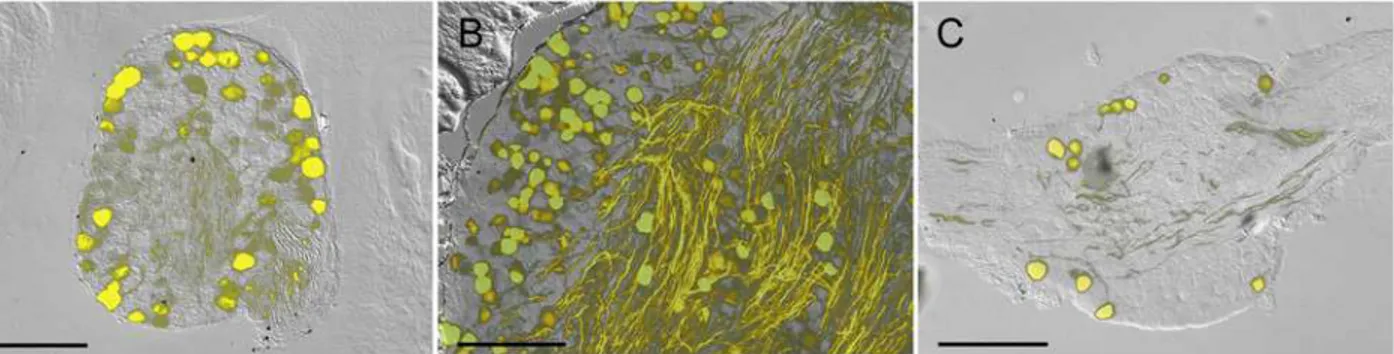
We began by determining the expression of YFP in sensory ganglia in the Thy1.2 YFP-16 mice. Serial frozen sections from DRG, trigeminal and vagal ganglia showed that YFP was present in neuronal cell bodies and axons but not in other cell types. Contrary to previous studies [1] we found that not all sensory neurons expressed YFP. Approximately half of DRG and trigeminal neurons and less than 10% of vagal neurons expressed YFP (Fig. 1). YFP-expressing axons were visible in all ganglia, but were particularly prevalent in the trigeminal. Some of these tri-geminal axons may be motor fibers [1], derived from cranial motor cell bodies that do not re-side in the trigeminal ganglia.
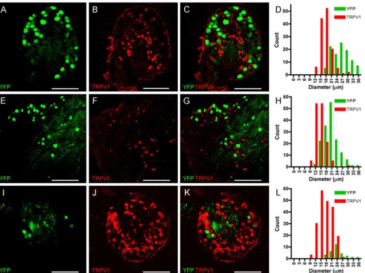
We hypothesized that the selective expression of YFP in subpopulations of sensory nerves was correlated to sensory nerve phenotype. In general, nociceptive nerves have small diameter cell bodies and respond to capsaicin (due to their selective expression of TRPV1), but do not express significant levels of heavy chain neurofilament (NF200). Non-nociceptive nerves have large diameter cell bodies, have significant expression of NF200 but do not express TRPV1. First, we performed confocal microscopy of sensory ganglia from Thy1.2 YFP-16 mice and in-vestigated the expression of TRPV1 using immunohistochemistry. As before YFP was express-ed in many DRG and trigeminal neurons, but only a few vagal neurons. Anti-TRPV1
antibodies labeled a completely distinct population of sensory neurons in all sensory ganglia (Fig. 2), which was absent in slides processed without primary antibody against TRPV1 (data not shown). We did not detect a single neuron that expressed both YFP and TRPV1 in the
sensory ganglia. In DRG we counted 105 YFP-expressing neurons and 130 TRPV1-expressing neurons (in 4 ganglia from 3 animals,Fig. 2A-D). Consistent with previously published corre-lations of nociceptive vs. non-nociceptive neuronal diameters, the YFP-expressing neurons were significantly larger than the TRPV1-expressing neurons (26.6 ± 0.47μm vs. 17.7 ± 0.27 μm, p<0.0001). In trigeminal ganglia we counted 156 YFP-expressing neurons and 140 TRPV1-expressing neurons (in 3 ganglia from 3 animals,Fig. 2E-H). Again, the YFP-express-ing neurons were significantly larger than the TRPV1-expressYFP-express-ing neurons (20.6 ± 0.31μm vs.
14.4 ± 0.24μm, p<0.0001). Finally, in vagal ganglia we counted 28 YFP-expressing neurons and 204 TRPV1-expressing neurons (in 4 ganglia from 3 animals,Fig. 2I-L). As before, the YFP-expressing neurons were significantly larger than the TRPV1-expressing neurons (24.0 ± 0.7μm vs. 17.5 ± 0.26μm, p<0.0001).
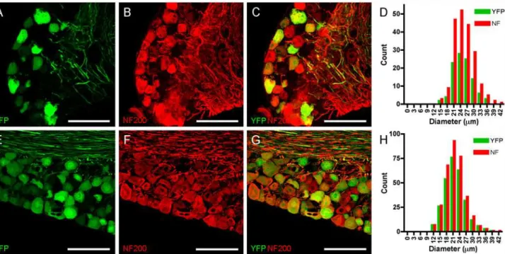
We also investigated the expression of the non-nociceptive marker NF200 in confocal mi-croscopy of DRG and trigeminal ganglia from Thy1.2 YFP-16 mice and compared it with YFP expression. Anti-NF200 antibodies labeled many large diameter sensory neurons in both sen-sory ganglia (Fig. 3), which was again absent in slides processed without primary antibody against NF200 (data not shown). Almost all YFP-expressing neurons were also positive for NF200. In DRG we counted 105 YFP-expressing neurons, and all but one of these neurons also expressed NF200 (in 7 ganglia from 3 animals,Fig. 3A-D). In total, we counted 202 NF200-expressing DRG neurons of which 51% expressed YFP. YFP- and NF200-NF200-expressing DRG neu-rons were not significantly different in diameter compared to the other NF200-expressing DRG neurons (25.3 ± 0.41μm vs. 25.3 ± 0.46μm, p>0.2). Similarly, in trigeminal ganglia we counted 280 YFP-expressing neurons, and all but 6 of these neurons also expressed NF200 (in 3 ganglia from 3 animals,Fig. 3E-H). In total, we counted 334 NF200-expressing trigeminal neurons of which 82% expressed YFP. YFP- and NF200-expressing DRG neurons were not sig-nificantly different in diameter compared to the other NF200-expressing DRG neurons (21.8 ± 0.28μm vs. 21.9 ± 0.46μm, p>0.2). In summary, the immunohistochemical analysis indicates that YFP expression is restricted to a subset of large-diameter NF200-expressing neurons that do not express TRPV1.
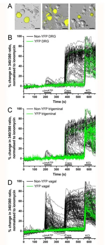
We next studied the correlation of YFP expression in sensory ganglia with functional re-sponses to sensory nerve stimuli. Using Fura 2AM Ca2+imaging in dissociated neurons we tested the sensitivity of these sensory neurons to the selective TRPV1 agonist capsaicin (1μM)
and the selective P2X2/3agonistα,βmethylene ATP (10μM). Both these channels are Ca2+
per-meable when activated. Capsaicin identifies nociceptive neurons [13], whereasα,βmethylene ATP identifies a subset of non-peptidergic sensory neurons [14,15] which, in the case of the vagal ganglia, are derived embryologically from the cranial placodes [16].
Fig 1. Fluorescence imaging of YFP in sensory ganglia of Thy1.2 YFP-16 mice.A, DRG.B, Trigeminal ganglion.C, vagal ganglion. YFP expression is shown in yellow overlaid upon bright field image. Scale bar represents 200μm.
doi:10.1371/journal.pone.0119538.g001
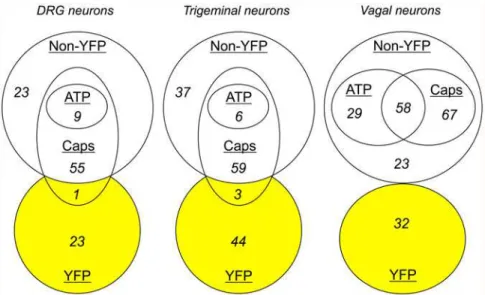
In experiments with dissociated DRG neurons (from 9 ganglia from 3 animals), we recorded from 24 YFP-expressing neurons and 87 non-fluorescent neurons (Figs.4Aand5). Only 1 out of 24 YFP-expressing neurons responded to capsaicin, and none responded toα,βmethylene ATP. The majority of non-fluorescent neurons responded to capsaicin (64 out of 87), with only 9 of these also responding toα,βmethylene ATP. Consistent with our confocal imaging of the DRG slices, the dissociated YFP-expressing DRG neurons were larger than both the entire non-fluorescent DRG population (34.4 ± 1.1μm vs. 23.9 ± 0.53μm, p<0.0001) and the sub-population of non-fluorescent DRG that failed to respond to either capsaicin orα,βmethylene ATP (vs. 25.5 ± 1.2μm, p<0.0001).
In experiments with dissociated trigeminal neurons (from 6 ganglia from 3 animals), we re-corded from 47 YFP-expressing neurons and 102 non-fluorescent neurons (Figs.4Band5). Only 3 out of 47 YFP-expressing neurons responded to capsaicin, and none responded toα,β methylene ATP. Again, the majority of non-fluorescent neurons responded to capsaicin (65 out of 102), with only 6 of these also responding toα,βmethylene ATP. The dissociated YFP-expressing trigeminal neurons were larger than both the entire non-fluorescent trigeminal population (25.4 ± 0.77μm vs. 19.1 ± 0.26μm, p<0.0001) and the subpopulation of
non-Fig 2. YFP and TRPV1 expression do not co-localize in sensory neurons.A-D, DRG.E-H, Trigeminal ganglion.I-L, vagal ganglion. YFP expression is shown in green (A,E,I), TRPV1 expression is detected immunohistochemically and shown in red (B,F,J), overlay showing distinct expression patterns (C, G,K). Scale bar represents 200μm.D,H and L, histogram of neuronal diameters.
doi:10.1371/journal.pone.0119538.g002
fluorescent trigeminal that failed to respond to either capsaicin orα,βmethylene ATP (vs. 19.5 ± 0.47μm, p<0.0001).
With vagal neurons (from 8 ganglia from 4 animals), we recorded responses to capsaicin andα,βmethylene ATP from 32 YFP-expressing neurons and 196 non-fluorescent neurons (Figs.4Cand5). None of the YFP-expressing neurons responded to either capsaicin orα,β methylene ATP. The majority of non-fluorescent neurons responded to capsaicin (125 out of 196). Neuronal activation byα,βmethylene ATP was far more prevalent in vagal non-fluores-cent neurons compared to DRG or trigeminal neurons with significant responses in 46% of capsaicin-sensitive neurons and 41% of capsaicin-insensitive neurons. The dissociated YFP-expressing vagal neurons were larger than both the entire non-fluorescent vagal population (28.2 ± 0.81μm vs. 20.7 ± 0.34μm, p<0.0001) and the subpopulation of non-fluorescent vagal that failed to respond to either capsaicin orα,βmethylene ATP (vs. 21.4 ± 0.60μm, p<0.0001). We also determined the sensitivity of vagal neurons to 100μM menthol, the selective agonist
for the cold-sensitive receptor TRPM8. Menthol activated 0 out of 33 YFP-expressing vagal neurons. However, menthol activated 57 out of 137 non-fluorescent vagal neurons, 48 of which were also capsaicin-sensitive (data not shown).
Lastly, we investigated the structure of peripheral YFP-expressing sensory nerve terminals. Given that YFP-expression was prevalent in DRG and trigeminal neurons, but not in vagal neurons, we concentrated on structures known to be innervated by these types of sensory re-ceptors: cutaneous terminals in glabrous skin of the paw and the hairy skin of the dorsal skin and muscle spindles of skeletal muscle (all innervated by DRG); and the hairy skin of the ear (innervated by the trigeminal). Confocal imaging of serial frozen sections of these tissues (from 6 animals) showed YFP expression in distinct nerve terminal structures (Figs.6–8).
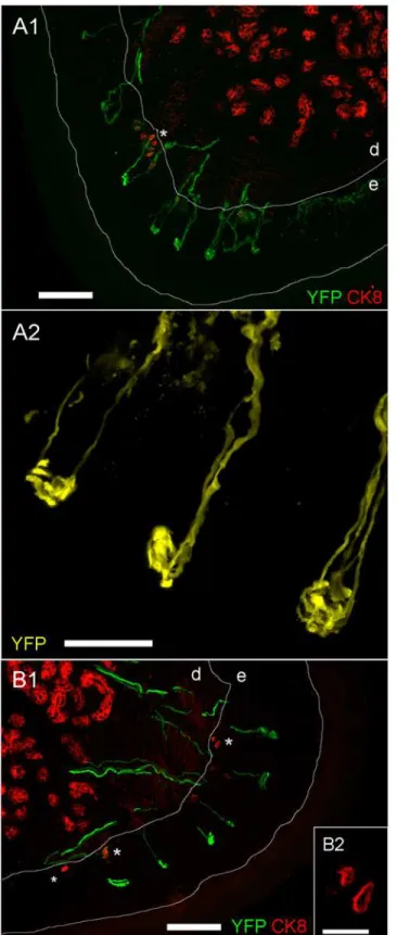
Numerous YFP-expressing terminals were found in glabrous skin of the hindpaw (Fig. 6A1,
6B1). The greatest density was found at the fingertips and on the central footpad dome, but
Fig 3. YFP co-localizes with NF200 in sensory neurons.A-D, DRG.E-H, Trigeminal ganglion. YFP expression is shown in green (A and E), NF200 expression is detected immunohistochemically and shown in red (B and F), overlay showing almost identical expression patterns (C and G). Scale bar represents 100μm.D and L, histogram of neuronal diameters.
doi:10.1371/journal.pone.0119538.g003
Fig 4. YFP-expressing sensory neurons are insensitive to capsaicin andα,βmethylene ATP.A, representative fluorescent images of YFP expression (yellow) in dissociated neurons overlaid upon bright field image: DRG (left), trigeminal (middle) and vagal (right). Scale bar represents 35μm.B-D, Ca2+
responses of individual dissociated DRG neurons (A), trigeminal ganglia neurons (B) and vagal ganglia neurons (C) in response toα,βmethylene ATP (10μM), capsaicin (Caps, 1μM,) and KCl (75 mM). Green lines for YFP neurons, black lines for non-YFP neurons. Blocked lines depict the duration of drug application.
doi:10.1371/journal.pone.0119538.g004
similar terminals were found across the full glabrous skin surface. The nerves uniformly termi-nated in endings in the middle of the epidermal layer, and these terminals were composed of complex corkscrew-like structures approximately 10 to 30μm in diameter (Fig. 6A2).
Cytoker-atin 8 (CK8) is a marker for Merkel cells [17,18]. As expected we found CK8-positive Merkel cells in small clusters at the basement layer of the epidermis. These small mechanotransductive cells (approximately 10μm in diameter) (Fig. 6B2) were not innervated by YFP-expressing
ter-minals. We also found significant CK8-positive staining deep in the dermis, which is consistent with the expression of CK8 in other cell types other than Merkel cells. These dermal cells were also not innervated by YFP-expressing terminals. We found identical YFP-expressing struc-tures in the glabrous skin of the forepaw (data not shown).
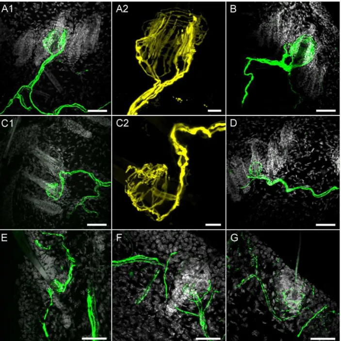
We found YFP-expressing terminals innervating the roots of some hair follicles in both dor-sal skin and ear skin. It was clear that the hair follicles were innervated by two distinct popula-tions of YFP-expressing nerves: terminals with longitudinal lanceolate endings (Fork-like structures;Fig. 7A, 7B and 7E) and terminals with circumferential lanceolate endings (Fig. 7C, 7D, 7F and 7G). In the dorsal skin we counted 1356 hairs (from 3 animals). Out of this total only 20 hairs (diameter 19.04 ± 0.84μm) were innervated by YFP-expressing longitudinal
lan-ceolate endings, and only 14 hairs (diameter 12.80 ± 0.78μm, p<0.01) were innervated by YFP-expressing circumferential lanceolate endings. The size and structure of these hairs indi-cate that YFP-expressing longitudinal lanceolate endings innervated tylotrich (guard) hairs and that non-tylotrich hairs (e.g. awl/auchene) were innervated by YFP-expressing circumfer-ential lanceolate endings. Both types of lanceolate endings had well-defined structures that completely encircled the hair follicle (Fig. 7A-D). These structures were approximately 25–55
μm in diameter, with longitudinal lanceolate endings being in general larger than
circumferen-tial lanceolate endings. Due to the rarity of YFP-expressing endings and the orientation and thickness of the sections it was not possible to determine if a single nerve innervated only a sin-gle hair, or a diffuse network of hairs.
In the ear skin, YFP-expressing terminal structures were less well-defined, particularly the circumferential lanceolate endings which often innervated only one side of the follicle (Fig. 7F and 7G). The diameter of the hair follicles innervated by YFP-expressing endings in the ear
Fig 5. Euler diagrams comparing sensory nerve populations with YFP expression.Populations ascribed by sensitivity toα,βmethylene ATP (10μM) and capsaicin (Caps, 1μM,).
doi:10.1371/journal.pone.0119538.g005
Fig 6. Confocal imaging of YFP expression in glabrous skin nerve terminals of the hind paw.A1 and B1, multiple YFP-expressing sensory terminals within the epidermis (e). CK8-positive Merkel cells (denoted by*) are located at the basal epidermal layer. Other CK8-positive cells are found within the dermis (d). YFP is shown in green and CK8 is shown in red, scale bar represents 100μm.A2, fine structure of YFP-expressing sensory terminals within the epidermis. YFP is shown in yellow, scale bar represents 25μm.B2, fine structure of CK8-positive Merkel cells, scale bar represents 20μm.
doi:10.1371/journal.pone.0119538.g006
skin was smaller than in dorsal skin (7.51 ± 0.79μm vs. 16.00 ± 0.76μm, p<0.001). Again, less than 3% of hairs were innervated by YFP-expressing endings.
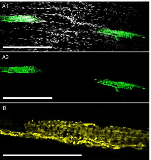
Lastly, we found YFP expression in muscle spindles throughout the tibialis anterior muscle (Fig. 8). These muscle spindles (approximately 150μm in length) were innervated by one or
more YFP-expressing axons. No other YFP-expressing sensory receptors were found in the skeletal muscle, including the tendons (data not shown).
Fig 7. Confocal imaging of YFP expression in hairy skin nerve terminals.A1,A2 and B, YFP-expressing longitudinal lanceolate endings innervating hair on dorsal skin.C1,C2 and D, YFP-expressing circumferential lanceolate endings innervating hair on dorsal skin.E, YFP-expressing longitudinal lanceolate ending innervating hair on ear skin.F and G, YFP-expressing circumferential lanceolate endings innervating hair on ear skin.A1,B,C1,D-G: YFP is shown in green and DAPI is shown in greyscale, scale bar represents 40μm.A2 and C2: YFP is shown in yellow, scale bar represents 10μm.
doi:10.1371/journal.pone.0119538.g007
Discussion
The Thy1.2 YFP-16 mouse model has been used in multiple studies of sensory nerve function [6,7,8,9]. The model was created by incorporation of YFP under the control of the neuronal Thy1.2 promoter [1]. Although the initial incorporation event was random, progeny from the mouse displayed consistent neuronal YFP expression. Originally this mouse model was re-ported to express YFP in all sensory nerves and somatic motor nerves but rarely in postgangli-onic autonomic nerves [1], thus making it a useful tool to study sensory nerves. Here, we have characterized the expression of YFP in sensory neurons from the DRG, trigeminal ganglia and vagal ganglia and we report that only a subset of these sensory nerves express YFP, based upon biochemical, functional and anatomical analysis.
YFP expression was observed in approximately 40–50% of DRG and trigeminal neurons based upon fluorescent and confocal imaging of frozen sections. In vagal ganglia, only a small number of neurons expressed YFP. Analysis of TRPV1 expression (nociceptor marker [13]), NF200 expression (non-nociceptive marker) and the size of the neuronal cell bodies [12] sug-gest that YFP was solely expressed in non-nociceptive sensory neurons. A significant
Fig 8. Confocal imaging of YFP expression in skeletal muscle spindles.A1, YFP-expressing muscle spindles in deep tissue of tibialis anterior skeletal muscle. YFP is shown in green and DAPI is shown in greyscale, scale bar represents 200μm.A2, asA1but without DAPI staining for clarity.B, fine structure of another YFP-expressing muscle spindle. YFP is shown in yellow, scale bar represents 100μm.
doi:10.1371/journal.pone.0119538.g008
population of large, NF200-positive sensory neurons failed to express YFP, suggesting that YFP was only expressed in a subset of non-nociceptive neurons.
Consistent with the fixed tissue studies, functional studies of dissociated sensory neurons suggest that YFP was indeed expressed in a subset of non-nociceptive neurons. Throughout all sensory ganglia we recorded from 135 YFP-expressing neurons and only 4 responded to capsa-icin (~3%), whereas 343 out of 522 non-fluorescent neurons were capsacapsa-icin-sensitive (~66%). Supporting this data, the YFP-expressing neurons were significantly larger than non-fluores-cent neurons. It is possible that the rare capsaicin-sensitivity of YFP-expressing neurons was due to culture-inducedde novoexpression of TRPV1 in neurons that would normally be TRPV1-negative in vivo. Such minor neuroplasticity has been shown previously to be sensitive to neurotrophins and culture conditions [19]. It should be noted that YFP expression may also be sensitive to culture conditions. Specific YFP expression in dissociated neurons and ganglion-ic slganglion-ices is diffganglion-icult to compare rigorously because only those neurons in the field of view are functionally assayed and selection of the field of view was biased by YFP expression (but not functional responses, which were reported for every neuron assayed).
We also studied the sensitivity of dissociated neurons toα,βmethylene ATP, the selective ag-onist of heteromeric P2X2/3channels. P2X2subunits are only expressed in a subset of non-peptidergic neurons [14,15]. P2X2subunits combine with P2X3in these neurons to form the het-eromeric P2X2/3channel. In the vagal ganglia, which comprises of two ganglia (nodose and jugu-lar) derived from different embryological sources (epibranchial placodes and neural crest, respectively), P2X2expression correlates with the nodose/placodal phenotype [16,20]. Given that many vagal neurons are nodose/placodal in origin, we were not surprised to find that a large number of vagal dissociated neurons (~44% in total) responded toα,βmethylene ATP, unlike the neural crest-derived DRG (~10%) and trigeminal (~6%). Despite the high percentage ofα,β methylene ATP-sensitivity in vagal capsaicin-insensitive neurons, YFP-expressing vagal neurons were insensitive to this P2X2/3agonist. This data suggests that YFP is only expressed in neural crest-derived non-nociceptive neurons and this may explain why YFP expression was only ob-served in a small number of vagal neurons (which is a combination of placodal and neural crest neurons) compared to DRG and trigeminal (which are neural crest in origin) (seeFig. 1).
Peripheral terminals of sensory nerves display different anatomical structures of various complexity and size that correlates with sensory function [11,21,22,23]. Non-nociceptive senso-ry nerves innervate structures throughout the body including the skin and skeletal muscle. In the skin, some sensory nerves serve as low-threshold mechanoreceptors (LTMRs) that convey mechanical information regarding brush, stretch, vibration, hair deflection and pressure. These diverse sensations are known to be triggered by activation of different classes of LTMRs includ-ing myelinated fast Aβfibers innervating Ruffini corpuscles, Pacinian corpuscles, Meissner cor-puscles, Merkel cells and thick Guard hair follicles; myelinated fast Aδfibers innervating lighter hair follicles; and unmyelinated slow C fibers innervating lighter hair follicles [11,23]. In skeletal muscle, myelinated sensory nerves innervate both the intrafusal fibers of muscle spindles (which senses muscle stretch) and the Golgi tendon organ (which senses muscle tension) [21].
In the glabrous skin of Thy1.2 YFP-16 mice, we found that only one type of ending expressed YFP: a characteristic and complex corkscrew-like ending innervating half-way through the epidermis, which were approximately 10–30μm in diameter. The location in the epidermis, size and shape of
these numerous YFP-expressing endings suggest that they are Meissner corpuscles [24,25]. CK8 (also known as Troma1) is a marker for Merkel cells [17,18] and, as expected, these were found individually or in small clusters throughout the basal layer of the epidermis of glabrous skin [24,26]. Merkel cells were not innervated by the YFP-expressing intraepidermal endings. In addition, we suggest that the YFP-expressing endings are not Ruffini corpuscles or Pacinian corpuscles, which are much larger and
are located in the dermis [24]. Sensory nerves innervating Meissner corpuscles in glabrous skin are thought to be rapidly-adapting LTMRs that sense dynamic skin deformation [11,23].
We failed to observe expressing corkscrew sensory endings in hairy skin of Thy1.2 YFP-16 mice, consistent with the fact that Meissner corpuscles are not found in hairy skin [11,23]. Nevertheless we observed two distinct subtypes of YFP-expressing endings innervating a small percentage of hair follicles in dorsal skin. Firstly, we found YFP-expressing longitudinal lanceo-late endings innervating the follicle of large guard hairs. These structures invariably surrounded the entire follicle and they are very similar in structure to longitudinal lanceolate endings as-cribed to Aβfibers innervating rapidly-adapting LTMR that sense hair displacement [7,18]. The second type of YFP-expressing terminal were circumferential lanceolate endings, which also surrounded the entirety of the follicle. The size of these particular follicles suggest that they are non-tylotrich hairs (likely to be awl/auchene hairs). Such endings have been described previously [7,27,28], but their function is presently unknown [11]. Comparisons between the YFP-express-ing endYFP-express-ings found in dorsal skin and ear skin suggest that the structure of trigeminal LTMR end-ings is more disorganized than DRG LTMR endend-ings. The reason for this is unknown, but the hairs innervated by YFP-expressing endings on the ear were much thinner than those found on the dorsal skin, consistent with the lack of thick guard hairs on the ear.
In skeletal muscle we found muscle spindles (and their innervating axons) expressed YFP. Muscle spindles are located in intrafusal fibers of skeletal muscle in parallel with the main contractile extrafusal muscle fibers. Muscle spindles detect skeletal muscle stretch and they respond in a rapidly adapting manner [21]. The location, shape and size of the YFP-expressing sensory structures found in the tibia-lis anterior muscle of Thy1.2 YFP-16 mice are identical to muscle spindle structures identified in other mice strains [21,29,30]. We failed to observe YFP-expressing afferents or structures in the tendons, thus we suggest that the Golgi tendon organ afferents [31] do not express YFP in this mouse model.
In our neuronal studies it was clear that some TRPV1-negative/capsaicin-insensitive/NF200-positive neurons did not express YFP. This is consistent with our nerve terminal studies that showed that some LTMR subtypes were not fluorescent (e.g. fibers innervating Ruffini corpus-cles, Pacinian corpuscorpus-cles, Merkel cells and Golgi tendon organs) and not many hair follicles were innervated by YFP-expressing endings. It is not clear why only a few LTMR subtypes express YFP, given that LTMR share the same characteristic of lack of TRPV1 and P2X2expression.
The original characterization of the expression of Thy1.2 YFP-16 mice was part of a massive study involving 25 individual Thy1.2 transgenic models [1]. Expression of fluorescent proteins in sensory neurons was reported for only the DRG. The authors reported“all”DRG neurons in the Thy1.2 YFP-16 mice were fluorescent, but no images were published for this data. Nevertheless, images of fluorescent protein expression in the DRG was reported for the YFP-G, GFP-I, CFP-4 and GFP-O models. Although these models had different levels of expression, it is clear from the images that large neurons were preferentially fluorescent. This is consistent with our data that YFP is expressed preferentially in TRPV1-negative/capsaicin-insensitive/NF200-positive neurons.
In summary, we have characterized the sensory nerve expression of YFP in the Thy1.2 YFP-16 mouse. Contrary to previous reports, we have found that only certain TRPV1-negative neu-rons express YFP. YFP expression was robust and allowed for detailed analysis of specific peripheral terminal structures. However, this mouse model is not suitable for the study of noci-ceptive nerves or the function of such nerves in pain and neuropathies.
Materials and Methods
Animals
All experiments were conducted with male 5–7 week old Thy1.2 YFP-16 mouse. Breeding pairs of the B6.Cg-Tg(Thy1-YFP)16Jrs/J strain were purchased from Jackson Laboratory (Maine) and
their progeny were used for all studies in accordance with and approved by the University of South Florida Institutional Animal Care and Use Committee. Prior to tissue harvest, mice were killed by CO2asphyxiation followed by exsanguination. DRG, trigeminal ganglia and vagal ganglia were immediately isolated prior to fixation with 4% paraformaldehyde (in phosphate-buffered sa-line (PBS), 2 hours, 4°C) or enzymatic dissociation (see below). In addition, the skin from the fore-paw and hindfore-paw, 5 mm square clips of mice ears, 8 mm square sections of dorsal skin and the tibialis anterior muscle were dissected and fixed in 4% paraformaldehyde (in PBS, 2 hours, 4°C).
Tissue processing
Following fixation, tissue was cryoprotected overnight in 18% sucrose (in PBS, 4°C) then immo-bilized in Tissue Freezing Medium (Triangle Biomedical Services, North Carolina). Ganglia were sectioned in 20μm slices, skin samples were sectioned in 40–80μm slices, skeletal muscle
were sectioned in 40μm slices. For immunohistochemical detection of specific proteins, sections
were blocked with donkey serum (10% in PBS with 1% bovine serum albumin (BSA), 0.5% Tween 20, 2 h) and incubated overnight (4°C) with appropriate primary. The primary antibody was omitted in 1 slide in 5 as a negative control. Sections were rinsed with PBS containing 0.3% Triton X and 1% BSA and incubated with appropriate secondary antibody for 2 hours at room temperature. TRPV1 was detected with a goat polyclonal antibody to TRPV1 (P-19 (sc-12498), Santa Cruz Biotechnology, Inc., Santa Cruz, CA; 1:150) and Alexa Fluor 594 rabbit anti-goat IgG (Molecular Probes, Inc., Eugene, OR, USA; 1:300). NF200 was detected with a rabbit poly-clonal antibody to NF200 (AB1989, EMD Millipore Corporation, Temecula, CA; 1:1000) and Cy5 goat anti-rabbit IgG (Molecular Probes, Inc., Eugene, OR, USA; 1:500). CK8 was detected with a rat monoclonal antibody to CK8 (also known as Troma1) (Developmental Studies Hy-bridoma Bank (University of Iowa); 1:100) and Alexa Fluor 594 rabbit anti-rat IgG (Molecular Probes, Inc., Eugene, OR, USA; 1:500). Ganglionic sections were coverslipped with VECTA-SHIELD HardSet Mounting Medium (Vector laboratories, CA, USA). Skin and skeletal muscle sections were coverslipped with VECTASHIELD HardSet Mounting Medium with DAPI.
Fluorescent YFP imaging
YFP expression was determined using 470 nm excitation (40 nm range) and 525 nm emission (50 nm range) and compared with bright field differential interference contrast measured by digital microscopy (CoolSnap HQ2; Photometrics, Surrey, BC, Canada) with a 10X objective and analyzed by Nikon Elements (Nikon, NY, USA).
Confocal imaging
Sections were viewed using an Olympus FV1000 laser scanning microscope and Olympus Fluoview 3.0 software with 10X, 20X and 40X objectives where appropriate. DAPI was excited by a 405 nm laser (emission 415–480 nm), YFP was excited by a 515 nm laser (emission 530–620 nm), Alexa Fluor 594 (TRPV1, CK8) was excited by a 543 nm laser (emission 620–720 nm) and Cy5 (NF200) was excited by a 633 nm laser (emission 650–750 nm). Z-stack images were created from individual sections ranging from 0.75 to 3μm in depth. Neuronal
diameters were measured in ImageJ.
Dissociation of mouse ganglia
Following isolation sensory ganglia were immediately enzymatically dissociated using previously described methods [32]. Isolated neurons were plated onto poly-D-lysine and laminin-coated coverslips, incubated at 37°C in L-15 (supplemented with 10% FBS) and used within 24 hours.
Fluorescent Ca2+ imaging
Neurons were studied for changes in [Ca2+]iwith Fura-2AM. Neuron-covered coverslips were incubated (37°C) with Fura-2AM (4μM, for 30 min) in L-15 media containing 10% FBS. For
imaging, the coverslip was placed in a custom-built heated chamber (bath volume of 300μL)
and superfused by gravity at 8 ml/min with HEPES-buffered solution (composition (mM)): 154 NaCl, 4.7 KCl, 1.2 MgCl2, 2.5 CaCl2, 10 HEPES, 5.6 dextrose adjusted to pH 7.4 with NaOH) for 10 minutes before and throughout each experiment. YFP expression was deter-mined using 470 nm excitation (40 nm range) and 525 nm emission (50 nm range) and com-pared with bright field differential interference contrast measured by digital microscopy (CoolSnap HQ2; Photometrics, Surrey, BC, Canada) and analyzed by Nikon Elements (Nikon, Melville, NY, USA). Fields of view were chosen to maximize the number of recorded YFP-expression neurons, thus this population is overrepresented in the dissociated neuron func-tional studies. Changes in [Ca2+]iwere monitored by sequential dual excitation, 340 and 380 nm (emission 510 nm). The ratio images were acquired every 6 seconds. At the end of each study neurons were exposed to KCl (75 mM, 60 sec) to confirm voltage sensitivity, and iono-mycin (5μM, 60 sec) to obtain a maximal response.
For the analysis of [Ca2+]i, we used the excitation ratio 340nm/380nm and related all mea-surements to the peak positive response in each cell. This approach bypasses the conversion of ratiometric responses into absolute [Ca2+]iusing Tsien parameters, thus we avoided the re-quirements for calibrating the Fura-2 Rmin, Rmax, Kdandβvalues for each recorded cell. Given that mammalian cells are heterogeneous with respect to their de-esterification of Fura-2AM following uptake, raw ratiometric responses should not be compared quantitatively between in-dividual cells. Thus, as before [33], we have chosen to normalize ratiometric responses at each time point in each cell to its maximum [Ca2+]i(evoked by the Ca2+ionophore ionomycin)— data was presented as the percentage change in 340nm/380nm ratio (R): response at time (x) = 100 X (Rx-Rbl)/(Rmax-Rbl), where Rxwas the 340nm/380nm ratio of the cell at a given time point, Rblwas the cell’s mean baseline 340nm/380nm ratio measured over 60s, and Rmaxwas the cell’s peak 340nm/380nm ratio. This normalization allows for quantitative comparisons be-tween cells. Only cells that had low [Ca2+]iat baseline (R<1.3) and yielded a robust response to the positive control were included in analyses.
A neuron was determined as responding positively to a given ligand if Rligand>Rbaseline+ (2 x SDbaseline), where Rligandis the mean ratio over the 60s of ligand challenge, Rbaselineis the mean ratio over the 60s preceding ligand challenge and SDbaselineis the standard deviation of the ratio over the 60s preceding ligand challenge.
Statistical analysis
Data was analyzed using Microsoft Excel and GraphPad software. Where appropriate unpaired Student’s T-test were used. A p value less than 0.05 was taken as significant. All values are mean ± SEM, unless otherwise stated.
Author Contributions
Conceived and designed the experiments: TTC. Performed the experiments: TTC KYW JAT KY PB. Analyzed the data: TTC KYW JMA. Wrote the paper: TTC.
References
1. Feng G, Mellor RH, Bernstein M, Keller-Peck C, Nguyen QT, et al. Imaging neuronal subsets in trans-genic mice expressing multiple spectral variants of GFP. Neuron. 2000; 28: 41–51. PMID:11086982
2. Mishra SK, Tisel SM, Orestes P, Bhangoo SK, Hoon MA. TRPV1-lineage neurons are required for ther-mal sensation. Embo J. 2011; 30: 582–593. doi:10.1038/emboj.2010.325PMID:21139565
3. Badea TC, Williams J, Smallwood P, Shi M, Motajo O, et al. Combinatorial expression of Brn3 transcrip-tion factors in somatosensory neurons: genetic and morphologic analysis. J Neurosci. 2012; 32: 995–1007. doi:10.1523/JNEUROSCI.4755-11.2012PMID:22262898
4. Belle MD, Pattison EF, Cheunsuang O, Stewart A, Kramer I, et al. Characterization of a thy1.2 GFP transgenic mouse reveals a tissue-specific organization of the spinal dorsal horn. Genesis. 2007; 45: 679–688. PMID:17987661
5. Porrero C, Rubio-Garrido P, Avendano C, Clasca F. Mapping of fluorescent protein-expressing neu-rons and axon pathways in adult and developing Thy1-eYFP-H transgenic mice. Brain Res. 2010; 1345: 59–72. doi:10.1016/j.brainres.2010.05.061PMID:20510892
6. Chen YS, Chung SS, Chung SK. Noninvasive monitoring of diabetes-induced cutaneous nerve fiber loss and hypoalgesia in thy1-YFP transgenic mice. Diabetes. 2005; 54: 3112–3118. PMID:16249433
7. Suzuki M, Ebara S, Koike T, Tonomura S, Kumamoto K. How many hair follicles are innervated by one afferent axon? A confocal microscopic analysis of palisade endings in the auricular skin of thy1-YFP transgenic mouse. Proc Jpn Acad Ser B Phys Biol Sci. 2012; 88: 583–595. PMID:23229751
8. Yan Y, Sun HH, Mackinnon SE, Johnson PJ. Evaluation of peripheral nerve regeneration via in vivo se-rial transcutaneous imaging using transgenic Thy1-YFP mice. Exp Neurol. 2011; 232: 7–14. doi:10. 1016/j.expneurol.2011.06.013PMID:21763310
9. Unezaki S, Yoshii S, Mabuchi T, Saito A, Ito S. Effects of neurotrophic factors on nerve regeneration monitored by in vivo imaging in thy1-YFP transgenic mice. J Neurosci Methods. 2009; 178: 308–315. doi:10.1016/j.jneumeth.2008.12.022PMID:19150631
10. Patapoutian A, Tate S, Woolf CJ. Transient receptor potential channels: targeting pain at the source. Nat Rev Drug Discov. 2009; 8: 55–68. doi:10.1038/nrd2757PMID:19116627
11. Abraira VE, Ginty DD. The sensory neurons of touch. Neuron. 2013; 79: 618–639. doi:10.1016/j. neuron.2013.07.051PMID:23972592
12. Lawson SN, Perry MJ, Prabhakar E, McCarthy PW. Primary sensory neurones: neurofilament, neuro-peptides, and conduction velocity. Brain Res Bull. 1993; 30: 239–243. PMID:7681350
13. Caterina MJ, Schumacher MA, Tominaga M, Rosen TA, Levine JD, et al. The capsaicin receptor: a heat-activated ion channel in the pain pathway. Nature. 1997; 389: 816–824. PMID:9349813
14. Burnstock G. P2X receptors in sensory neurones. Br J Anaesth. 2000; 84: 476–488. PMID:10823099
15. Dunn PM, Zhong Y, Burnstock G. P2X receptors in peripheral neurons. Prog Neurobiol. 2001; 65: 107–134. PMID:11403876
16. Nassenstein C, Taylor-Clark TE, Myers AC, Ru F, Nandigama R, et al. Phenotypic distinctions between neural crest and placodal derived vagal C-fibres in mouse lungs. J Physiol. 2010; 588: 4769–4783. doi:
10.1113/jphysiol.2010.195339PMID:20937710
17. Fradette J, Godbout MJ, Michel M, Germain L. Localization of Merkel cells at hairless and hairy human skin sites using keratin 18. Biochem Cell Biol. 1995; 73: 635–639. PMID:8714683
18. Li L, Rutlin M, Abraira VE, Cassidy C, Kus L, et al. The functional organization of cutaneous low-thresh-old mechanosensory neurons. Cell. 2011; 147: 1615–1627. doi:10.1016/j.cell.2011.11.027PMID:
22196735
19. Simonetti M, Fabbro A, D’Arco M, Zweyer M, Nistri A, et al. Comparison of P2X and TRPV1 receptors in ganglia or primary culture of trigeminal neurons and their modulation by NGF or serotonin. Mol Pain. 2006; 2: 11. PMID:16566843
20. Kwong K, Kollarik M, Nassenstein C, Ru F, Undem BJ. P2X2 Receptors Differentiate Placodal vs Neu-ral Crest C-fiber Phenotypes Innervating Guinea Pig Lungs and Esophagus. Am J Physiol Lung Cell Mol Physiol. 2008; 295: L858–865. doi:10.1152/ajplung.90360.2008PMID:18689601
21. Bewick GS, Banks RW. Mechanotransduction in the muscle spindle. Pflugers Arch. 2014.
22. Kollarik M, Carr MJ, Ru F, Ring CJ, Hart VJ, et al. Transgene expression and effective gene silencing in vagal afferent neurons in vivo using recombinant adeno-associated virus vectors. J Physiol. 2010; 588: 4303–4315. doi:10.1113/jphysiol.2010.192971PMID:20736420
23. Roudaut Y, Lonigro A, Coste B, Hao J, Delmas P, et al. Touch sense: functional organization and mo-lecular determinants of mechanosensitive receptors. Channels (Austin). 2012; 6: 234–245. doi:10. 4161/chan.22213PMID:23146937
24. Luo W, Enomoto H, Rice FL, Milbrandt J, Ginty DD. Molecular identification of rapidly adapting mecha-noreceptors and their developmental dependence on ret signaling. Neuron. 2009; 64: 841–856. doi:10. 1016/j.neuron.2009.11.003PMID:20064391
25. Honma Y, Kawano M, Kohsaka S, Ogawa M. Axonal projections of mechanoreceptive dorsal root gan-glion neurons depend on Ret. Development. 2010; 137: 2319–2328. doi:10.1242/dev.046995PMID:
20534675
26. Niu J, Vysochan A, Luo W. Dual innervation of neonatal Merkel cells in mouse touch domes. PLoS One. 2014; 9: e92027. doi:10.1371/journal.pone.0092027PMID:24637732
27. Millard CL, Woolf CJ. Sensory innervation of the hairs of the rat hindlimb: a light microscopic analysis. J Comp Neurol. 1988; 277: 183–194. PMID:3230157
28. Wu H, Williams J, Nathans J. Morphologic diversity of cutaneous sensory afferents revealed by geneti-cally directed sparse labeling. Elife. 2012; 1: e00181. doi:10.7554/eLife.00181PMID:23256042
29. Shenton F, Bewick GS, Banks RW. A study of the expression of small conductance calcium-activated potassium channels (SK1–3) in sensory endings of muscle spindles and lanceolate endings of hair folli-cles in the rat. PLoS One. 2014; 9: e107073. doi:10.1371/journal.pone.0107073PMID:25191752
30. Shneider NA, Brown MN, Smith CA, Pickel J, Alvarez FJ. Gamma motor neurons express distinct ge-netic markers at birth and require muscle spindle-derived GDNF for postnatal survival. Neural Dev. 2009; 4: 42. doi:10.1186/1749-8104-4-42PMID:19954518
31. Gregory JE, Proske U. The responses of Golgi tendon organs to stimulation of different combinations of motor units. J Physiol. 1979; 295: 251–262. PMID:521931
32. Taylor-Clark TE, Ghatta S, Bettner W, Undem BJ. Nitrooleic acid, an endogenous product of nitrative stress, activates nociceptive sensory nerves via the direct activation of TRPA1. Mol Pharmacol. 2009; 75: 820–829. doi:10.1124/mol.108.054445PMID:19171673
33. Nesuashvili L, Hadley SH, Bahia PK, Taylor-Clark TE. Sensory nerve terminal mitochondrial dysfunc-tion activates airway sensory nerves via transient receptor potential (TRP) channels. Mol Pharmacol. 2013; 83: 1007–1019. doi:10.1124/mol.112.084319PMID:23444014