Apo pto sis o f se nso ry ne uro ns and
sate llite ce lls afte r sciatic ne rve
transe ctio n in C5 7 BL/6 J m ice
Departamento de Anatomia, Instituto de Biologia, Universidade Estadual de Campinas, Campinas, SP, Brasil A.L.R. O liveira
Abstract
The rate of axonal regeneration, after sciatic nerve lesion in adult C57BL/6J mice, is reduced when compared to other isogenic strains. It was observed that such low regeneration was not due just to a delay, since neuronal death was observed. Two general mechanisms of cell death, apoptosis and necrosis, may be involved. By using the terminal deoxynucleotidyl transferase dUTP nick end labeling (TUNEL) tech-nique, we demonstrated that a large number of sensory neurons, as well as satellite cells found in the dorsal root ganglia, were intensely labeled, thus indicating that apoptotic mechanisms were involved in the death process. Although almost no labeled neurons or satellite cells were observed one week after transection, a more than ten-fold increase in TUNEL labeling was detected after two weeks. The results obtained with the C57BL/6J strain were compared with those of the A/J strain, which has a much higher peripheral nerve regeneration potential. In A/J mice, almost no labeling of sensory neurons or satellite cells was observed after one or two weeks, indicating the absence of neuronal loss. Our data confirm previous observations that approximately 40% of C57BL/6J sensory neurons die after sciatic nerve transection, and indicate that apoptotic events are involved. Also, our observations reinforce the hypothesis that the low rate of axonal regeneration occurring in C57BL/6J mice may be the result of a mismatch in the timing of the neurons need for neurotrophic sub-stances, and production of the latter by non-neuronal cells in the distal stump.
Co rre spo nde nce
A.L.R. O liveira
Departamento de Anatomia Instituto de Biologia, Unicamp Caixa Postal 6109
13083-970 Campinas, SP Brasil
Fax: + 55-19-3289-3124 E-mail: alroliv@ unicamp.br
Publication supported by FAPESP.
Received August 16, 2000 Accepted January 30, 2001
Ke y wo rds
·TUNEL
·Apoptosis
·Axotomy
·Neuronal death
·Satellite cells
·Isogenic mice
Subsequent to a peripheral lesion, a broad range of events is initiated, both in the proxi-mal and distal stump of the nerve (1). In the distal stump, a process called Wallerian de-generation starts as a local inflammation, being characterized by an extensive recruit-ment of macrophages which scavenge axon and myelin debris, stimulate Schwann cell proliferation and produce several signaling molecules such as cytokines (2-4). At the
dys-function and passive cell disassembly (7). The products of cytoplasmic leakage acti-vate the immune system, leading to macro-phage invasion and local inflammation. In contrast, apoptosis is an active process, ex-tensively identified under electron micros-copy by a series of morphological alterations (7). It is characterized by early chromatin condensation followed by internucleosomal DNA cleavage, cell shrinkage, reorganiza-tion of the cytoskeleton, organelle relocareorganiza-tion and production of apoptotic bodies. Apopto-sis also requires the transcription of certain genes, protease and endonuclease activation and the expression of phagocytic signals on the cell surface. The entire process ends as a silent and quick cell self destruction, without detectable inflammation (8-10).
It has been reported in the literature that C57BL/6J mice have a lower axonal regen-eration potential after a crush lesion when compared to other isogenic strains. Although Lu et al. (11) proposed that such a defect would be a delay rather than a permanent neuronal impairment, Lainetti et al. (12) ob-served a large percentage of sensitive neuron death four weeks after axotomy. It has been proposed that such neuronal death may be the result of a mismatch in the timing of the neuronal need for trophic substances and their production by the non-neuronal cells in the nerve (13). In this respect, apoptotic mechanisms may be involved, and may be related to the previously described loss of C57BL/6J dorsal root ganglion (DRG) neu-rons. Taking into account the facts reported above, the aim of the present study was to characterize neuronal loss in C57BL/6J mice using the terminal deoxynucleotidyl trans-ferase dUTP nick end labeling (TUNEL) method for the detection of apoptosis.
Six adult male mice of the C57BL/6J and A/J strains were used. The animals were anesthetized (0.25 g ketamine and 0.02 g xylazine dissolved in 5 ml of distilled water, 0.2 ml/25 g body weight, ip) and the left sciatic nerve was exposed and transected at
the midthigh level. The proximal stump was ligated with an 8-0 Ethicon stitch and a 2-mm segment of the distal stump was resected in order to avoid regeneration. After one and two weeks (N = 3 for each strain and survival time) the animals were sacrificed with an overdose of chloral hydrate (0.6 mg/kg, ip) and perfused transcardially with 150 ml of 4% paraformaldehyde in 0.1 M PBS. The L4, L5 and L6 spinal cord segments and L5 DRG were dissected out, left in the same fixative for 24 h, washed in PBS and pro-cessed for paraplast embedding. Transverse sections of the spinal cords (7 µm) and DRG were obtained, transferred to albumin-coated slides and stored until use. After deparaffini-zation, endogenous peroxidase was inacti-vated with 3% H2O2 in distilled water for 5
min at room temperature. The slides were transferred to a humidified chamber, the equilibration buffer solution was applied (Oncor, s7110-1) and the slides were incu-bated for 5 min at room temperature. The equilibration buffer was shaken off and the TdT enzyme solution (Oncor, s7110-2 and 3) applied for 60 min at 37o
C. The reaction was stopped by applying the stop/wash solu-tion (Oncor, s7110-4) for 30 min at 37oC.
Cybernet-ics, Baltimore, MD, USA). The total number of neurons and satellite cells of each section was obtained as the sum of the number counted in the six sample areas of each section.
For the spinal motoneurons, alternate sec-tions of the lumbar intumescence were used and processed as described above. The nu-merical data are reported as mean ± SD.
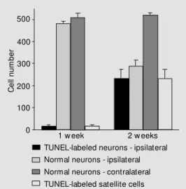
One week after sciatic nerve transection, almost no TUNEL labeling was detected in the DRG sections from A/J mice. Also, coun-terstaining revealed no signs of neuronal or satellite cell degeneration. The same pattern was observed two weeks after lesion (Figure 1). With respect to the spinal motoneurons found in the ventral horn of the lumbar spi-nal cord, no labeling was observed after one or two weeks.
Analysis of sections from C57BL/6J mice obtained one week after transection revealed the absence of neuronal labeling on the con-tralateral side. Also, a reduced number of TUNEL-positive neurons (18.7 ± 6.1) and satellite cells (16.7 ± 6.1) was found in the lesioned side. For the neurons, the number of labeled profiles represented 3% of the sampled cells. However, two weeks after lesion, a marked increase in TUNEL-labeled sensory neurons (237.3 ± 13.6) was observed on the ipsilateral side, representing approxi-mately 45% of the population studied. Such an increase in labeling occurred simulta-neously in the satellite cells (232.7 ± 41.7; Figures 1 and 2). No labeling was detected on the contralateral side and the number of normal neurons (508.0 ± 21.2) was similar to that found one week after lesion (509.1 ± 10.1).
Although a number of labeled cells showed the basic characteristics of apopto-sis, i.e., chromatin condensation, cell shrink-age and fragmentation, some of the TUNEL-positive neurons and satellite cells were mor-phologically normal. No TUNEL labeling was found in the motor nuclei of the lumbar intumescence from C57BL/6J mice after one
or two weeks.
Peripheral nerve transection in neonatal rats induces an extensive death of lesioned motoneurons in the spinal cord and primary afferent sensory neurons in the DRG (14,15). However, this kind of lesion in adult animals does not result in extensive neuronal death, although a series of alterations known as chromatolysis take place in the cell body (14). In fact, Ekström (16) reported that only a small proportion of the lesioned neurons became apoptotic after sciatic nerve crush in vivo. Also, Schwann cells at the site of injury became TUNEL labeled in the first 2 h after injury.
The survival rate of adult neurons after disconnection from the target is probably due to the relative independence of certain neurotrophic molecules produced by the non-neuronal cells and target organ. This also means that the neurons from adult individu-als have an intrinsic regenerative potential, which is triggered by their disconnection from the target. In this respect, after lesion, adult neurons are able to switch from a trans-mitting to a growth mode, producing cyto-skeleton proteins such as neurofilaments, CGRP and GAP-43 (17). Later, the timing between axonal elongation and the genera-tion of a supportive microenvironment dis-tally to the lesion seems to be essential for neuronal survival and the success of regen-eration (18).
factor and ciliary neurotrophic factor to as-sure cell survival during the critical period without contact with the distal stump and the target organ.
Despite the fact that, in general, sensory neuron death is unlikely after peripheral
dam-age in adult animals, it has been proposed as a possible explanation for the observations made in C57BL/6J mice, based on horserad-ish peroxidase retrograde labeling (12). The present data confirm this hypothesis and show that it is a characteristic of C57BL/6J
mice, since almost no DRG neuron death was identified after A/J sciatic nerve tran-section, as reported in the literature for NMRI mice (16). Also, our observations indicate that a major proportion of cell death is the result of apoptosis and that satellite cells are also affected. By using the TUNEL tech-nique, it was possible to identify cells with early chromatin fragmentation before any morphological sign of cell death could be noticed. Also, neuronal damage was detected prior to cell death, at least four weeks before the previous report (12), showing that neu-ronal and satellite cell death occurs around the second week post-lesion. Such timing is consistently different from that reported by Ekström (16) and reinforces the fact that adult C57BL/6J mice have a greater neu-ronal loss after peripheral lesion compared to other strains.
The low level of TUNEL labeling one week after axotomy may be explained by the fact that, soon after lesion, neurons are able to survive disconnected from the target. Con-versely, after this period there is an increase in the need to obtain a certain amount of trophic factors, such as nerve growth factor (NGF), synthesized by non-neuronal cells in the distal stump (19). If such substances are not available, neuronal death can be trig-gered, also resulting in the loss of satellite cells, as observed two weeks after axotomy. Interestingly, at this point, we found that approximately 45% of the DRG neurons were TUNEL positive, indicating that a large number of labeled neurons would probably die within a short period of time. This fact can be confirmed by an analysis of the re-sults after horseradish peroxidase tracing, where only about 60% of the neurons sur-vived (12).
Taken together, our results support the hypothesis that DRG neuron loss is the result
of a putative mismatch between the neuronal need for trophic factors and their production by the cells in the distal stump. Also, the possible lack of trophic support results in apoptotic neuronal death, which also includes the satellite cells in the DRG. This hypo-thesis is in agreement with a previous report which showed that if a predegenerated nerve was grafted to the proximal stump, C57BL/ 6J regenerative performance was greatly en-hanced (13). Also, if exogenous NGF is administered after sciatic nerve transection, sensory neuron death can be partially re-duced (20). Other studies are underway in order to identify possible apoptotic mechan-isms involved in C57BL/6J DRG cell loss after peripheral nerve lesion.
Ackno wle dgm e nts
The author is grateful to Prof. Glauce A. Pinto and Prof. José Vassallo (Laboratory of Experimental Pathology and Department of Anatomic Pathology, Unicamp) for their sup-port with the TUNEL technique, and also to Cristiano A. Chagas for excellent technical assistance.
C
e
ll
n
u
m
b
e
r
500
400
300
200
100
0
1 w eek 2 w eeks TUNEL-labeled neurons - ipsilateral Normal neurons - ipsilateral Normal neurons - contralateral TUNEL-labeled satellite cells
Re fe re nce s
1. Lunn ER, Brow n M C & Perry VH (1990). The pattern of axonal degeneration in the peripheral nervous system varies w ith dif-ferent types of injury. Journal of Neuro-science, 35: 157-165.
2. Bruck W, Bruck C, M arushak B & Fried RL (1995). M echanisms of macrophage re-cruitment in Wallerian degeneration. Acta Neuropathologica, 89: 363-367.
3. Hall SM (1993). Observations on the prog-ress of Wallerian degeneration in tran-sected peripheral nerves of C57BL/Wld mice in the presence of recruited macro-phages. Journal of Neurocytology, 22: 480-490.
4. Taskinen HS, Olsson T, Bucht A, Khademi M , Svelander L & Royotta M (2000). Pe-ripheral nerve injury induces endoneural expression of IFN-gamma, IL-10 and TNF-alpha mRNA. Journal of Neuroimmunolo-gy, 102: 17-25.
5. Peters A, Palay SL & Webster HF (1991).
The Fine Structure of the Nervous Sys-tem Neurons and their Supporting Cells. 3rdedn. Oxford University Press, New York.
6. Walker NI, Harmon BV, Gobe GC & Kerr JF (1988). Patterns of cell death. M ethods and Achievements in Experimental Pa-thology, 13: 18-54.
7. M cConkey DJ, Zhivotovsky B & Orrenius
S (1996). Apoptosis - molecular mechan-isms and biomedical implications. M olec-ular Aspects of M edicine, 17: 1-110. 8. Adams JM & Cory Z (1998). The BCL-2
protein family: arbiters of cell survival. Sci-ence, 281: 1322-1326.
9. Ashkenazi A & Dixt VM (1998). Death re-ceptors: signaling and modulation. Sci-ence, 281: 1305-1308.
10. Thornberry NA & Lazebnik Y (1998). Cas-pases: enemies w ithin. Science, 281: 1312-1316.
11. Lu X, Richardson PM , Gervais F & Skaeme E (1990). A deficiency of axonal regenera-tion in C57BL/6J mice. Brain Research, 510: 144-146.
12. Lainetti RD, Pereira FC & Da-Silva CF (1995). Reduced sensory neuron regen-eration by C57BL/6J mice. Brazilian Jour-nal of M edical and Biological Research, 28: 781-785.
13. Oliveira ALR & Langone F (2000). Non-neuronal cells are not the limiting factor for the low axonal regeneration in C57BL/ 6J mice. Brazilian Journal of M edical and Biological Research, 33: 1467-1475. 14. Low rie M B & Vbrová G (1992).
Depend-ence of postnatal motoneurons on their targets: review and hypothesis. Trends in Neurosciences, 15: 80-84.
15. W hit eside G, Doyle CA, Hunt SP &
M unglani R (1998). Dif f erent ial t im e course of neuronal and glial apoptosis in neonatal rat dorsal root ganglia after sci-atic nerve axotomy. European Journal of Neuroscience, 10: 3400-3408.
16. Ekström AR (1995). Neurones and glial cells of the mouse sciatic nerve undergo apoptosis after injury in vivo and in vitro.
NeuroReport, 6: 1029-1032.
17. Fu SY & Gordon T (1997). The cellular and molecular basis of peripheral nerve re-generation. M olecular Neurobiology, 14: 67-116.
18. Taira E, Takaha N & M iki N (1993). Extra-cellular matrix proteins w ith neurite pro-moting activity and their receptors. Neu-roscience Research, 17: 1-8.
19. Funakoshi H, Frisén J, Barabany G, Timmusk T, Zachrisson O, Verge VM K & Persson H (1993). Expression of mRNAs for neurotrophins and their receptors af-ter axotomy of the sciatic nerve. Journal of Cell Biology, 123: 455-464.